


Introduction
The Cyathocotylidae Mühling, 1896 is a globally distributed family of digeneans parasitic as adults in fishes, birds, reptiles, and mammals. After the revision by Achatz and Tkach (Reference Achatz and Tkach2025), it is 1 of 2 families belonging to the superfamily Diplostomoidea Poirier, 1886. Unlike the other family of diplostomoideans (Diplostomidae Poirier, 1886), cyathocotylids possess a cirrus sac. At present, the family includes 17 genera (Achatz and Tkach Reference Achatz and Tkach2025; Niewiadomska Reference Niewiadomska, Gibson, Jones and Bray2002; Young et al. Reference Young, Tkach, Bell, Ludovica, Gillett and Achatz2026). Recent molecular phylogenetic studies have led to re-evaluation of cyathocotylid systematics, predominantly by focusing on the use of partial sequences of the large ribosomal subunit (28S) rDNA gene. However, only 11 of the 17 cyathocotylid genera have been included so far in 28S-based phylogenetic analyses (see the phylogeny in Achatz et al. (Reference Achatz, Von Holten, Calhoun, Johnson, Pritchard, Keel and Tkach2025)).
Two of the remaining 6 genera, Prohemistomum Odhner, 1913 and Linstowiella Szidat, 1933, share closely intertwined taxonomic and systematic histories with Paracoenogonimus Katsurada, 1914; the latter has been included in recent phylogenetic analyses (Achatz et al. Reference Achatz, Von Holten, Calhoun, Johnson, Pritchard, Keel and Tkach2025; Sokolov et al. Reference Sokolov, Vlasenkov, Bugmyrin, Kalmykov and Lebedeva2024). Different authors routinely transferred species among the 3 genera (Dubois Reference Dubois1951, Reference Dubois1989; Mehra Reference Mehra1947; Sudarikov Reference Sudarikov and Skrjabin1961; Yamaguti Reference Yamaguti1971). Sudarikov (Reference Sudarikov and Skrjabin1961) and Dubois (Reference Dubois1989) considered Linstowiella to be a junior synonym of Paracoenogonimus. However, Yamaguti (Reference Yamaguti1971) and Niewiadomska (Reference Niewiadomska, Gibson, Jones and Bray2002) rejected the synonymisation. Achatz and Tkach (Reference Achatz and Tkach2025) did not comment on the history of these genera but included both Linstowiella and Paracoenogonimus in their key to species. None of these authors previously considered Linstowiella and Paracoenogonimus to be synonymous with Prohemistomum.
As part of a biodiversity survey of bird helminths in the USA, we collected a number of cyathocotylid digeneans from ospreys, Pandion haliaetus (L.). Here we provide descriptions of a new Prohemistomum species and Neogogatea kentuckiensis (Cable, 1935). The latter is included because the molecular phylogenetic work by Achatz et al. (Reference Achatz, Von Holten, Calhoun, Johnson, Pritchard, Keel and Tkach2025) provided the background on Neogogatea taxonomic history but did not contain a description. Based on morphological and molecular evidence, we reassess the validity of Linstowiella and Paracoenogonimus.
Materials and methods
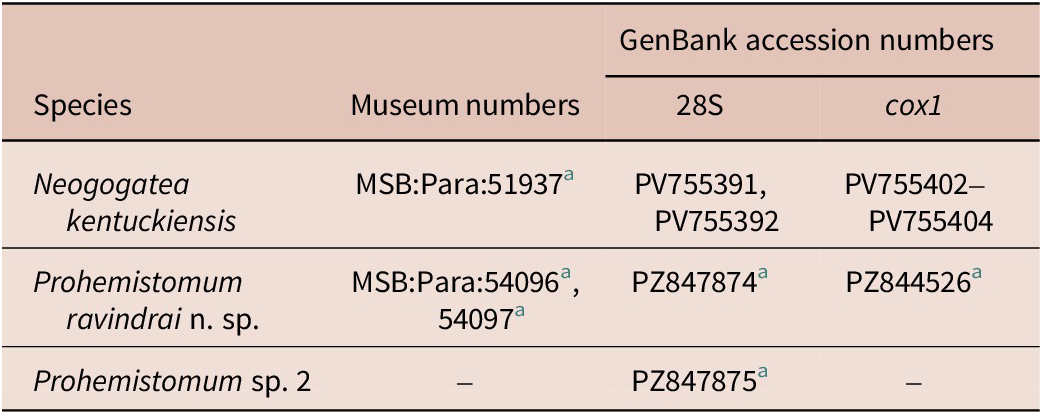
Cyathocotylid specimens were obtained from ospreys collected from Lake Seminole, Silver Lake Wildlife Management Area, Decatur County, Georgia (30°48′55.6″N 84°45′28.9″W) during July 2025 and a juvenile male osprey from retention ponds in Orange County, Florida (28°33′51.012″N 81°35′8.159″W) during June 2022. Live digeneans were rinsed in saline, killed in hot water, and immediately fixed in 80% ethanol; dead digeneans were immediately placed in 80% ethanol. Specimens studied using light microscopy were stained with aqueous alum carmine according to the protocol of Lutz et al. (Reference Lutz, Tkach, Weckstein and Webster2017). Slides were examined and illustrated using Olympus BX53 compound microscope (Tokyo, Japan) equipped with differential interference contrast optics, a digital imaging system and drawing tube. All measurements are provided in micrometres. The type series and voucher specimens are deposited in the Museum of Southwestern Biology Division of Parasites (MSB:Para), University of New Mexico, Albuquerque, New Mexico (Table 1). In addition, the morphology of N. kentuckiensis specimens sequenced by Achatz et al. (Reference Achatz, Von Holten, Calhoun, Johnson, Pritchard, Keel and Tkach2025) (MSB:Para:51937) was re-examined.
GenBank and Museum of Southwestern Biology Division of Parasites (MSB:Para) accession numbers of cyathocotylids described from Pandion haliaetus in the present study

Table 1. Long description
The table consists of four columns and four rows including the header.
Column 1: Species.
Column 2: Museum numbers.
Column 3: GenBank accession numbers for 28 S.
Column 4: GenBank accession numbers for c o x 1.
Row 1: Neogogatea kentuckiensis. Museum number M S B Para 51937 super a. 28 S numbers P V 755391 and P V 755392. c o x 1 numbers P V 755402 through P V 755404.
Row 2: Prohemistomum ravindrai n. sp. Museum numbers M S B Para 54096 super a and 54097 super a. 28 S number P Z 847874 super a. c o x 1 number P Z 844526 super a.
Row 3: Prohemistomum sp. 2. Museum numbers are not listed. 28 S number P Z 847875 super a. c o x 1 numbers are not listed.
Footnote a indicates new sequences or slides.
a New sequences or slides.
Genomic DNA was extracted from entire specimens according to the protocol provided by Tkach and Pawlowski (Reference Tkach and Pawlowski1999). The fragments of 28S rDNA and cytochrome c oxidase subunit I (cox1) mtDNA genes were amplified by polymerase chain reaction (PCR). The PCR amplifications, purification, and sequencing were performed as stated in Achatz et al. (Reference Achatz, Von Holten, Calhoun, Johnson, Pritchard, Keel and Tkach2025). Contiguous sequences were assembled using Sequencher 4.2 software (GeneCodes Corp., Michigan, USA) and deposited in GenBank (Table 1).
New 28S sequences of Prohemistomum spp. were aligned with 30 previously published sequences of cyathocotylids using ClustalW as implemented in MEGA7 software (Kumar et al. Reference Kumar, Stecher and Tamura2016). The alignment was trimmed to the length of the shortest sequence included in the analysis (1,086 bp); 39 nucleotide positions with ambiguous homology were excluded from the analyses. Harmotrema laticaudae Yamaguti, 1933 was selected as the outgroup in the analysis, based on the tree topology published by Pérez-Ponce de León and Hernández-Mena (Reference Pérez-Ponce de León and Hernández-Mena2019). The general time-reversible model with estimates of invariant sites and gamma-distributed among-site variation (GTR + G + I) was determined to be the best-fitting nucleotide substitution model using MEGA7. The phylogenetic analysis was conducted using Bayesian inference as implemented in MrBayes v3.2.6 software (Ronquist and Huelsenbeck Reference Ronquist and Huelsenbeck2003) with Markov chain Monte Carlo (MCMC) chains run for 3,000,000 generations with a sampling frequency set at 1,000. The number of generations was considered sufficient because the standard deviation stabilised below 0.01. Log-likelihood scores were plotted and only the final 75% of trees were retained to produce the consensus trees.
Results
Molecular phylogeny
The 28S alignment for the analysis was 1,086 bp long; 39 bases were excluded from the analysis due to ambiguous homology. The topology and branch support values in the phylogeny were generally consistent with those of Achatz et al. (Reference Achatz, Von Holten, Calhoun, Johnson, Pritchard, Keel and Tkach2025). Therefore, we only focus on the 100% supported Prohemistomum clade (Fig. 1). This clade positioned Prohemistomum sp. 1 from a hooded merganser, Lophodytes cucullatus (L.) (=Neogogatea sp. of Achatz et al. Reference Achatz, Pulis, Junker, Binh, Snyder and Tkach2019; characterised by the lack of ventral sucker) as a sister group to an 86% supported subclade comprising all remaining congeners. The subclade included a 4-branch polytomy consisting of Prohemistomum sp. 3 (cercaria), Prohemistomum sp. 4 (cercaria), Prohemistomum ovatus n. comb. (Katsurada, 1914) represented by sequences from cercaria (PX600339) and an adult digenean (PP093049) from a western marsh harrier, Circus aeruginosus (L.), and a 100% supported subclade uniting the new species with Prohemistomum sp. 2 (both from ospreys and lacking a ventral sucker).
Phylogenetic interrelationships among cyathocotylids based on Bayesian inference analysis of partial 28S rDNA gene sequences. Posterior probabilities are provided above internodes; values below 80% are not shown. The new 28S sequences generated in this study are in bold. The scale bar indicates the number of substitutions per site. GenBank accession numbers are provided after names of taxa. Abbreviations: A – ventral sucker absent; P – ventral sucker present; FW – freshwater; SW – saltwater. * – sequence originally published as Paracoenogonimus ovatus; † – sequences of cercariae originally published as Cyathocotylidae sp.

Figure 1. Long description
The tree originates from a root on the left and splits into two primary lineages.
Top Lineage:
Branching at a node with 100 percent probability, it divides into several sub-clades.
- The uppermost group includes Mesostephanus species (M F 3 9 8 3 2 6, O Q 2 1 1 1 1 0, M K 6 5 0 4 4 5, M K 6 5 0 4 4 6) all from saltwater environments.
- Below this are Braunina cordiformis and Braunina species from saltwater.
- Glandocirrus crikey and Neogogatea kentuckiensis follow as single branches.
- A large clade with 100 percent probability contains Gogatea species (G. mehri, G. serpentum, G. anacetabulata, G. acrochordi, and Gogatea sp.) all from freshwater.
- The next major clade includes the bolded new sequences Prohemistomum sp. 2 P Z 8 4 7 8 7 5 and Prohemistomum ravindrai n. sp. P Z 8 4 7 8 7 4, both freshwater and lacking a ventral sucker. They cluster with other Prohemistomum species and Holostephanoides ictaluri.
- A final large group in this lineage contains various Cyathocotylidae species, Cyathocotyle bushiensis, Holostephanus dubinini, and Georduboisia cf. teganuma, predominantly from freshwater.
Bottom Lineage:
Branching at a node with 100 percent probability, it contains Suchocyathocotyle crocodili and Suchocyathocotyle fraterna from freshwater.
The outgroup at the very bottom is Harmotrema laticaudae O L 4 1 3 0 0 9.
A scale bar at the bottom left indicates 0.04 substitutions per site.
Descriptions
Prohemistomum ravindrai n. sp. Achatz, Young, Ramsingh et Tkach (Fig. 2)
Prohemistomum ravindrai n. sp. (a) Holotype, entire, ventral view. (b) Holotype, entire, ventral view, vitellarium omitted, holdfast organ not coloured. (c) Paratype, entire, ventral view. (d) Paratype, entire, ventral view, vitellarium omitted, holdfast organ not coloured.

Figure 2. Long description
A four-panel scientific illustration of a trematode species. Each panel includes a vertical scale bar representing 200 micrometers.
* Panel a: Holotype in ventral view. At the anterior tip is a circular oral sucker followed by a short pharynx. Two intestinal caeca extend down the sides of the body. The center contains a large, circular holdfast organ. Surrounding the lower half of the body are numerous small, dark, granular clusters representing the vitellarium.
* Panel b: Holotype in ventral view with the vitellarium omitted. This view clarifies the reproductive organs. Below the intestinal bifurcation is a small, circular ovary containing several ova, positioned next to a larger, oval-shaped testis. A second, elongated testis is located further down toward the posterior.
* Panel c: Paratype in ventral view. Similar to panel a, it shows the full distribution of the granular vitellarium clusters concentrated in the posterior two-thirds of the body, partially overlapping the holdfast organ.
* Panel d: Paratype in ventral view with the vitellarium omitted. The internal organs are visible, including the oral sucker, pharynx, and the arrangement of the ovary and tandem testes in the posterior half of the body.
Taxonomic summary
Host: Pandion haliaetus (L.) (Accipitriformes: Pandionidae).
Site of infection: Small intestine.
Locality: Decatur County, Georgia (30°48′55.6″N 84°45′28.9″W).
Type material: Holotype: MSB:Para:54096, ex. Pandion haliaetus, small intestine, Decatur Co., Georgia, USA, 21 July 2025, coll. T.J. Achatz. Paratypes: 6 slides, MSB:Para:54097, labelled identically to the holotype.
Representative DNA sequences: 28S: PZ847874; cox1: PZ844526.
Etymology: The new species is named in honour of Ravindra Ramsingh for his support of parasitological research in Georgia.
ZooBank registration: urn:lsid:zoobank.org:act:33379C2A-21F3-4862-8060-F0F740293F84.
Description
Based on 6 adult specimens. Measurements of the holotype provided in text; measurements of entire series provided in Table 2. Body elongate-oval, 623 × 300, consisting of indistinctly separated prosoma and opisthosoma; prosoma widest at level of holdfast organ, with strong ventral concavity; opisthosoma short, not strongly apparent. Tegument armed with fine spines throughout entire body. Oral sucker subterminal, 55 × 51. Ventral sucker absent. Holdfast organ oval or subspherical, with longitudinal opening, 168 × 229. Prepharynx absent. Pharynx oval, 34 × 30; pharynx similar in size to or smaller than oral sucker. Oral sucker length:pharynx length ratio 0.6, oral sucker width:pharynx width ratio 0.6. Oesophagus 53 long, representing 9% of body length. Caecal bifurcation in anterior third of body length. Caeca slender, extending to at least anterior margin of posterior testis and not reaching its posterior margin. Exact ends of caeca were obscured by internal organs.
Morphometric characters of the cyathocotylids described in the present study. Range provided first, followed by mean in parentheses

Table 2. Long description
The table compares Paracoenogonimus ravindrai n. sp. and Neogogatea kentuckiensis across 29 morphometric characters. Measurements are presented as range followed by mean in parentheses.
* Body length: P. ravindrai 455–623 (545); N. kentuckiensis 774–1,105 (926).
* Prosoma width: P. ravindrai 247–345 (287); N. kentuckiensis 220–333 (271).
* Opisthosoma width: P. ravindrai 213–286 (243); N. kentuckiensis 107–249 (184).
* Forebody: P. ravindrai is not recorded; N. kentuckiensis 254–492 (388), representing 28–48% (42%) of body length.
* Oral sucker length: P. ravindrai 38–55 (46); N. kentuckiensis 45–66 (57).
* Oral sucker width: P. ravindrai 41–63 (53); N. kentuckiensis 46–70 (57).
* Ventral sucker: Not recorded for P. ravindrai. For N. kentuckiensis, length is 28–41 (36) and width is 35–47 (40).
* Oral sucker to ventral sucker width ratio: 1.2–1.6 (1.4) for N. kentuckiensis.
* Holdfast organ: P. ravindrai length 90–173 (148), width 98–229 (160); N. kentuckiensis length 112–235 (155), width 93–153 (121).
* Pharynx: P. ravindrai length 33–39 (35), width 21–31 (27); N. kentuckiensis length 43–65 (52), width 34–50 (39).
* Oesophagus length: P. ravindrai 41–53 (47) or 8–9% of body; N. kentuckiensis 15–109 (66) or 2–12% of body.
* Anterior testis: P. ravindrai 67–75 (71) by 41–64 (53); N. kentuckiensis 80–177 (138) by 66–155 (113).
* Posterior testis: P. ravindrai 93–153 (126) by 71–138 (95); N. kentuckiensis 81–164 (124) by 84–163 (126).
* Cirrus sac: P. ravindrai 164 by 26 (single specimen); N. kentuckiensis 182–348 (225) by 41–109 (73).
* Ovary: P. ravindrai 64–75 (70) by 52–67 (60); N. kentuckiensis 60–104 (74) by 50–69 (60).
* Eggs: P. ravindrai 0–3 (1) eggs, 85–87 (86) length by 64–72 (68) width; N. kentuckiensis 0–6 (2) eggs, 86–106 (91) length by 55–72 (61) width.
a Excludes the 2 specimens without ventral sucker.
b Only 1 specimen with entire cirrus sac visible.
Testes 2, tandem or diagonal, rounded; anterior testis 75 × 64; posterior testis 131 × 71. Cirrus sac extending slightly anterior to level of posterior testis, strongly obscured by uterus and vitellarium, containing seminal vesicle and short cirrus, 164 × 26. Genital pore terminal.
Ovary oval, 64 × 52; overlapping (or mostly anterior to) and ventral to anterior testis. Oötype and Mehlis’ gland not observed. Vitelline follicles large, distributed between near levels of holdfast organ and posterior testis. Vitelline reservoir intertesticular. Uterus ventral to gonads, difficult to observe, opening into shallow genital atrium. Vaginal sphincter not observed. Uterus contains up to 3 eggs; holotype had one egg which was not depicted due to the overlap with other structures. Eggs 85–87 × 64–72. Excretory pore subterminal, ventral.
Remarks
Because we consider Linstowiella and Paracoenogonimus to be junior synonyms of Prohemistomum (see Discussion below), we compare the new species with species from all 3 genera.
The lack of the ventral sucker in the new species readily distinguishes it from its congeners possessing the ventral sucker, namely Prohemistomum fregatae (Tubangui et Masiluñgan, 1941), Prohemistomum indicum (Mehra, 1947), P. ovatus, Prohemistomum ussuriensis (Besprozvannykh et Ermolenko, 2009) n. comb., Prohemistomum vidyarthii Yamaguti, 1958, and Prohemistomum vivax (Sonsino, 1892).
Only two other members of the genus, Prohemistomum szidati (Anderson, 1944) n. comb. and Prohemistomum viviparae (von Linstow, 1877) n. comb., also lack the ventral sucker. The elongate-oval body shape of P. ravindrai is quite different from the subspherical or weakly oval body shape of both P. szidati and P. viviparae. The ventral concavity extends along most of the prosoma in the new species but is not apparent in P. viviparae. The oesophagus of the new species is about 1.5–2.0 times longer than the pharynx. In contrast, the oesophagus in P. szidati and P. viviparae is extremely short. The eggs of the new species (85–87 × 64–72) are much smaller than those in P. szidati (108–120 × 76–82) and P. viviparae (120 × 70).
Neogogatea kentuckiensis (Cable, 1935) (Fig. 3)
Neogogatea kentuckiensis. (a) Voucher, entire, ventral view. (b) Opisthosoma of voucher, ventral view, vitellarium omitted. (c) Posterior end of opisthosoma with the terminal part of the female reproductive tract shown (after Achatz et al. Reference Achatz, Von Holten, Calhoun, Johnson, Pritchard, Keel and Tkach2025). (d) Opisthosoma of voucher, partial, lateral view, ovary omitted. (e) Opisthosoma of voucher, lateral view, ovary and vitellarium omitted.

Figure 3. Long description
A multi-panel set of line drawings labeled a through e.
Panel a: A full ventral view of the voucher specimen. At the anterior end is a circular oral sucker followed by a pharynx. A long esophagus leads to a central region containing a large, shaded organ. Below this, numerous small, stippled vitelline follicles are clustered. The posterior region shows large oval testes and a shaded genital atrium. A 200 micro m scale bar is on the right.
Panel b: A ventral view of the opisthosoma. It focuses on the posterior half, showing the arrangement of the testes and the shaded genital atrium leading to the posterior pore. The vitellarium is omitted. A 200 micro m scale bar is on the right.
Panel c: A high-magnification view of the posterior end. It shows the terminal part of the female reproductive tract as a shaded tube narrowing into a small, ringed structure before the exit pore. A 50 micro m scale bar is on the right.
Panel d: A partial lateral view of the opisthosoma. The ovary is omitted. It shows a vertical chain of stippled vitelline follicles on the left side, large oval testes in the center, and a shaded genital duct on the right. A 200 micro m scale bar is on the right.
Panel e: A lateral view of the opisthosoma with both the ovary and vitellarium omitted. This view clearly shows the three-dimensional stacking of the large oval testes and the shaded reproductive duct leading to the posterior tip. A 200 micro m scale bar is on the right.
Taxonomic summary
Host: Pandion haliaetus (L.) (Accipitriformes: Pandionidae).
Site of infection: Small intestine.
Locality: Decatur County, Georgia (30°48′55.6″N 84°45′28.9″W).
Described material: MSB:Para:51937 (11 slides with 22 specimens total).
Representative DNA sequences: 28S: PV755391; cox1: PV755402, PV755403.
Description
Based on 21 mature adult specimens. Measurements of the specimen illustrated in Fig. 3a provided in the text of description; measurements of entire series provided in Table 2. Body elongate, 853 long, consisting of weakly separated prosoma and opisthosoma; prosoma usually widest at level of holdfast organ, concave, 271; opisthosoma cylindrical, 150. Forebody 44% of body length. Tegument armed with fine spines. Oral sucker subterminal, 59 × 55. Ventral sucker present but may be obscured by holdfast organ, smaller than oral sucker, not strongly developed, 34 × 37. Oral sucker:ventral sucker width ratio 1.5. Pseudosuckers absent. Holdfast organ oval or subspherical, with longitudinal opening, 132 × 93. Prepharynx absent. Pharynx oval, well-developed, 50 × 40; oral sucker length:pharynx length ratio 0.8, oral sucker width:pharynx width ratio 0.7. Oesophagus 66 long, representing 8% of body length. Caecal bifurcation at some distance after pharynx. Caeca slender, extending to level of posterior testis.
Testes 2, tandem, rounded; anterior testis 142 × 120; posterior testis 85 × 139. Cirrus sac extends to or slightly anterior to level of posterior testis, containing seminal vesicle and short cirrus, 216 × 57. Genital pore terminal.
Ovary oval, 72 × 50; anterior to or diagonal with anterior testis. Oötype and Mehlis’ gland intertesticular. Vitelline follicles large, distributed between levels of holdfast organ and posterior testis. Vitelline reservoir intertesticular. Uterus ventral to gonads, voluminous, extends anteriorly beyond level of ovary before turning and extending posteriorly as metraterm, opening into shallow genital atrium. Weakly developed vaginal sphincter present. Uterus contains few eggs. Eggs 86–106 × 55–72. Excretory pore subterminal, ventral.
Remarks
Neogogatea kentuckiensis was originally described based on cercariae from Pleurocera semicarinata (Say) (referred to as Goniobasis semicarinata) in Kentucky (Cable Reference Cable1935). Chandler and Rausch (Reference Chandler and Rausch1948) described adults of Neogogatea pandionis Chandler et Rausch, 1948 from naturally infected ospreys in Wisconsin. Hoffman (Reference Hoffman1959) experimentally obtained adults referred to as N. pandionis. Myer (Reference Myer1960) experimentally obtained adults referred to as Mesostephanus kentuckiensis (Cable, Reference Cable1935). Hoffman and Dunbar (Reference Hoffman and Dunbar1963) experimentally obtained adults that were morphologically consistent with the specimens reported by the three prior authors. Our described specimens, the first associated with DNA sequences, are consistent with these prior descriptions.
Chandler and Rausch (Reference Chandler and Rausch1948) stated that N. kentuckiensis lacked a ventral sucker, whereas Hoffman and Dunbar (Reference Hoffman and Dunbar1963) described the ventral sucker as present but feebly developed in adult specimens. In our specimens, the ventral sucker is present but typically feebly developed and sometimes difficult to observe. The specimens from Georgia sequenced by Achatz et al. (Reference Achatz, Von Holten, Calhoun, Johnson, Pritchard, Keel and Tkach2025) included a mixture of individuals with a typical ventral sucker (GenBank accession PV755402, PV755404) and with a feebly developed ventral sucker (GenBank accession PV755403). The 2 forms only varied by 0.9–1.1% in partial cox1 sequences (Achatz et al. Reference Achatz, Von Holten, Calhoun, Johnson, Pritchard, Keel and Tkach2025). This small difference in cox1 is likely not enough to consider these to be separate species considering the lack of other morphological differences. Achatz et al. (Reference Achatz, Von Holten, Calhoun, Johnson, Pritchard, Keel and Tkach2025) reported lack of variation in 28S sequences from the same individuals.
It is noteworthy that Prohemistomum chandleri Vernberg, 1952 was originally described based on immature adult specimens obtained from experimental infection of the common watersnake Nerodia sipedon L. Myer (Reference Myer1960) suggested that P. chandleri represents N. kentuckiensis based on immature adult and cercariae morphology. Our observations of immature adult N. kentuckiensis support this synonymisation.
Discussion
In the present study, we collected 3 cyathocotylid species, representing Prohemistomum and Neogogatea, from ospreys in the USA. We described Prohemistomum ravindrai n. sp. as a new species, provided a supplementary description of Neogogatea kentuckiensis based on newly collected material, and sequenced two species of Prohemistomum (Table 1). The molecular phylogeny suggests that Prohemistomum originated in the Nearctic and subsequently dispersed to the Palearctic (Fig. 1). Unfortunately, only a single Neogogatea sp. has an available 28S sequence, which prevents any remarks on the biogeographic history of the genus. The current diagnosis of Neogogatea available in the Keys to the Trematoda, Volume 1 (Niewiadomska Reference Niewiadomska, Gibson, Jones and Bray2002) does not accurately reflect the genus. Therefore, we provide an amended diagnosis below.
Neogogatea Chandler et Rausch, 1947
Diagnosis
Cyathocotylidae: Body weakly bipartite; prosoma with deep concavity; opisthosoma much shorter, cylindrical. Oral sucker well-developed. Ventral sucker present but may be very weakly developed. Pseudosuckers absent. Holdfast organ rounded, with slit-like median opening. Pharynx larger than or similar in size to oral sucker. Testes 2, rounded, tandem. Cirrus sac well-developed. Ovary anterior to testes. Weakly developed vaginal sphincter present. Vitelline follicles large, distributed around, or dorsal to, holdfast organ, in a horseshoe-shaped manner or confluent throughout most of vitellarium length. Genital pore terminal. Excretory pore subterminal, ventral. In birds (ospreys and eagles). Holarctic (USA, Russia).
Type species: Neogogatea bubonis Chandler et Rausch, 1947.
Other species: Neogogatea kentuckiensis (Cable, 1935), Neogogatea rauschi Zazornova, 1995.
The taxonomic and systematic histories of Linstowiella, Paracoenogonimus, and Prohemistomum are closely intertwined. Different authors (Dubois Reference Dubois1951, Reference Dubois1989; Mehra Reference Mehra1947; Sudarikov Reference Sudarikov and Skrjabin1961; Yamaguti Reference Yamaguti1971) considered the constituents of these genera to vary with species transferred among the 3 genera. Sudarikov (Reference Sudarikov and Skrjabin1961) and Dubois (Reference Dubois1989) considered Linstowiella to be a junior synonym of Paracoenogonimus. However, Yamaguti (Reference Yamaguti1971) and Niewiadomska (Reference Niewiadomska, Gibson, Jones and Bray2002) rejected the synonymisation. Achatz and Tkach (Reference Achatz and Tkach2025) did not comment on the history of these genera but included both Linstowiella and Paracoenogonimus in their key to species. None of these authors previously considered Linstowiella and Paracoenogonimus to be synonymous with Prohemistomum.
Prohemistomum spp. have a prosoma concavity that opens ventrally, albeit at different levels of development. The specimens of L. szidati illustrated by Anderson and Cable (Reference Anderson and Cable1950) had a well-developed ventral concavity. However, the type species L. viviparae lacks ventral concavity based on illustrations by Yamaguti (Reference Yamaguti1971) and Niewiadomska (Reference Niewiadomska, Gibson, Jones and Bray2002). It cannot be ruled out, however, that a weakly developed ventral concavity could have been overlooked as it can easily be missed when specimens are fixed on microscope slides under pressure. Both Paracoenogonimus ovatus (the type species) and P. ussuriensis have a ventral concavity (Besprozvannykh and Ermolenko Reference Besprozvannykh and Ermolenko2009; Sokolov et al. Reference Sokolov, Vlasenkov, Bugmyrin, Kalmykov and Lebedeva2024). Previous keys to genera (Achatz and Tkach Reference Achatz and Tkach2025; Niewiadomska Reference Niewiadomska, Gibson, Jones and Bray2002) relied on the well-developed prosoma concavity as a feature to distinguish these genera.
Niewiadomska (Reference Niewiadomska, Gibson, Jones and Bray2002) states that (i) the oral sucker, ventral sucker, and pharynx are of similar size and the cirrus sac is well-developed in Prohemistomum spp., (ii) the oral sucker, pharynx, and cirrus sac are well developed in Linstowiella spp., and (iii) the oral sucker, pharynx, and cirrus sac are ‘relatively small’ in Paracoenogonimus spp. Our specimens of the new species have an oral sucker that is larger than the pharynx and have a cirrus sac that is well-developed (albeit difficult to observe due to other structures). We do not consider the size of the oral sucker, pharynx, and cirrus sac to be systematically meaningful for separating these genera, as variation in these features has been routinely demonstrated amongst diplostomoideans (e.g., Achatz et al. Reference Achatz, Chermak, Martens, Pulis, Fecchio, Bell, Greiman, Cromwell, Brant, Kent and Tkach2021, Reference Achatz, Von Holten, Kipp, Fecchio, LaFond, Greiman, Martens and Tkach2023, Reference Achatz, Von Holten, Binh and Tkach2024, Reference Achatz, Von Holten, Calhoun, Johnson, Pritchard, Keel and Tkach2025; Young et al. Reference Young, Orlofske, Jadin, Tkach, Greiman, Locke, Fernandes, Brant, Bates, Michalski and Achatz2025).
Prohemistomum spp. and Paracoenogonimus spp. possess a ventral sucker, while Linstowiella spp. and the new species lack a ventral sucker (Anderson and Cable Reference Anderson and Cable1950; Dubois Reference Dubois1938; Niewiadomska Reference Niewiadomska, Gibson, Jones and Bray2002; Sokolov et al. Reference Sokolov, Vlasenkov, Bugmyrin, Kalmykov and Lebedeva2024). However, Cyathocotyle Mühling, 1896 and Gogatea Lutz, 1935 include species that have or lack ventral suckers, which is supported by molecular phylogenetic analyses (Achatz et al. Reference Achatz, Pulis, Junker, Binh, Snyder and Tkach2019, Reference Achatz, Von Holten, Calhoun, Johnson, Pritchard, Keel and Tkach2025; Achatz and Tkach Reference Achatz and Tkach2025; Dubois Reference Dubois1938; Vlasenkov et al. Reference Vlasenkov, Lipkovich and Sokolov2025). It is clear that the presence/absence of a ventral sucker is not suitable for differentiation among cyathocotylid genera.
The key provided by Achatz and Tkach (Reference Achatz and Tkach2025) separates the genera based on the presence (Paracoenogonimus) and absence (Linstowiella) of an opisthosoma. However, Anderson and Cable (Reference Anderson and Cable1950) noted that the opisthosoma (referred to as a caudal appendage) was visible in live specimens of L. szidati but not in killed specimens. Prohemistomum spp. have a short opisthosoma (see illustrations and descriptions in Dubois Reference Dubois1938; Niewiadomska Reference Niewiadomska, Gibson, Jones and Bray2002). The new species has a short opisthosoma that is indistinctly separated from the prosoma.
Our specimens of P. ravindrai share features with the diagnoses of the 3 genera. The ventral sucker of the new species is absent, similar to Linstowiella spp. The new species has a short opisthosoma, similar to Paracoenogonimus and Prohemistomum spp. The molecular phylogeny based on 28S (Fig. 1) positioned the new species in a strongly supported clade with P. ovatus, the type species of Paracoenogonimus. This clade also included Prohemistomum sp. 1 from hooded merganser and Prohemistomum sp. 2 from osprey (Fig. 1), both of these species lacking a ventral sucker as well. Considering the lack of meaningful morphological differences and the molecular phylogeny, we consider Linstowiella and Paracoenogonimus to be junior synonyms of Prohemistomum and transfer all members of both genera to Prohemistomum. Considering the major systematic changes to Prohemistomum, we provide an amended diagnosis with species list below. Until a new revision of the complete identification key for the Diplostomoidea is available, the keys provided by Achatz and Tkach (Reference Achatz and Tkach2025) remain usable when it comes to identification of Prohemistomum, but all three genera (Prohemistomum, Linstowiella, and Paracoenogonimus) in that key should read Prohemistomum in part.
Prohemistomum Odhner, 1913
[Syns. Paracoenogonimus Katsurada, 1914; Linstowiella Szidat, 1933]
Diagnosis
Cyathocotylidae: Body indistinctly bipartite, with or without ventral concavity. Oral sucker present, variable in size. Ventral sucker present or absent. Pseudosuckers absent. Holdfast organ rounded, with slit-like median opening. Pharynx variable in size compared to oral sucker. Testes 2, rounded, tandem or diagonal. Cirrus sac present, typically well-developed. Ovary anterior to testes or opposite/diagonal to anterior testis. Vitelline follicles large, distributed around and dorsal to holdfast organ. Genital pore terminal or subterminal. Excretory pore terminal. In birds from freshwater habitats. Holarctic and Indomalaya.
Type species: Prohemistomum vivax (Sonsino, 1892).
Other species: Prohemistomum fregatae (Tubangui et Masiluñgan, 1941), Prohemistomum indicum (Mehra, 1947), Prohemistomum ovatus n. comb. (Katsurada, 1914), Prohemistomum ravindrai n. sp., Prohemistomum szidati (Anderson, 1944) n. comb., Prohemistomum ussuriensis (Besprozvannykh et Ermolenko, 2009) n. comb., Prohemistomum vidyarthii Yamaguti, 1958, Prohemistomum viviparae (von Linstow, 1877) n. comb.
The 28S sequence of Cyathocotylidae sp. 7 of Kudlai et al. (Reference Kudlai, Binkienė, Rakauskas and Baker2026) is essentially identical to P. ovatus of Sokolov et al. (Reference Sokolov, Vlasenkov, Bugmyrin, Kalmykov and Lebedeva2024) except for a single base. At the only variable site, Cyathocotylidae sp. 7 (and all other cyathocotylids) has 4 adenines in a row, while P. ovatus has 5 adenines. While it cannot be excluded that this insertion in P. ovatus may be real, caution is needed.
It is worth noting that the 28S phylogeny demonstrated the non-monophyly of Cyathocotyle Mühling, 1896. Members of this genus were positioned within a 100% supported polytomy with Holostephanus Szidat, 1936 and several unknown cyathocotylid cercariae (Fig. 1). Unfortunately, our analysis only included 1 morphologically identified species of Cyathocotyle, which is not the type species of the genus. Another morphologically identified adult, Cyathocotyle fulicae, also has a 28S sequence available (Vlasenkov et al. Reference Vlasenkov, Lipkovich and Sokolov2025; GenBank PQ189083). Unfortunately, that sequence is short, and its inclusion would require further trimming of the alignment used for the present analysis and loss of phylogenetically informative sites. The 28S sequence PQ189083 of Vlasenkov et al. (Reference Vlasenkov, Lipkovich and Sokolov2025) is nearly identical to the sequence of the cercarial stage of Cyathocotyle sp. (MN726943) in our analysis. However, without sequences of a more variable DNA region, a conclusion regarding possible conspecificity of these forms would be premature.
As noted by Vlasenkov et al. (Reference Vlasenkov, Lipkovich and Sokolov2025), the presently available data suggest that Holostephanus and Cyathocotyle may be synonymous. However, additional sequence data from morphologically identified adults remain needed to adequately address the potential synonymy of these genera.
In the ITS2-based phylogeny of Sokolov et al. (Reference Sokolov, Vlasenkov, Bugmyrin, Kalmykov and Lebedeva2024), the sequences of Prohemistomum vivax (Sonsino, 1892) appeared separately from P. ovatus. However, the majority of available DNA sequences of P. vivax originated from larval stages from snails and fishes (e.g., Wiroonpan et al. Reference Wiroonpan, Chontananarth and Purivirojkul2021) and unpublished data available in GenBank (e.g., GenBank accession PP747818). Some available sequences were generated from adult specimens (e.g., OP348897), but the studies lack rigorous morphology components (see Abuzeid et al. Reference Abuzeid, Hefni, Huang, Zhuang and Li2024). The photographed voucher specimens of Abuzeid et al. (Reference Abuzeid, Hefni, Huang, He, Zhuang and Li2022), which were also sequenced by Abuzeid et al. (Reference Abuzeid, Hefni, Huang, Zhuang and Li2024), clearly represent a Mesostephanus sp. These specimens differ strongly from P. vivax (see Dubois Reference Dubois1938). Collection, morphological identification, and sequencing of P. vivax, preferably from the type host (yellow-billed kite, Milvus aegyptius (Gmelin)), remain necessary to reveal the phylogenetic position of the type species of Prohemistomum.
While we did not collect any additional Mesostephanus spp. in the present study, the genus includes members morphologically similar to Prohemistomum spp. (e.g., Mesostephanus longisaccus Chandler, 1950). These genera can be distinguished based on the presence (Mesostephanus spp.) or absence (Prohemistomum spp.) of a vaginal sphincter. Based on the currently sequenced species, these genera can also be separated based on habitat: Prohemistomum spp. are found in freshwater systems, while Mesostephanus spp. are found in marine systems (Fig. 1; Achatz et al. Reference Achatz, Pulis, Junker, Binh, Snyder and Tkach2019; Hernández-Mena et al. Reference Hernández-Mena, García-Varela and Pérez-Ponce de León2017; Kudlai et al. Reference Kudlai, Binkienė, Rakauskas and Baker2026; Sokolov et al. Reference Sokolov, Vlasenkov, Bugmyrin, Kalmykov and Lebedeva2024; present study).
While recent studies, including the present work, have made substantial advances in the knowledge of cyathocotylid systematics and diversity, it is clear that a number of questions in cyathocotylid systematics (e.g., the status of Holostephanus and Cyathocotyle or the phylogenetic relationships among Mesostephanus spp.) are still awaiting their answers. Additional morphological and molecular studies based on quality adult specimens are needed to adequately address these questions.
Acknowledgements
We are grateful to the Georgia Department of Natural Resources and the Middle Georgia State University parasitology research group (Jaipreet Raina, Caley Chun, Caitlin Chun, Marissa Hampton, and Nathaniel Hornung) for their assistance in collecting specimens. We would also like to thank the Audubon Center for Birds of Prey, specifically Samantha Little, for collecting the osprey.
Financial support
This work was financially supported by the University System of Georgia Stem Initiative IV (Middle Georgia State University), the Center for Middle Georgia Studies, Eppley Foundation (CU Boulder) and grant OCE 2546962 (CU Boulder).
Competing interests
The authors declare that they have no known competing financial interests or personal relationships that could have appeared to influence the work reported in this paper.


 Open access

