Paediatric cardiomyopathy is characterised by serious and potentially fatal heart muscle disorders, affecting at least one in every 100,000 children under 18 years.Reference Lipshultz, Sleeper and Towbin 1 Hypertrophic cardiomyopathy accounts for 40% of all childhood cardiomyopathies.Reference Lipshultz, Sleeper and Towbin 1 , Reference Colan 2 Significant morbidity and mortality is associated with hypertrophic cardiomyopathy, making it the most common cause of sudden cardiac death in the young, with lowest survival rates for those diagnosed under 20 years.Reference Maron, Casey, Poliac, Gohman, Almquist and Aeppli 3 However, detailed data about healthcare utilisation arising from this deadly disorder among the children remains sparse. The Pediatric Cardiomyopathy Registry is a clinical database established in 1994 by the National Heart, Lung and Blood Institute, currently numbering more than 3500 patients at more than 100 participating paediatric cardiology centres in the United States of America, Canada, and Puerto Rico. The inclusion and exclusion criteria for the registry were based on the results from echocardiograms, pathological diagnosis, and clinical information, including whether diagnosed cardiomyopathies were associated with other malformation syndromes, chemotherapy, or drug use. Accordingly, given the well-phenotyped analyses generated, the Pediatric Cardiomyopathy RegistryReference Grenier, Osganian and Cox 4 provides useful information about children with hypertrophic cardiomyopathy and is widely used for studies. For example, Lipschultz et alReference Lipshultz, Sleeper and Towbin 1 combined Pediatric Cardiomyopathy Registry data with population estimates from the United States Census Bureau, estimating population-based incidence for paediatric cardiomyopathy between 1996 and 1999. Targeting large geographic regions, the Central Southwest and New England, incidence of paediatric cardiomyopathy per year was estimated at 1.13 cases per 100,000 children, with the incidence of hypertrophic cardiomyopathy estimated to be 0.47 per 100,000 children.Reference Lipshultz, Sleeper and Towbin 1 Notably, cardiomyopathy incidence between the two regions – 1.44 in New England and 0.98 in Central Southwest – was statistically different (p<0.001). In addition, the study found higher incidence in Black children as compared to White children (1.48 versus 1.06, p=0.02). A study in Australia reported incidence of paediatric cardiomyopathy per 100,000 children at 0.12 cases, with incidence of hypertrophic cardiomyopathy at 0.03.Reference Nugent, Daubeney and Chondros 5
Our objective was to examine hospitalisation rates in children with hypertrophic cardiomyopathy, and patient characteristics of paediatric hospitalisation for hypertrophic cardiomyopathy using a nationwide dataset of hospital discharge data. We also explored the association of the patient’s age group, sex, and race/ethnicity with clinical factors, such as admission from the emergency department, need for surgical intervention, and associated diagnoses. On the basis of variations in incidence of paediatric cardiomyopathy and hypertrophic cardiomyopathy among different regions and demographics, we hypothesised that the healthcare utilisation among patients with hypertrophic cardiomyopathy also varies by regions and demographics, necessitating a nationally representative analysis. In this study, we focussed on hospitalisation, which carries the greatest burden in healthcare utilisation. Characteristics of hospitalisation have been evaluated in order to better understand the current state and distribution of healthcare utilisation, and, in turn, maximise healthcare value for patients while optimising the use of limited resources.Reference Hasegawa, Tsugawa, Brown and Camargo 6 – Reference Nandi, Lin and O’Connor 8 For example, Nandi et alReference Nandi, Lin and O’Connor 8 assessed characteristics of paediatric heart failure hospitalisations, and demonstrated the increasing cost burden by hospitalisation over time. Understanding where the higher numbers of hospitalisations are occurring can guide more proactive measures to evaluate disease progression, using clinical, outpatient, and inpatient resources more effectively to reduce and manage disease progression, and suggesting future research directions that are most beneficial.
Materials and methods
We conducted a serial cross-sectional analysis of hospitalisations for children with hypertrophic cardiomyopathy from 2001 through 2014, using the Healthcare Cost and Utilization Project’s Nationwide Inpatient Sample, compiled by the Agency for Healthcare Research and Quality (AHRQ). The Nationwide Inpatient Sample is one of the largest all-payer, inpatient-care databases in the United States, enabling calculation of national estimates and variances for all United States hospitalisations by providing weight and study design variables. Additional details of the Nationwide Inpatient Sample can be found elsewhere. 9
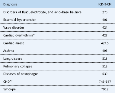
Patients with hypertrophic cardiomyopathy were identified using the International Classification of Diseases, 9th revision, Clinical Modification (ICD-9-CM) diagnostic codes, such as 425.1, which includes 425.11: hypertrophic obstructive cardiomyopathy and 425.18: other hypertrophic cardiomyopathies. All hospitalisations for which hypertrophic cardiomyopathy was the principal, secondary, or tertiary diagnosis between 2001 and 2014 were identified for patients <18 years of age at the time of admission. For our study, cases with a secondary or tertiary diagnosis of hypertrophic cardiomyopathy were included, but only if diagnoses in higher diagnostic positions were one of the cardiac-related diagnostic codes shown in Table 1. Our inclusion criteria minimised the impact of misclassification bias for cases with hypertrophic cardiomyopathy that were hospitalised for other primary reasons, such as asthma. Among cases identified in the above-mentioned inclusion criteria, we excluded those that were “syndrome of infant of a diabetic mother” (775.0) or live birth (V30) with glycogenosis (271.0), as these were likely infants of a diabetic mother.
Detailed information about the cardiac-related diagnostic codes.

* Cardiac dysrhythmia includes, but is not be limited to, atrial tachycardia (427.0), ventricular tachycardia (427.1), atrial fibrillation (427.3), ventricular fibrillation (427.4), and cardiac arrest (427.5)
** ICD-9-CM code 745 is bulbus cordis anomalies and anomalies of cardiac septal closure, which includes, but is not be limited to, ventricular septal defect (745.4) and atrial septal defect (745.5)
ICD-9-CM code 746 is other congenital anomalies of heart, which includes, but is not be limited to, congenital stenosis of aortic valve (746.3)
ICD-9-CM code 747 is other congenital anomalies of circulatory system, which includes, but is not be limited to, coarctation of aorta (747.1) and pulmonary atresia (747.3)
*** ICD-9-CM code 759.89 includes Noonan syndrome and Leopard syndrome, which could be associated with hypertrophic cardiomyopathy
The primary outcome of interest was yearly hospitalisation rate of patients with hypertrophic cardiomyopathy. Other outcomes of interest included death during hospitalisation, admission via emergency department, need for surgical intervention during hospitalisation, and associated diagnoses.
The rationale to use admission via emergency department is that it is more expensive than planned admission, and is therefore an extra burden in healthcare utilisation.Reference Muñoz, Soldano, Laughlin, Margolis and Wise 10 – Reference Baker and Baker 12
Surgical interventions were identified using the procedure codes operations on the cardiovascular system (35.x–39.x) in ICD-9-CM and excluding diagnostic procedures on heart and pericardium (37.2). We also individually identified ventricular septal myectomy (37.33), implantation of automatic cardioverter defibrillator (ICD: 37.94–37.98), heart transplantation (37.51), and extracorporeal membrane oxygenation (39.65) during the hospitalisation.
The primary predictors of interest were age group, sex, and race/ethnicity. Ages were grouped into three categories (<1, 1–9, and ⩾10 y/o) in order to show how descriptive features differed between age groups. Cut-off points were determined based on critical periods of infancy (<1 year of age) and puberty (⩾10 years of age), since puberty causes significant hormonal changes, which will affect the manifestation of hypertrophic cardiomyopathy.
The Nationwide Inpatient Sample contains information on both patient- and hospital-level characteristics such as age, sex, race, primary insurance type (payer), and hospital teaching status and size. Race/ethnicity was grouped into non-Hispanic White, non-Hispanic Black, Hispanic, and others. Primary payer was grouped into public insurance such as Medicaid and Medicare, private insurance, and others. A high-risk medical condition was defined as history of prematurity, that is ⩽36 weeks of gestational age, or at least one complex medical condition, previously defined using ICD-9-CM codes in nine illness categories, such as neuromuscular, cardiovascular excluding hypertrophic cardiomyopathy, respiratory, renal, gastrointestinal, haematology and immunodeficiency, metabolic, other congenital or genetic defect, and malignancy.Reference Feudtner, Hays, Haynes, Geyer, Neff and Koepsell 13
Hospital characteristics included United States regions – Northeast, South, Midwest, and West as defined according to the United States Census Bureau boundaries, 14 urban–rural status, teaching status (teaching versus non-teaching institution), and size of hospital by number of beds (small, medium, and large), which was categorised based on number of hospital beds, and is specific to the hospital’s location and teaching status. 15
In the Nationwide Inpatient Sample, the database maintained fields for 14–24 diagnoses besides the primary diagnosis, including patient co-morbidities, if relevant. We examined these diagnostic codes co-existing with hypertrophic cardiomyopathy.
We calculated 2001–2014 hospitalisation rates using the United States Census Bureau denominator population estimates. 16 Hospitalisation rates were expressed as number of estimated hospitalisations per 100,000 population in the corresponding age group. Yearly data were used for national hospitalisation rates, and stratified into three age groups as noted earlier.
Descriptive statistics of continuous variables were presented as estimated means with 95% confidence intervals, while categorical variables were presented as percentages. A logistic regression model tested for differences between age groups.
To examine how death during hospitalisation, admission numbers from the emergency department, and the need for surgical intervention during hospitalisation might be related to age group, sex, and race/ethnicity, we fit the following analytical models. First, we developed unadjusted logistic regression models with only primary predictors of interest, first one including age group, second one including sex, and the third one including race/ethnicity, as independent variables. Second, we examined the independent association between primary predictors of interest and each outcome using multivariable regression. We adjusted for both patient-level variables, such as primary payer and high-risk medical conditions, and hospital-level variables, such as region, urban and rural distinction, hospital teaching status, and bed size. To control for general secular and country-wide trends, year fixed effects were included in models. Owing to low numbers of identified hypertrophic cardiomyopathy cases reported yearly, time spans were categorised into four groups: 2001–2003, 2004–2006, 2007–2009, 2010–2012, 2013–2014, and race was categorised into three groups: non-Hispanic White, non-Hispanic Black, and other.
An ad hoc analysis was conducted to examine the interaction between sex and age. Based on the descriptive characteristics, age was categorised into two groups: ⩾10 and <10 y/o to examine how gender differentially affects outcomes among older and younger children. A logistic regression model tested for differences between older ⩾10 y/o and younger <10 y/o children in the males-to-females ratio diagnosed with hypertrophic cardiomyopathy. Multivariable logistic regression was conducted to test the interaction between sex and age on in-hospital mortality, admission from the emergency department, and the need for surgical intervention, controlling for hospital- and patient-level variables described earlier.
Sample weights were applied at the individual level using AHRQ’s weighted patient discharge values to obtain nationally representative estimates of inpatient characteristics. All analyses were conducted using the survey analysis command sets such as SURVEYMEANS, SURVEYFREQ, and SURVEYLOGISTIC of SAS, version 9.4 (SAS Institute Inc., Cary, NC, United States of America) in order to account for sample weights in variance calculations, in compliance with analysis recommendations from Healthcare Cost and Utilization Project. A two-sided p-value <0.05 was considered statistically significant.
Results
We identified 2317 patients under age 18, who met the diagnostic criteria for hypertrophic cardiomyopathy. Among which, 15 were excluded due to “syndrome of infant of a diabetic mother”, and none were excluded due to live birth with glycogenosis, yielding a final weighted sample size of 2302. Hypertrophic cardiomyopathy was the primary diagnosis for 1749 (76.0%), secondary diagnosis for 440 (19.1%) with a cardiac-related primary diagnosis, and the tertiary diagnosis for 118 (5.1%) with a cardiac-related primary and secondary diagnosis.
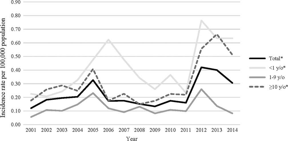
Hospitalisation rates during the study period were 0.22 per 100,000 population, with the lowest in 1–9 y/o (0.13). The youngest (<1 y/o) and oldest (⩾10 y/o) age groups had the higher hospitalisation rates at 0.41 and 0.31, respectively. Hospitalisation rates fluctuated over time with a notable spike observed after 2012, which is likely due to the change in weighting method in the database 17 (Fig 1).
Hospitalisation rates per 100,000 population due to hypertrophic cardiomyopathy by year, 2001–2014.

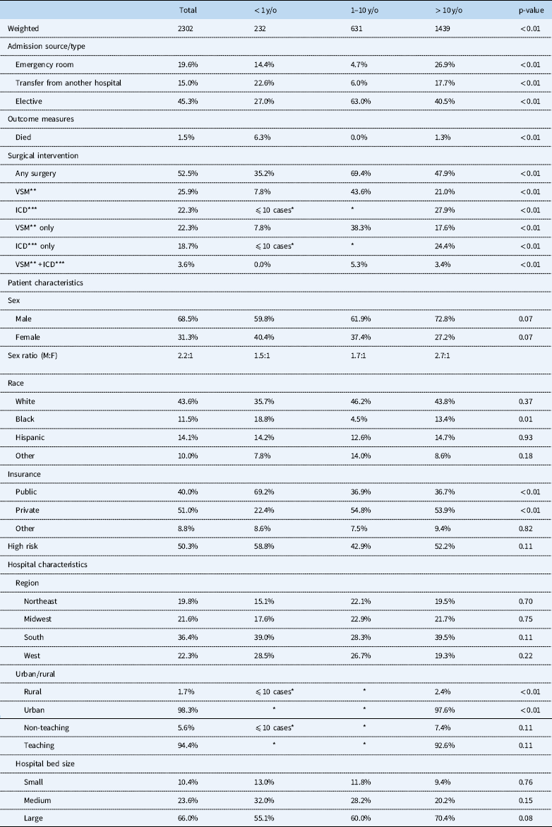
The overall admission rate from the emergency department was 19.6%, with a higher rate (26.9%) in the oldest age group. In-hospital mortality during the study period was 1.5%, with the highest rate in the youngest age group (6.3%). Ventricular septal myectomy was the most common surgical procedure for hospitalised patients with hypertrophic cardiomyopathy (25.9%) followed by implantation of automatic cardioverter-defibrillator (22.3%). Among those who had implantation of automatic cardioverter-defibrillator during the hospitalisation, <10 cases had automatic cardioverter-defibrillator before the hospitalisation, which was captured by v code (v 45.0). Heart transplantation was performed in 54 (2.4%) hospitalisations during the study period, and extracorporeal membrane oxygenation was used in 29 (1.3%).
The overall male/female ratio in hospitalisation for patients with hypertrophic cardiomyopathy was 2.2 to 1, with lower ratios for <1 y/o and 1–9 y/o ages (1.5:1 and 1.7:1, respectively) and a higher ratio for ⩾10 y/o ages (2.7:1). Nearly half of the cases were White (43.6%). Most cases were treated at urban (98.3%) and teaching (94.4%) hospitals, with over half of cases treated in large hospitals (66.0%) (Table 2).
Descriptive statistics of hospitalised children with hypertrophic cardiomyopathy, 2001–2014.

* Cells with ten or less observation values could not be reported for HCUP IRB human subject research protections
** Ventricular septal myectomy
*** Implantation of automatic cardioverter defibrillator
*Not reported to protect subgroups with ten or fewer cases
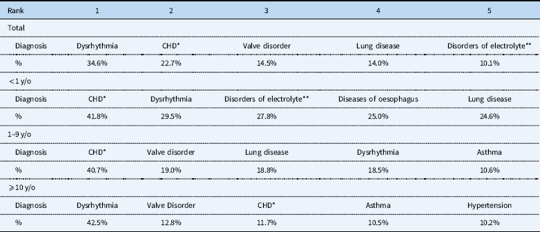
Table 3 shows the top 5 most common associated diagnoses in total number and by age groups, with dysrhythmia (34.6%), followed by CHDs (22.7%), valve disorder (14.5%), lung diseases (14.0%), and disorders of fluid, electrolyte, and acid–base balance (10.1%). In the younger age groups from new-born to 9 y/o, CHD was the most common associated diagnosis, whereas in the older age group of ⩾10 y/o, dysrhythmia was the most common diagnosis. Supplementary Table S1 shows the detail breakdown of CHDs. Table 4 provides the ICD-9-CM diagnostic code in detail.
Top 5 common co-diagnoses by age groups.

* Congenital Heart Diseases. Detail breakdown of CHD is provided in Supplementary Table S1
** Disorders of fluid, electrolyte, and acid–base balance
Detailed information about co-existing diagnoses.

* Cardiac dysrhythmia includes, but is not be limited to, atrial tachycardia (427.0), ventricular tachycardia (427.1), atrial fibrillation (427.3), ventricular fibrillation (427.4), and cardiac arrest (427.5)
** ICD-9-CM code 745 is bulbus cordis anomalies and anomalies of cardiac septal closure, which includes, but is not be limited to, transposition of great vessels (745.1), tetralogy of Fallot (745.2), ventricular septal defect (745.4), and atrial septal defect (745.5)
ICD-9-CM code 746 is other congenital anomalies of heart, which includes, but is not be limited to, Ebstein’s anomaly (746.2), congenital stenosis of aortic valve (746.3), congenital mitral stenosis (746.5), and hypoplastic left heart syndrome (746.7)
ICD-9-CM code 747 is other congenital anomalies of circulatory system, which includes, but is not be limited to, coarctation of aorta (747.1), pulmonary atresia (747.3), and total anomalous pulmonary venous return (747.41)
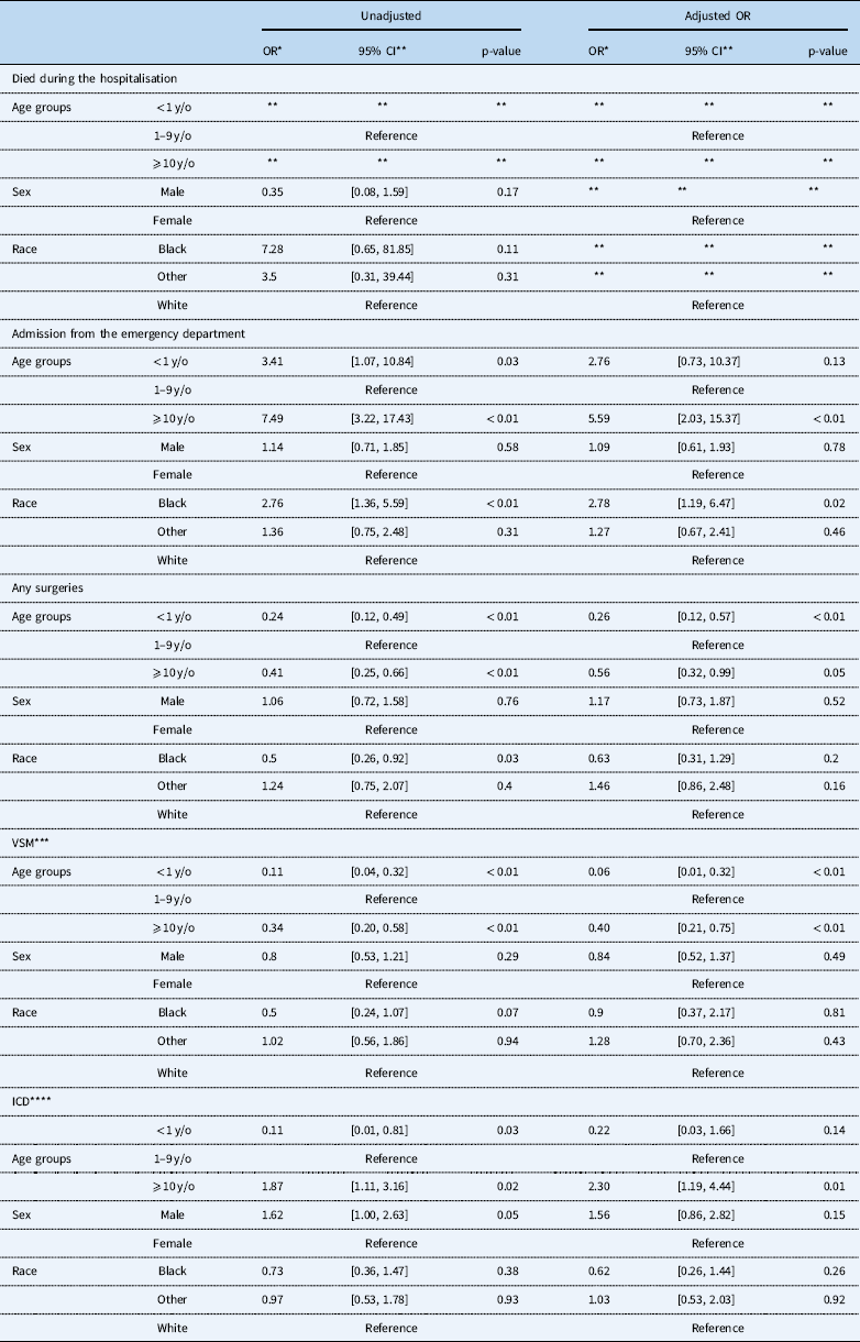
Odds of in-hospital mortality, admission from the emergency department, and surgery among paediatric hypertrophic cardiomyopathy patients.

**Parameter estimates could not be calculated due to rareness of in-hospital mortality, causing monotone likelihood in the model
* Odds ratio
** Confidence interval
*** Ventricular septal myectomy
**** Implantation of automatic cardioverter defibrillator
Owing to the rareness of in-hospital mortality, the regression models suffered from monotone likelihood, and parameter estimates could not be calculated. The oldest age group was associated with increased risk for admission from the emergency department compared to the mid age group which has odds ratio 5.59 with 95% confidence interval (2.03–15.37). Black population was more likely to be admitted from the emergency department compared to White whose odds ratio 2.78 with 95% confidence interval (1.19–6.47).
As for surgical intervention during hospitalisation, the youngest and oldest age groups were associated with decreased risk with odds ratio 0.26 and 95% confidence interval (0.12–0.57), and odds ratio 0.56 with 95% confidence interval (0.32–0.99) compared with the age group of 1–9 y/o.
The youngest and the oldest age groups were less likely to have ventricular septal myectomy compared with the 1–9 y/o age group, whose odds ratios was 0.06 at 95% confidence interval (0.01–0.32) and 0.40 at 95% confidence interval (0.21–0.75), respectively. The oldest age group was also more likely to have implantation of automatic cardioverter-defibrillator during the hospitalisation compared with 1–9 y/o because odds ratio was 2.30 at 95% confidence interval (1.19–4.44). These results suggested that ventricular septal myectomy was performed more for the 1–9 y/o age group, and implantation of automatic cardioverter-defibrillator was performed more for the age group ⩾10 y/o.
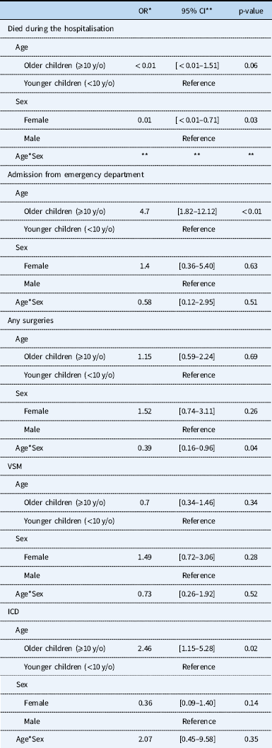
When age category was regrouped as a dichotomous variable in the ad hoc analysis (⩾10 and <10 y/o), significant sex-based differences were observed in the hospitalisation rate of patients with hypertrophic cardiomyopathy between younger and older children, with a male-to-female diagnosis ratio of 1.6 for younger children (<10 y/o) and 2.7 for older children (⩾10 y/o) (p=0.02) (Table 6). Difference in risk of surgical intervention was also magnified among males and females ⩾10 y/o, with older girls having lower risk of surgery compared with older boys – odds ratio 0.39 with 95% confidence interval (0.16–0.96) (Table 7).
Result from ad hoc analysis to compare the hospitalisation rate between younger children (<10 y/o) and older children (⩾10 y/o) children.

Results from ad hoc analysis assessing interaction of age and sex on in-hospital mortality, admission from the emergency department, and surgery.

**Parameter estimates could not be calculated due to rareness of in-hospital mortality, causing monotone likelihood in the model
* Odds ratio
** Confidence interval
Discussion
With a nationally representative data cohort of children, our study identified several characteristics of hospitalisations for patients with hypertrophic cardiomyopathy. We found that hospitalisation rate for patients with hypertrophic cardiomyopathy was 0.22 per 100,000 per year in the United States paediatric population between 2001 and 2014, and hospitalisation rate for patients with hypertrophic cardiomyopathy differed between age groups and sex.
We observed a higher rate of hospitalisation for patients with hypertrophic cardiomyopathy among the youngest and oldest age groups. Previous literature reported higher incidence, morbidity, and mortality of hypertrophic cardiomyopathy in the first year of life.Reference Colan, Lipshultz and Lowe 18 – Reference Nugent, Daubeney and Chondros 20 Lipschultz et alReference Lipshultz, Sleeper and Towbin 1 also presented a higher rate in infants and 12–18 y/o.Reference Gersh, Maron and Bonow 21 , Reference Gersh, Maron and Bonow 22 Our finding of higher hospitalisation in the youngest and oldest paediatric age groups, namely infants and adolescents, was in agreement with the earlier studies showing higher incidence in these groups.
Hypertrophic cardiomyopathy has been known to be male-dominant, and incidence differences by sex increase with age.Reference Colan, Lipshultz and Lowe 18 , Reference Ostman-Smith, Wettrell and Keeton 23 – Reference Becton, Cheng and Nieman 26 Colan et alReference Colan, Lipshultz and Lowe 18 used the Pediatric Cardiomyopathy Registry data in the United States, and compared infant (<1 y/o) with non-infant (⩾1 y/o) hypertrophic cardiomyopathy. They reported that there was a male predilection in non-infant hypertrophic cardiomyopathy compared to infant hypertrophic cardiomyopathy. We divided age into smaller categories, establishing that sex differences were more pronounced after 10 y/o in terms of hospitalisation. Evidence suggests that exposure of cardiac myocytes to androgens results in hypertrophy.Reference Marsh, Lehmann, Ritchie, Gwathmey, Green and Schiebinger 27 The study by Ostman-Smith et alReference Ostman-Smith, Wettrell and Keeton 23 reported that increased incidence of sudden death between 1 and 7 years of age was not observed, but increased markedly after puberty. They suggested that higher levels of androgens could be associated with rapid disease progression in hypertrophic cardiomyopathy, which is the most common cause of sudden cardiac death in the young.Reference Maron, Haas, Ahluwalia, Murphy and Garberich 28 Lind et alReference Lind, Chiu and Ingles 29 also reported male-dominant prevalence in 200 adult patients with hypertrophic cardiomyopathy. They stated the possibility that males present more symptoms and are more often diagnosed with hypertrophic cardiomyopathy than females, because higher levels of androgens could be associated with rapid disease progression in hypertrophic cardiomyopathy. Given the higher male-to-female ratios in hospital admissions after 10 y/o, when male patients start to develop higher levels of androgens, our results support previous studiesReference Marsh, Lehmann, Ritchie, Gwathmey, Green and Schiebinger 27 – Reference Lind, Chiu and Ingles 29 suggesting that increased exposure to androgens can lead to androgen receptors mediating hypertrophy in cardiac myocytes.
Maron et alReference Maron, Haas, Ahluwalia, Murphy and Garberich 28 reported that 97% of athletes in the US National Registry of Sudden Death in Athletes whose deaths were associated with hypertrophic cardiomyopathy were males. They argued that potential determinants of gender disproportionality included intensity of training, with more demanding conditioning regimens more common in males. Similarly, males in our cohort could also have been exposed to more intense training, thus developing symptoms due to hypertrophic cardiomyopathy.
We found the youngest age group to have a higher in-hospital mortality rate (6.3%) than other age groups. Studies using the Pediatric Cardiomyopathy Registry data in the United States of AmericaReference Colan, Lipshultz and Lowe 18 and Australia,Reference Nugent, Daubeney and Chondros 20 both showed lower survival rates if the diagnosis of hypertrophic cardiomyopathy was made at <1 year of age. Ostman-Smith et alReference Ostman-Smith, Wettrell and Keeton 23 reported a higher number of deaths in patients with hypertrophic cardiomyopathy <1 year of age. Colan et alReference Colan, Lipshultz and Lowe 18 reported that 25% of their cohort was diagnosed with an inborn error of metabolism, malformation syndrome, or neuromuscular diseases, and patients with syndromic or “secondary hypertrophic cardiomyopathy” were found to have higher mortality compared with those with primary hypertrophic cardiomyopathy.
We observed age differences in the admission source. The oldest age group had the highest rate of admission from the emergency department. After adjusting for patient- and hospital-level characteristics, the oldest age group was more likely to be admitted from the emergency department compared to the age group of 1–9 y/o. A possible explanation might be that seemingly healthy people, who unknowingly had asymptomatic hypertrophic cardiomyopathy, developed symptoms such as dysrhythmia or syncope, and were diagnosed with hypertrophic cardiomyopathy in the emergency department, subsequently requiring hospitalisation. A large population study for young adults suggested that most young patients identified with hypertrophic cardiomyopathy had no previous symptoms.Reference Maron, Gardin, Flack, Gidding, Kurosaki and Bild 30 A recent report by Maron et alReference Maron, Haas, Ahluwalia, Murphy and Garberich 28 using the US National Registry of Sudden Death in Athletes demonstrated that hypertrophic cardiomyopathy was the single most common cause of sudden death in athletes, accounting for one-third of these events. In our cohort, almost nobody in the oldest age group (<1%) was admitted from emergency department with CHD, and most common associated diagnoses in this group were dysrhythmia (42.5%) followed by valve disorder (12.8%). Our database did not indicate if the symptoms occurred during exercise; however, more strenuous activity in the oldest age group may have exacerbated development of symptoms from hypertrophic cardiomyopathy for previously asymptomatic patients.
Differences in admission source also existed across race/ethnicity, that is Blacks were more likely to be admitted from the emergency department compared to Whites. One possible explanation for these differences is the higher likelihood of undiagnosed hypertrophic cardiomyopathy among the Black children. Movahed et alReference Movahed, Strootman, Bates and Sattur 31 performed screening echocardiography among 2066 students between the ages of 13 and 19 years, and demonstrated that African-American teenagers had higher prevalence of hypertrophic cardiomyopathy than teenagers of other races. Another possible explanation is that differences could be attributed to poor medical follow-up among the Black children with hypertrophic cardiomyopathy. Previous studies have reported that Black patients were less likely to receive care from board-certified physicians and to have access to newer medical technology.Reference Dimick, Ruhter, Sarrazin and Birkmeyer 32 – Reference Birkmeyer, Stukel, Siewers, Goodney, Wennberg and Lucas 34 In fact, at the screening event Movahed et alReference Movahed, Strootman, Bates and Sattur 31 conducted, Blacks represented 9% of the screening participants among those who documented race demography (n=551), despite Blacks representing 16% of the United States population. Racial disparities exist in many settings and contexts, and we demonstrated that the children with hypertrophic cardiomyopathy was no exception. Further studies are needed to clarify the mechanisms behind this racial disparity in admission source among paediatric hypertrophic cardiomyopathy patients.
This study has the following limitations. First, Nationwide Inpatient Sample observations were de-identified; therefore, patients who were discharged and subsequently readmitted could be double-counted if a diagnosis of hypertrophic cardiomyopathy was present on both records.Reference Bor, Woolhandler, Nardin, Brusch and Himmelstein 35 , Reference Pant, Patel and Deshmukh 36 For example, it is possible that a patient was diagnosed as having hypertrophic cardiomyopathy and admitted to the hospital in an earlier age group, and then re-admitted for ventricular septal myectomy or implantation of automatic cardioverter-defibrillator in an older age group. Since some patients were double-counted, and the database only included patients admitted to the hospital, we were unable to estimate incidence rates or prevalence of hypertrophic cardiomyopathy. Instead, we provided the nationally representative hospitalisation rate among patients with hypertrophic cardiomyopathy.
Second, diagnostic coding practices vary among hospitals for the same conditions, leading to misclassification,Reference Pant, Patel and Deshmukh 36 with sampling methods for aggregating Nationwide Inpatient Sample adding data bias in unknown ways. In particular, we observed a sharp increase in hypertrophic cardiomyopathy after 2012 compared to previous years, which could be attributed to a change in the database’s sampling methods. Methods were revised in 2012 to draw a sample of discharges from all hospitals, whereas prior years included a sample of hospitals from which all discharges were retained. 17 Weights from 2001 to 2011 were converted to be consistent with the new sampling methods, however, minor discrepancies may still exist.
Third, Nationwide Inpatient Sample is an administrative database of discharge-level data, which lacks depth of clinical information. Therefore, we were not able to distinguish between the morphological subtypes of hypertrophic cardiomyopathy that could lead to diagnosis of cause, such as familial hypertrophic cardiomyopathy and hypertrophic cardiomyopathy secondary to hypertension.
Because of the limitation in the ICD-9-CM, we were not able to distinguish patients with hypertrophic cardiomyopathy associated with other clinical syndromes, such as Noonan syndrome, LEOPARD syndrome, Costello syndrome, cardio-facial-cutaneous syndrome, which are classified as 759.89 (other specified congenital anomalies) or 799.89 (other ill-defined condition).
Lastly, owing to low numbers of identified hypertrophic cardiomyopathy cases reported yearly, time spans and race were categorised into smaller groups in the regression models than in the descriptive statistics. We were not able to provide the specific numbers for all descriptive statistic variables due to HCUP IRB human subject research protections. However, we believe that hospitalisation rate could be a surrogate for overall health and well-being of patients with active cases of hypertrophic cardiomyopathy. Gender disproportionality could be due to a higher probability of females with hypertrophic cardiomyopathy being asymptomatic.
Conclusions
By using a nationally representative database of United States of America hospitalisations during 2001–2014, we found that there was a male dominance of hospitalisations for cases with hypertrophic cardiomyopathy that increases with age. Infants with hypertrophic cardiomyopathy had higher in-hospital mortality. The oldest age group and Blacks are more likely to be admitted from the emergency department. Further studies are needed to clarify the mechanisms behind age and racial disparity in hospitalisation, especially admission source, among children with hypertrophic cardiomyopathy.
Supplementary material
To view supplementary material for this article, please visit https://doi.org/10.1017/S1047951118002421
Acknowledgements
The authors would like to thank Mr Scott Friedlander, MPH, at Los Angeles Biomedical Research Institute at Harbor-UCLA Medical Center for statistical advice.
Financial support
No external funding for this manuscript.
Conflicts of interest
None.
Ethical approval
This article does not contain any studies with human participants or animals performed by any of the authors. The Institutional Review Board at Los Angeles Biomedical Research Institute at Harbor-UCLA Medical Center confirmed that this study does not meet the Department of Health and Human Services (DHHS) definition of human subjects’ research, as defined in 45 CFR 46.102(f), and approved the study under the “exempt” category.