Monochorionic twins often present unique challenges in prenatal management. The single placenta can create an unbalanced haemodynamic exchange through vascular connections leading to twin-to-twin transfusion syndrome.
Pulmonary artery calcifications in the recipient twin are an extremely rare but known entity. They are generally hypothesised to be secondary to chronic right ventricular pressure and volume overload in the recipient twin. Idiopathic infantile arterial calcification is another rare entity, where widespread calcium deposits are seen in vessels, soft tissues, and organs. Most case reports of pulmonary artery calcifications in the recipient twin in twin-to-twin transfusion syndrome are likely not related to calcifications found in idiopathic infantile arterial calcification and not as widespread.Reference Lougheed, Sinclair and Fung 1 – Reference Cha, Kim, Kwon, Bae, Noh and Kim 4
Case report
Our patient was a male monochorionic recipient twin of twin-to-twin transfusion syndrome, delivered by caesarean section at 33 3/7 weeks of gestation owing to preterm labour to a 35-year-old mother. During fetal life, there had been two laser ablations of placental vessels and two amnio-reductions due to twin-to-twin transfusion syndrome.
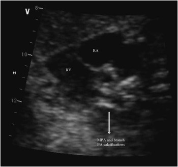
A fetal echocardiogram at 31 6/7 weeks of gestation showed diffuse brightness of the main pulmonary artery and the ductus arteriosus (Fig 1) with antegrade flow in both. Qualitatively, the pulmonary valve appeared mildly hypoplastic without insufficiency/stenosis and with normal Doppler flow velocity. Moderate holosystolic tricuspid regurgitation was noted with mild right atrial enlargement and right ventricular hypertrophy with normal biventricular function and a small pericardial effusion. The cardiovascular profile score was 8/10. A repeat fetal echocardiogram at 33 1/7 weeks showed similar findings. In addition, the ascending aorta also appeared echo bright, and the pericardial effusion was smaller.
Close up still image of the calcifications in the wall of the main pulmonary artery and proximal branch pulmonary arteries on fetal echocardiogram.

At delivery, the baby was vigorous and plethoric with a hematocrit of 60%. Vital signs and physical examinations were otherwise unremarkable. A postnatal echocardiogram showed the prominent main and branch pulmonary artery echogenicity as well as milder echogenicity of the proximal ascending aorta. All had non-obstructive forward flow. Pulmonary hypertension was noted at about two thirds systemic level. The coronary arteries were not clearly visualised. The remainder of the echocardiogram was normal.
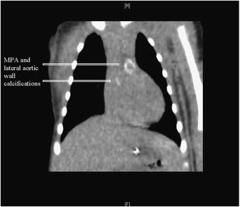
Chest X-ray was normal, but a chest CT scan showed calcifications in the main and branch pulmonary arteries to the hilum and minimal calcification of the lateral aortic wall with no involvement of the aortic sinuses or coronary vessels (Fig 2). Genetic testing for idiopathic infantile arterial calcification [ectonucleotide pyrophosphatase/phosphodiesterase 1(ENPP1) and ABCC6 (ATP binding cassette subfamily C member 6) gene mutations] as well as Williams syndrome were negative, and the calcifications noted were significantly less than those reported for idiopathic infantile arterial calcification in the literature. Therefore, we elected not to treat our patient with bisphosphonates.
Circumferential calcifications seen in the wall of main pulmonary artery as well as lateral aortic wall on post natal chest CT scan.

After 2 months since discharge, our patient’s right ventricular pressure had increased to supra-systemic levels and he underwent surgical main pulmonary artery plasty, which improved the pressure to 2/3 systemic. An intra-operative angiogram showed diffuse hypoplasia of his branch pulmonary arteries beyond the hilum, which improved with subsequent balloon angioplasty on two separate occasions. After 6 months, his right ventricular pressure is normal and he is clinically thriving.
Discussion
Pulmonary valve and infundibular stenosis has been documented in the past with twin-to-twin transfusion syndrome and attributed to right ventricular volume and pressure overload in the recipient twin. Right ventricular hypertrophy may cause varying degrees of right ventricular outflow tract obstruction and lead to hypoplasia of the pulmonary valve and pulmonary artery branches.Reference Lougheed, Sinclair and Fung 1 , Reference Nicosia, Krouse and Mobini 5 The three earliest reported in utero cases of pulmonary artery calcifications in the recipient twin of twin-to-twin transfusion syndrome were those by Popek et al in 1993Reference Popek, Strain, Neumann and Wilson 3 and all occurred in the volume-overloaded twin. In addition, there might have been a relationship between pulmonary artery calcifications and the severity of the cardiac volume overload based on the weight difference between the twins in each pair. Saxena et alReference Saxena and Soni 2 and Cha et alReference Cha, Kim, Kwon, Bae, Noh and Kim 4 reported one and three similar cases, respectively, with the same hypothesis for causation.
The etiology for the pulmonary, and rarely aortic,Reference Saxena and Soni 2 calcifications in recipients of twin-to-twin transfusion syndrome has been largely speculative to date. Intimal thickening without calcification has been reported in the aorta in recipient twins in twin-to-twin transfusion syndrome,Reference Nicosia, Krouse and Mobini 5 possibly secondary to volume overload. In addition, recipient fetuses have been shown to have increased pulmonary and aortic velocities compared with their donor co-twins.Reference Murakoshi, Yamamori and Tojo 6 Endothelin-1, a potent vasoconstrictor, has been found to be higher in recipient twins and even higher in those with right ventricular outflow tract obstruction than in those with normal cardiac function. The mitogenic and vasoconstrictor effects of endothelin-1 may contribute indirectly to ventricular hypertrophy and hypertension.Reference Bassil Eter, Dulac and Acar 7 Increased resistance in the monochorionic placenta may lead to systemic hypertension and increased right ventricular afterload in the recipient twin. However, there have been reports of right ventricular outflow tract obstruction without systemic hypertension.Reference Nizard, Bonnet, Fermont and Ville 8
Idiopathic infantile arterial calcification is a rare entity (<200 cases described), inherited in an autosomal recessive manner, most cases of which are fatal before 6 months of age and usually diagnosed at autopsy. Most sporadic case reports of pulmonary artery calcifications in the recipient twin in twin-to-twin transfusion syndrome have not been likened with the calcifications found in idiopathic infantile arterial calcification and not as widespread.Reference Saxena and Soni 2 – Reference Cha, Kim, Kwon, Bae, Noh and Kim 4 , Reference Bassil Eter, Dulac and Acar 7 Idiopathic infantile arterial calcification has been associated with inactivating mutations of the ENPP1Reference Rutsch, Ruf and Vaingankar 9 gene or loss of function mutations in the ABCC6 gene. Both are potent inhibitors of calcification – hydroxyapatite deposition – in soft tissue and vessel walls. These mutations lead to unregulated and widespread calcium deposition. Loss-of-function mutations of ENPP1 cause a wide spectrum of phenotypes, ranging from lethal idiopathic infantile arterial calcification to hypophosphatemic rickets along with early-onset hearing loss, and progressive diabetic nephropathy. ABCC6 has also been associated with pseudoxanthoma elasticum.
Our patient was negative for both ENPP1 and ABCC6 loss-of-function mutations seen in idiopathic infantile arterial calcification. Severely calcified aortic valve stenosis has been noted in Williams–Beuren syndrome.Reference Yetkin, Bal, Bayata and Gürbüz 10 Our patient was negative for this as well. His clinical picture was more consistent with previously reported recipient twins in twin-to-twin transfusion syndrome with arterial calcifications. He did, however, have aortic calcifications as well as pulmonic calcifications, the former being reported only once previously in twin-to-twin transfusion syndrome.Reference Saxena and Soni 2 Therefore, the diagnosis of idiopathic infantile arterial calcification was considered initially until his genetic testing returned negative. The various theories discussed above for the development of pulmonary artery and aortic calcifications are promising but may need further study to elucidate the exact mechanism and develop possible treatment strategies in the future.
Acknowledgements
The authors wish to thank Dr. Tarek S. Husayni, Pediatric Cardiologist, Advocate Children’s Hospital, Oak Lawn, Illinois.
Financial Support
This research received no specific grant from any funding agency or from commercial or not-for-profit sectors.
Conflicts of Interest
None.
Ethical Standards
The authors assert that all procedures contributing to this work comply with the ethical standards of the relevant national guidelines on human experimentation [NIH HHS Regulations: 45 CFR part 46; HHS Regulations for the Protection of Human Subjects and 45 CFR parts 160 and 164; Health Insurance Portability and Accountability Act (HIPAA) Regulations for Standards for Privacy of Individually Identifiable Health Information] and with the Helsinki Declaration of 1975, as revised in 2008.