Implications
Reproductive disease in the first 2 months after calving is very common and spans severe clinical illness to unapparent yet substantial impairment of fertility. These conditions result from the interaction between infection and stimulus for inflammation and effective immune response and regulation of inflammation. Recent advances underline links among metabolism, inflammation and health that were previously thought to be separate processes.
Introduction
Available data indicate that almost all dairy cows have bacterial contamination of the uterus in the 2 to 3 weeks after calving (Sheldon et al., Reference Sheldon, Cronin, Goetze, Donofrio and Schuberth2009a). Because of this, as well as the substantial required repair of the endometrium, uterine inflammation is a normal and necessary component of involution. However, the growth of pathogenic bacteria may overcome innate immune defences, which are the main protection for the uterus or the severity or duration of inflammation may impair rather than enhance fertility. Between 5% and 20% may experience metritis (overt systemic illness), and if examined systematically, 5% to 25% have purulent vaginal discharge (PVD) at 4 to 5 weeks postpartum, and 30% to over 50% of cows have subclinical inflammation of the uterus (endometritis) and/or cervix at 4 to 8 weeks postpartum that is associated with reduced reproductive performance. The documented direct and indirect impacts make these conditions costly for the affected individual cows (Overton and Fetrow, Reference Overton and Fetrow2008; Dubuc et al., Reference Dubuc, Duffield, Leslie, Walton and LeBlanc2011) and their high incidence risks make them costly for herds and the industry. The high incidence of disease is attributable in part to reduced innate immune function and impaired regulation of inflammation from ∼2 weeks before to 3 weeks after calving. Insulin resistance (IR) and adaptation to negative energy balance (NEB) contribute to the degree and duration of reduced immune defence. Essentially, all cows experience NEB in early lactation with IR as part of the homeorhetic adaptations to allow NEB in support of lactation. As with uterine inflammation, the concern is for the cows that experience a severity of NEB or IR that goes beyond adaptive to being a contributor to pathology or impaired reproduction. The points at which NEB, IR or reproductive tract inflammation shift from physiologic to undesirable remain to be quantified and likely include numerous interactions. The determinants of effective immune and inflammatory response v. chronic reproductive tract infection and/or inflammation are not well understood. However, there are emerging lines of investigation that promise to improve understanding of these determinants and the ability to treat and ultimately prevent these conditions. This paper discusses factors that may contribute to postpartum reproductive tract inflammatory disease in dairy cows and its diagnosis, impacts and treatment.
Current concepts in metabolic health and inflammation
There has been rapid emergence of data about interactions among metabolism (specifically related to insulin and fat), inflammation and immune function in humans and laboratory animals (Osborn and Olefsky, Reference Osborn and Olefsky2012; McArdle et al., Reference McArdle, Finucane, Connaughton, McMorrow and Roche2013). Very recently, fascinating evidence has been provided about the role of intestinal microflora in interacting with regulation of inflammation and extra-intestinal metabolism (Henao-Mejia et al., Reference Henao-Mejia, Elinav, Jin, Hao, Mehal, Strowig, Thaiss, Kau, Eisenbarth, Jurczak, Camporez, Shulman, Gordon, Hoffman and Flavell2012). It will be interesting to see whether similar relationships exist with ruminal or intestinal microbiota in cattle. It is already clear that dietary changes around calving may have the unintended consequence of inducing rumen acidosis, which in turn may increase the production and uptake of endotoxin (lipopolysaccharide; LPS) from gram-negative, or lipoteichoic acid (LTA) from gram-positive rumen bacteria. Endotoxin is a potent inducer of pro-inflammatory response in most cells (not only immune cells), and among its many effects, reduces feed intake (Mani et al., Reference Mani, Weber, Baumgard and Gabler2012). It is possible that certain intestinal bacteria (e.g. Prevotella and Porphorymonas species in mice; Henao-Mejia et al., Reference Henao-Mejia, Elinav, Jin, Hao, Mehal, Strowig, Thaiss, Kau, Eisenbarth, Jurczak, Camporez, Shulman, Gordon, Hoffman and Flavell2012) or their products (other than LPS) may also influence inflammation and metabolism through mechanisms that are not well explored in ruminants. Apparently, more as an effect than a cause, it is likely that LPS from the uterus contributes to the sickness behaviour associated with metritis.
Fat mobilization and inflammation
Adipose tissue, especially visceral fat, is more than storage for fatty acids (FAs) and glycerol: fat mobilization produces pro-inflammatory signals (tumor necrosis factor α (TNFα) and interleukin-6 (IL-6); Tilg and Moschen, Reference Tilg and Moschen2008), which block the intracellular signalling of insulin and so contribute to IR. IR in dairy cows has recently been well reviewed (DeKoster and Opsomer, Reference DeKoster and Opsomer2013). Although the focus in humans relates to obesity leading to chronic inflammation and IR, type 2 diabetes, and cardiovascular disease, there are similarities in periparturient dairy cows despite an environment of fat mobilization rather than obesity, and hypoglycaemia rather than hyperglycaemia. Both obese patients (Osborn and Olefsky, Reference Osborn and Olefsky2012) and high producing dairy cows are characterized by elevated circulating plasma non-esterified fatty acids (NEFA), IR and inflammation. The similarities relate to the links of energy and fat metabolism with inflammation, specifically loss of physiologic feedback against ongoing high rates of lipid mobilization mediated by the action of excessive TNFα from mobilized fat leading to IR and in turn to insulin-dependent inhibition of further release of NEFA (Sordillo and Raphael, Reference Sordillo and Raphael2013). This mechanism is at play in obese patients and is consistent with the strong associations of over-conditioning in dairy cows (e.g. BCS>3.5 out of 5 at calving) with increased risk of fatty liver and metabolic disease. The same mechanism seems likely to contribute to metabolic and infectious disease risk in cattle that are not visibly over-conditioned, but may have reduced feed intake due, for example, to inadequate feeding space or to previous disease.
Links between metabolic and inflammatory responses have recently been reviewed in detail (Ingvartsen and Moyes, Reference Ingvartsen and Moyes2013; Sordillo and Raphael, Reference Sordillo and Raphael2013). Fat mobilization increases the supply of NEFA, which brings along pro-inflammatory cytokines from fat (TNF and IL-6). These cytokines suppress intracellular signalling with the result of blocking the action of insulin (i.e. contributing to IR), which has the effect of potentially exacerbating release of NEFA from adipose. In addition, NEFA may contribute directly to greater inflammation by binding Toll-like receptor 4 (TLR4) (the pathogen-associated molecular patterns sensor for LPS) and initiating an inflammatory cascade through TNF (Hotamisligil and Erbay, Reference Hotamisligil and Erbay2008). NEFA may also impair polymorphonuclear (PMN) function; specific FAs more so than others, particularly saturated FA, which predominate in transition cows, appear to trigger TLR (Ingvartsen and Moyes, Reference Ingvartsen and Moyes2013), which provides an additional source of inflammatory stimulus (on top of LPS and perhaps LTA that may be coming from the uterus and/or the rumen) and further input into the vortex of hepatic or whole body inflammation. The saturated FA stearate and palmitate are predominant in NEFA mobilized from body fat in transition cows and based on evidence in other species, these have pro-inflammatory effects by activating NF-κB. Briefly, NEFA activate TLR4, a main receptor for LPS, which activates NF-κB and leads to secretion of TNFα, IL-1 and IL-8. TNFα and IL-1 act on intracellular messengers to up-regulate inflammation and increase IR. Specifically, TNFα from fat stimulates NF-κB (the same intracellular signal pathway stimulated by LPS binding to TLR4) leading to decreased downstream insulin signalling and therefore IR. Pro-inflammatory cytokines and saturated FA can increase expression of genes for ceramides that also impair insulin signalling, apparently through the TLR4 pathway (Osborn and Olefsky, Reference Osborn and Olefsky2012). In contrast, polyunsaturated FA bind peroxisome proliferator-activated nuclear receptors, which generally effect anti-inflammatory responses (Sordillo and Raphael, Reference Sordillo and Raphael2013). In particular, n-3 polyunsaturated fatty acid (PUFA) are associated with reduced inflammation. While PUFA are relatively scarce in the mixture of NEFA in transition cows, their amounts may be altered by dietary supplementation. While this avenue merits continued investigation, development of methods to maintain the overall magnitude of NEFA flux below the concentrations associated with compromised immune function (Ster et al., Reference Ster, Loiselle and Lacasse2012) or clinical disease (Dubuc et al., Reference Dubuc, Duffield, Leslie, Walton and LeBlanc2010b) is a high priority.
Inflammatory response and regulation
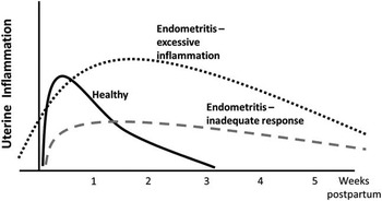
While PMN leukocytes (neutrophils) are the predominant cells in response to infection in the uterus and mammary gland, macrophages are important for detection of pathogen-associated molecular patterns (e.g. LPS) and initiation of the inflammatory cascade. The goal is to affect a prompt, robust and effective yet constrained inflammatory response as opposed to inflammation that is more severe or prolonged than beneficial to reproductive function. Loss of this regulation appears to be central to the pathogenesis of endometritis (Figure 1). This is exemplified in severe coliform mastitis in which, based on LPS challenge models, the severe inflammatory response induced by LPS appears to be responsible for most of the clinical signs. Recent evidence provides an explanatory mechanism for this, and for the observation that severe coliform mastitis is particularly common in early lactation. The inflammatory response of macrophages and endothelial cells to LPS is heightened in early lactation (Sordillo and Raphael, Reference Sordillo and Raphael2013). This effect is associated with the metabolic milieu of the transition period but may be further modulated by diet: cows fed 150% v. 100% of energy requirement for 45 days prepartum had greater increases in NEFA, haptoglobin and liver fat in response to LPS challenge (Graugnard et al., Reference Graugnard, Moyes, Trevisi, Khan, Keisler, Drackley, Bertoni and Loor2013). Given that gram-negative bacteria appear to play a role as uterine pathogens, diet (specifically the effects of energy status) may influence the response to LPS from the uterus and in turn, reproductive tract inflammation.
Schematic representation of concepts of the patterns of immune and inflammatory response in dairy cows in the postpartum period. ‘Excessive’ inflammation refers to inflammation that is severe or prolonged enough to impair rather than aid reproductive performance. ‘Inadequate response’ refers to innate immune function that fails to clear uterine pathogens within 3 weeks after calving.

It is well documented and obvious that the transition period in general, the effects of lactation, and more specifically the effects of the degree and duration of NEB or of markers of aspects of adaptation to NEB (e.g. NEFA and β-hydroxybutyrate (BHBA)) are associated with the risk of many metabolic and infectious diseases. Data are increasingly available to identify the mechanisms through which excessive NEB or maladaptation to NEB lead to suppressed immune efficacy or excessive inflammation (Ingvartsen and Moyes, Reference Ingvartsen and Moyes2013). Oxidative stress (excessive formation of reactive oxygen species (ROS) or lack of antioxidants) contributes to inflammation by activating the pro-inflammatory NF-κB pathway and by apparently heightening the production of TNF by immune (peripheral mononuclear) cells in cows (Sordillo et al., Reference Sordillo, Contreras and Aitken2009).
Probably all dairy cows experience IR and considerable fat mobilization in early lactation, and 30% to 35% have NEFA and 45% have BHBA above thresholds associated with metabolic disease or compromised production or reproduction (McArt et al., Reference McArt, Nydam, Oetzel, Overton and Ospina2013). It is clear that pathogens (LPS associated with intramammary infection is probably the best described in dairy cows) can provoke a vicious cycle of systemic inflammation, which exacerbates IR, further release of NEFA, fat accumulation in the liver and inflammation that impair immune defence mechanisms and increase the severity of the first disease and increase susceptibility to further infectious and metabolic disease, as is often observed. For example, in a large data set, 44% of cows that had at least one disease condition in early lactation, and 39% of these had two or more separate diseases (Santos et al., Reference Santos, Bisinotto, Ribeiro, Lima, Greco, Staples and Thatcher2010). Attention is now being directed to whether non-pathogen-associated (or at least clinically unapparent) inflammation can set off a similar if less severe cycle of inflammation, IR and elevated NEFA.
Metabolic inflammation
The phenomena of metabolic inflammation are being investigated in dairy cows where they appear to be central to health in the transition period (Trevisi et al., Reference Trevisi, Amadori, Cogrossi, Razzuoli and Bertoni2012). Trevisi et al. (Reference Trevisi, Amadori, Cogrossi, Razzuoli and Bertoni2012) suggest the hypothesis that greater inflammatory status, starting before calving, is reflected in a lower ‘Liver Functionality Index’ based on changes in concentrations of albumin, cholesterol and bilirubin in early lactation. Greater inflammation, as estimated by this index, was associated with increased circulating IL-6 and decreased serum lysozyme concentrations through the transition period, and higher haptoglobin after calving. The authors postulate that dysregulated inflammatory response starting ∼1 month prepartum, apparently associated with IL-6 and serum lysozyme, leads to an unhelpfully heightened acute phase response postpartum. This notion is supported by the repeated observation that elevated serum haptoglobin in the week after calving is associated with subsequent occurrence of metritis, PVD and endometritis (Dubuc et al., Reference Dubuc, Duffield, Leslie, Walton and LeBlanc2010b). A mechanism for this is suggested by experimental simulation of prepartum inflammation with interferon-α or TNF, which was associated with decreased glucose and increased BHBA, NEFA, ROS and liver fat accumulation (Bradford et al., Reference Bradford, Mamedova, Minton, Drouillard and Johnson2009; Trevisi et al., Reference Trevisi, Amadori, Bakudila and Bertoni2009). Given the concept that dysregulation of immunity and inflammation is a central determinant of reproductive tract disease, better understanding of variables associated with IR and inflammatory regulation in the liver, adipose and other tissues may link to reproductive tract health.
Regulation of innate immunity and inflammation in the uterus
The mechanisms of pathogen detection, immune response, and to a lesser degree, regulation of inflammation in the uterus of dairy cows in the postpartum have been described (Sheldon et al., Reference Sheldon, Cronin, Goetze, Donofrio and Schuberth2009a; LeBlanc, Reference LeBlanc2012). Innate immunity from PMN is the predominant mechanism of early immune defence in both the udder and the uterus. It is clear that there are interactions between energy status and immune function. For example, there is evidence that concentrations of NEFA as seen in circulation of cows around calving are associated with impaired PMN function (Scalia et al., Reference Scalia, Lacetera and Bernabucci2006; Ster et al., Reference Ster, Loiselle and Lacasse2012). However, the direct or indirect effects of concentrations of BHBA in cows with ketosis on PMN functions are less consistent (Suriyasathaporn et al., Reference Suriyasathaporn, Heuer, Noordhuizen-Stassen and Schukken2000; Ster et al., Reference Ster, Loiselle and Lacasse2012). The fuels used by bovine PMN are not well characterized but glucose appears to be crucial (Ingvartsen and Moyes, Reference Ingvartsen and Moyes2013). PMN glycogen stores were lower in cows that subsequently developed metritis or endometritis (Galvão et al., Reference Galvão, Flaminio, Brittin, Sper, Fraga, Caixeta, Ricci, Guard, Butler and Gilbert2010). It is hypothesized that reproductive tract inflammatory disease represents a failure of the immune system to shift from the down-regulated state necessary for maintenance of pregnancy to a heightened state of function for postpartum clearance of bacteria and tissue debris, and back to little inflammation 3 to 4 weeks later. A desirable response appears to be a prompt, substantial (and presumably effective) migration of PMN into the uterus after calving (Gilbert et al., Reference Gilbert, Santos, Galvão, Brittin and Roman2007). An excessive pro-inflammatory state early in the postpartum period appears to be a key feature of cows with endometritis about 1 month later (Herath et al., Reference Herath, Lilly, Santos, Gilbert, Goetze, Bryant, White, Cronin and Sheldon2009; Sheldon et al., Reference Sheldon, Cronin, Goetze, Donofrio and Schuberth2009a and 2009Reference Sheldon, Price, Cronin, Gilbert and Gadsbyb; LeBlanc, Reference LeBlanc2012). Generally, worse postpartum NEB is associated with more severe or prolonged uterine inflammation.
It is generally considered that bacterial infection with one or more recognized pathogens (see below) initiates inflammation of the uterus. This inflammation is a normal adaptive response but it may fail to clear bacteria or contribute to involution (insufficient response) or inflammation may be disproportionately severe or prolonged (excessive response). It is not clear if excessive or persistent inflammation is provoked by the type (species, strain or virulence factors – see below) or quantity of bacterial infection, by genetic or metabolic influences on immune function and regulation, or both. Presently, more data are available to support the importance of innate immune response as a critical variable in the development of reproductive tract disease. This underlines the importance of understanding what determines the variation in the effectiveness of the inflammatory response to parturient tissue trauma and postpartum pathogen challenges. It appears that metabolism outside the reproductive tract (i.e. adipose mobilization and hepatic function) establish conditions of systemic inflammation that are at least as important as the local interactions of bacteria and inflammatory response in the reproductive tract. However, this remains to be quantified. Specific variables that link metabolism, inflammation and reproductive health are reviewed by LeBlanc (Reference LeBlanc2012).
The spectrum of reproductive tract disease
Most cows have mixed bacterial infection of the uterus for several weeks after calving but the relative importance of infection (the stimulus for inflammation) v. immune response (regulation of inflammation such that the response is adequate but causes minimal damage) in producing disease is in question. The process of normal involution and current concepts of uterine inflammation and defence have been reviewed (Sheldon et al., Reference Sheldon, Cronin, Goetze, Donofrio and Schuberth2009a and 2009Reference Sheldon, Price, Cronin, Gilbert and Gadsbyb; LeBlanc, Reference LeBlanc2012). In most cows, this process leads to clearance of bacterial infection and eventual repair of the epithelium, at which point inflammation is down-regulated. Escherichia coli are particularly prevalent in the 1st week postpartum and are associated with metritis, with increased risk of infection with Trueperella pyogenes in weeks 2 and 3, and with endometritis (Gilbert et al., Reference Gilbert, Santos, Galvão, Brittin and Roman2007). Metritis and PVD are commonly associated with mixed bacterial infection of the uterus, often including anaerobes, notably Fusobacterium and Prevotella species. Conversely, endometritis is often not associated with concurrent bacterial infection (Gilbert et al., Reference Gilbert, Santos, Galvão, Brittin and Roman2007; Galvão et al., Reference Galvão, Greco, Vilela, Sá Filho and JEP2009a; McDougall et al., Reference McDougall, Hussein, Aberdein, Buckle, Roche, Burke, Mitchell and Meier2011), which raises the question of whether endometritis is initiated by infection but the inflammation persists well past active infection, or whether the driver of chronic inflammation is not local infection but a manifestation or residual effect of systemic inflammation. The debate is fuelled by some reports of benefits of antimicrobial treatment in cows with >15% to 18% PMN at 3 to 6 weeks postpartum (Kasimanickam et al., Reference Kasimanickam, Duffield, Foster, Gartley, Leslie, Walton and Johnson2005; Ghasemi, Reference Ghasemi2011). Recent studies have explored the potential for specific virulence factors or strains of bacteria to be associated with uterine disease and these data have been summarized (LeBlanc et al., 2011). Briefly, there are strains of E. coli that appear to be adapted uterine pathogens (Bicalho et al., Reference Bicalho, Machado, Bicalho, Gilbert, Teixeira, Caixeta and Pereira2010; Sheldon et al., Reference Sheldon, Rycroft, Dogan, Craven, Bromfield, Chandler, Roberts, Price, Gilbert and Simpson2010). Bicalho et al. (Reference Bicalho, Machado, Oikonomou, Gilbert and Bicalho2012) showed that specific virulence factors in E. coli, T. pyogenes and Fusobacterium necrophorum were associated with metritis and PVD. However, E. coli was not associated with endometritis (Sens and Heuwieser, Reference Sens and Heuwieser2013).
In metritis, the balance between bacterial infection and immune defence tips in favour of the pathogens. In cows with more chronic, localized reproductive tract inflammatory disease, the magnitude and/or duration of inflammation are excessive: it produces conditions that impair uterine or ovarian function. The distinction between physiologic and pathologic inflammation depends on the severity, timing and duration of inflammation and whether it contributes to or impairs fertility by the start of the breeding period. Differences in PMN oxidative burst capacity are key determinants of the risk of metritis and endometritis (Hammon et al., Reference Hammon, Evjen, Dhiman, Goff and Walters2006).
Metritis
Metritis is systemic illness with fetid vulvar discharge and fever, mostly between 3 and 9 days after calving. By definition, metritis is an obvious clinical disease, which reduces production and cow well-being in the short term. There are limited data on the longer-term (full lactation) effects of metritis on production, reproductive performance and culling (Overton and Fetrow, Reference Overton and Fetrow2008; Dubuc et al., Reference Dubuc, Duffield, Leslie, Walton and LeBlanc2011; Giulodori et al., Reference Giuliodori, Magnasco, Becu-Villalobos, Lacau-Mengido, Risco and de la Sota2013), and these effects are smaller than might be expected, given the clinical severity of at least some cases of metritis. Therefore, better criteria and methods are needed to identify cows with meaningful short- and long-term impacts and to select cows for treatment to mitigate these problems.
The pathophysiology and diagnostic criteria for metritis have been reviewed elsewhere (Sannmann et al., Reference Sannmann, Arlt and Heuwieser2012). Briefly, metritis may practically be identified based on at least two of fetid discharge, fever and signs of systemic illness (dullness, inappetance or decreased milk production). However, the evidence base for this working definition and the consistency of application of valid diagnostic criteria are weak (Sannmann et al., Reference Sannmann, Arlt and Heuwieser2012). Daily monitoring of rectal temperature for 7 to 10 days after calving may increase the rate of diagnosis of metritis, and if this practice is implemented it should not be the sole basis for treatment with antibiotics. Rather, vulvar discharge should also be assessed as well as cows’ demeanour and apparent appetite; mastitis shoud be ruled out as a cause of fever. Routine, systematic screening of fresh cows is likely useful to increase early detection of health problems, especially in large herds, but it is likely most useful if training and experience of personnel and facilities allow for assessment of the cows’ attitude, appetite, ketosis status (once or twice weekly), rumination and abomasal displacement.
Treatment of metritis is presently based on administration of systemic antibiotics. Recent studies have shown conflicting results on the efficacy of a single dose of long-acting ceftiofur (crystalline fee acid) to reduce the incidence of metritis in cows with risk factors at calving (Dubuc et al., Reference Dubuc, Duffield, Leslie, Walton and LeBlanc2011; McLaughlin et al., Reference McLaughlin, Stanisiewski, Risco, Santos, Dahl, Chebel, LaGrow, Daugherty, Bryson, Weigel, Hallberg and Lucas2013). There are data to support treatment of cows with metritis with ceftiofur or penicillin (LeBlanc, 2008; McLaughlin et al., Reference McLaughlin, Stanisiewski, Lucas, Cornell, Watkins, Bryson, Tena, Hallberg and Chenault2012), but clinical cure is only around 75% to 80% over 10 to 14 days as compared with 62% in untreated cases (Chenault et al., Reference Chenault, McAllister, Chester, Dame, Kausche and Robb2004). The impacts of treatment for metritis on subsequent health and reproductive performance are unclear and studies with longer follow-up are needed. Concentrations of drug are not maintained consistently above the target levels in the uterus in cows with metritis (von Krueger et al., Reference von Krueger, Scherpenisse, Roiger and Heuwieser2013). Spontaneous clinical resolution occurs in some cases (Chenault et al., Reference Chenault, McAllister, Chester, Dame, Kausche and Robb2004; McLaughlin et al., Reference McLaughlin, Stanisiewski, Lucas, Cornell, Watkins, Bryson, Tena, Hallberg and Chenault2012) and preliminary data suggest that delay of a few days in the onset of treatment of metritis – a ‘wait-and-see’ approach to allow for spontaneous resolution in perhaps 20% of cases – is worth testing in larger randomized clinical trials.
Diagnosis of and relationships between PVD and endometritis
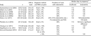
PVD (muco-purulent or purulent discharge in the cranial vagina, formerly called clinical endometritis) typically affects 15% to 20% of cows at 4 to 5 weeks postpartum and is consistently, though not unanimously, associated with impaired reproductive performance (LeBlanc et al., Reference LeBlanc, Duffield, Leslie, Walton and Johnson2002a; McDougall et al,. Reference McDougall, Macaulay and Compton2007; Santos et al., Reference Santos, Bisinotto, Ribeiro, Lima, Greco, Staples and Thatcher2010; Dubuc et al., Reference Dubuc, Duffield, Leslie, Walton and LeBlanc2010a; Lima et al., Reference Lima, Bisinotto, Ribeiro, Greco, Ayres, Favoreto, Carvalho, Galvão and JEP2013). Subclinical endometritis is inflammation assessed by endometrial cytology that is associated with impaired reproductive performance (Table 1). Accurate diagnosis of PVD requires examination of discharge in the vagina after a minimum of 3 weeks postpartum (LeBlanc et al., Reference LeBlanc, Duffield, Leslie, Walton and Johnson2002a), which may be done with a vaginoscope, clean gloved hand or a Metricheck device. Subclinical endometritis is diagnosed by endometrial cytology obtained trans-cervically either by uterine lavage or cytobrush (Barlund et al., Reference Barlund, Carruthers, Waldner and Palmer2008). Neither technique is sufficiently rapid or practical for widespread use in clinical practice, although rapid cow-side tests have been explored (Cheong et al., Reference Cheong, Nydam, Galvão, Crosier, Ricci, Caixeta, Sper, Fraga and Gilbert2012).
Summary of studies on diagnosis, prevalence and impact of endometritis assessed by uterine cytology to assess the cut-point proportion of PMN associated with time to pregnancy (updated from LeBlanc et al., 2011)

PMN=polymorphonuclear leukocytes; DIM=days in milk; PVD=purulent vaginal discharge.
* All differences P<0.05 in time-to-event (survival) analysis, except where indicated.
** Multiparous cows only; no effect in primiparous cows.
It is clear that endometritis often occurs without clinical signs and that PVD can occur in the absence of concurrent endometritis (Dubuc et al., Reference Dubuc, Duffield, Leslie, Walton and LeBlanc2010a; Lima et al., Reference Lima, Bisinotto, Ribeiro, Greco, Ayres, Favoreto, Carvalho, Galvão and JEP2013). The conditions are separate and each of PVD and endometritis (>5% to 8% PMN in an endometrial smear at 4 to 6 weeks postpartum) is associated with substantial reductions in subsequent reproductive performance and their effects are additive (Dubuc et al., Reference Dubuc, Duffield, Leslie, Walton and LeBlanc2011). In contrast with endometritis, PVD is commonly associated with concurrent bacterial uterine infection (Williams et al., Reference Williams, Fischer, Pfeiffer, England, Noakes, Dobson and Sheldon2005), in particular with T. pyogenes, although in one study only 41% of cows with PVD had positive uterine bacterial culture at diagnosis (Galvão et al., Reference Galvão, Greco, Vilela, Sá Filho and JEP2009a). Recently, uterine infection with α-haemolytic streptococci (AHS) or T. pyogenes at 10 DIM was associated with elevated PMN percentage 2 weeks later. Interestingly, infection with AHS was more prevalent (20% v. 6% of cows, respectively), and was independently associated with worse reproductive performance (Sens and Heuwieser, Reference Sens and Heuwieser2013).
Impacts of PVD and endometritis on reproductive performance
In a study in which 357 cows were examined by Metricheck and endometrial cytology, Lima et al. (Reference Lima, Bisinotto, Ribeiro, Greco, Ayres, Favoreto, Carvalho, Galvão and JEP2013) had few cases of only PVD (n=22 at 25 DIM) but showed that cows with both PVD and endometritis had reduced probability of pregnancy at first synchronized insemination than those with endometritis alone. Notably, they also documented substantially higher pregnancy loss from day 32 to day 60 in cows with endometritis that was present at 46 DIM or in cows with both PVD and endometritis at 25 DIM (43% and 30%, respectively, compared with 9% to 10% in unaffected cows). Conversely, cows with endometritis at 25 or 32 DIM, but not at 46 DIM had similar reproductive performance to unaffected cows. Two large studies have found no effect of subclinical endometritis at ∼5 weeks postpartum on pregnancy at first artificial insemination (AI) (whether all by synchronization and timed AI at 75 DIM (Lima et al., Reference Lima, Bisinotto, Ribeiro, Greco, Ayres, Favoreto, Carvalho, Galvão and JEP2013), or largely by detection of oestrus at a median of 74 DIM (Dubuc et al., Reference Dubuc, Duffield, Leslie, Walton and LeBlanc2011)). This is intriguing because many studies (Table 1) consistently show substantial increases in time to pregnancy in cows with endometritis at 3 to 6 weeks postpartum. Taken together, these data indicate that more investigation using longitudinal sampling of cows from 3 to 9 weeks postpartum is needed to better understand the time at which endometritis is associated with worse achievement of the economic goal of pregnancy by 100 to 150 DIM, to optimize the timing of diagnosis, and to probe the mechanisms by which endometritis affects longer-term reproductive performance.
Treatment of PVD and endometritis
Treatment of PVD and endometritis has been reviewed (Lefebvre and Stock, Reference Lefebvre and Stock2012). There is consistent evidence that cows with PVD have improved reproductive performance when treated with a single intrauterine (IU) infusion of cephapirin ∼1 month before first insemination, relative to receiving no treatment (LeBlanc et al., Reference LeBlanc, Duffield, Leslie, Bateman, Keefe, Walton and Johnson2002b; Runciman et al., Reference Runciman, Anderson and Malmo2009). IU infusion of ceftiofur at ∼6 weeks postpartum between two injections of PGF, 2 weeks apart, reduced the prevalence of uterine bacterial infection with E. coli from 10% to 2% and with T. pyogenes from 6% to 1% among cows with PVD but did not improve the probability of pregnancy at first insemination among cows that also received PGF twice before that insemination (Galvão et al., Reference Galvão, Greco, Vilela, Sá Filho and JEP2009a).
Numerous studies reported that one or two injections of prostaglandin F2α (PGF) improved reproductive performance or produced clinical outcomes similar to IU antibiotics. However, in studies of cows with risk factors for, or with endometritis, PGF consistently did not improve reproductive performance, but many of these studies lacked valid case definitions, statistical power or both. In a clinical trial in over 2000 cows, including over 600 with PVD, cytological endometritis or both, cows were randomly assigned to receive PGF at weeks 5 and 7 postpartum, or not (Dubuc et al., Reference Dubuc, Duffield, Leslie, Walton and LeBlanc2011). Overall, or among cows with reproductive tract disease, there was no difference in time to pregnancy between PGF-treated and control cows, which is similar to the findings of Galvão et al. (2009b) for cytological endometritis. However, the data from Dubuc et al. (Reference Dubuc, Duffield, Leslie, Walton and LeBlanc2011) were re-analysed to examine cows with PVD specifically and without regard to endometritis status (i.e. to address the clinical question of treatment of cows examined only for PVD (which is practical) but without diagnosis of endometritis by cytology (which is well validated, but impractical for routine clinical application)). Among 323 cows with PVD at 5 weeks postpartum, clinical resolution (absence of PVD) at 8 weeks postpartum was 72% in cows that received PGF at weeks 5 and 7 and 58% in untreated controls (P=0.01, not accounting for other variables). Among cows with PVD, 43% had a corpus luteum (CL) (serum progesterone>1 ng/ml) at week 5 and 63% had a CL week 7; 69% had a CL at least one of the times when PGF was administered. Accounting for parity, BCS at calving, occurrence of dystocia, retained placenta, twins and herd, cows with PVD that received two injections of PGF tended (P=0.07) to become pregnant sooner than untreated cases (hazard ratio=1.2, 95% confidence interval: 0.95 to 1.6). There was no interaction of the effect of PGF with the presence of a CL. Therefore, these results join others (LeBlanc et al., Reference LeBlanc, Duffield, Leslie, Bateman, Keefe, Walton and Johnson2002b) pointing to an equivocal effect of PGF for treatment of PVD. Subsequently, a systematic review of prostaglandin treatment trials for PVD/endometritis confirmed weaknesses and heterogeneity in the quality of diagnostic criteria, study design and outcome assessment (Haimerl et al., Reference Haimerl, Arlt and Heuwieser2012). These authors concluded that there is limited evidence in support of the efficacy of PGF as therapy for PVD/endometritis. The same research group built on this with a meta-analysis that similarly identified no improvement on reproductive performance from treatment of endometritis with PGF (Haimerl et al., Reference Haimerl, Heuwieser and Arlt2013). Subsequent to these syntheses, a trial showed that among cows with PVD, a programme of treating those with a palpable CL with PGF produced equivalent reproductive performance as treating all with IU cephapirin (McDougall et al., Reference McDougall, de Boer, Compton and LeBlanc2013). Lima et al. (Reference Lima, Bisinotto, Ribeiro, Greco, Ayres, Favoreto, Carvalho, Galvão and JEP2013) showed that PGF at 25 DIM reduced the prevalence of endometritis 1 week later, but PGF at 25 or 25 and 39 DIM did not improve reproductive performance. Different strategies for PGF as therapy for reproductive tract inflammatory disease merit further investigation.
Taken together, it appears that IU cephapirin is beneficial for reproductive performance in cases of PVD (which may be associated with cervicitis or endometritis) but that the benefit of PGF as commonly employed as therapy for PVD is unclear. While there is evidence of benefits to reproductive performance of either PGF or IU cephapirin (Kasimanickam et al., Reference Kasimanickam, Duffield, Foster, Gartley, Leslie, Walton and Johnson2005; Ghasemi, Reference Ghasemi2011) relative to no treatment, further investigation is needed of rapid ‘cow-side’ diagnostic tests to identify cases and of treatment for endometritis. Development of more effective treatments for reproductive tract inflammatory disease will require a better understanding of the factors that initiate and sustain endometrial inflammation, but investigation of modulators of inflammation for prevention and treatment is of interest.
Cervicitis
It was assumed that discharge found in the cranial vagina, or less commonly, observed externally on the vulva or tail, resulted from endometritis. The nature of vaginal content is associated with the density of putative bacterial pathogens in the uterus (Sheldon, 2004), but agreement between PVD and endometritis is weak (Dubuc et al., Reference Dubuc, Duffield, Leslie, Walton and LeBlanc2010a). This leads to the question of the source of the pus in the vagina if it is not always from the uterus. Cervicitis (inflammation within the cervix based on >5% PMN by cytobrush cytology ∼1 month after calving) affects 15% to 40% of cows and exists as a distinct condition, which is associated with both separate and additive impaired reproductive performance (Deguillaume et al., Reference Deguillaume, Geffré, Desquilbet, Dizien, Thoumire, Vornière, Constant, Fournier and Chastant-Maillard2012). Approximately half of cows with PVD have cervicitis and vice versa, and 50% to 75% of cows with endometritis have cervicitis and vice versa (Deguillaume et al., Reference Deguillaume, Geffré, Desquilbet, Dizien, Thoumire, Vornière, Constant, Fournier and Chastant-Maillard2012; Osawa and LeBlanc, unpublished observations). Treatment of cervicitis has not been studied directly. One hypothesis is that the observed benefit of treatment of PVD with IU cephapirin may include a benefit to at least some cows with cervicitis but not endometritis.
Risk factors for and relationships among reproductive tract diseases
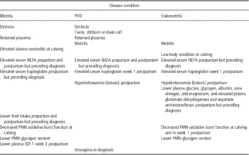
Numerous risk factors have been documented for each of metritis, PVD and endometritis. Complications at calving, retained placenta, more severe NEB or poorer adaptation to it, and more severe reductions in PMN oxidative burst function are common and shared elements among these diseases. Because no study measured all possible or even all known risk factors, and because many studies had limited statistical power to detect effects of moderate magnititude, it is impossible to accurately quantify or rank all the documented risk factors for reproductive tract inflammatory disease. However, cow-level clinical and metabolic risk factors for these conditions are summarized in Table 2. There were inconsistent or contradictory reports of parity, season of calving and milk yield as risk factors for these diseases. Differences in expression of genes related to inflammation in cows with endometritis were reviewed by LeBlanc (Reference LeBlanc2012).
Cow-level risk factors associated with reproductive tract inflammatory disease

PVD=purulent vaginal discharge; NEFA, non-esterified fatty acids; PMN=polymorphonuclear leukocytes.
Metritis is systemic illness with fetid vulval discharge and fever in the first 2 weeks postpartum; PVD is muco-purulent or purulent discharge in the cranial vagina after 3 weeks postpartum; and endometritis is inflammation diagnosed by uterine cytology between 3 and 9 weeks postpartum (detailed in Table 1).
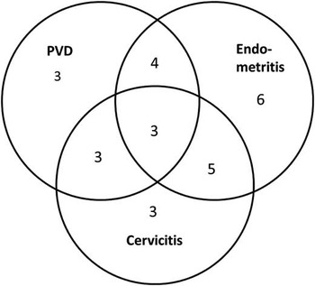
Although reproductive tract diseases may occur alone, affected cows commonly have more than one of these problems in the postpartum period, such that when studied together in the same cows, 37% had at least one of metritis, PVD or endometritis (Dubuc et al., Reference Dubuc, Duffield, Leslie, Walton and LeBlanc2011) and 56% had endometritis or cervicitis (Deguillaume et al., Reference Deguillaume, Geffré, Desquilbet, Dizien, Thoumire, Vornière, Constant, Fournier and Chastant-Maillard2012) by 5 weeks postpartum. We have explored the relationships among the chronic, localized forms of reproductive tract inflammatory disease and confirmed their overlapping nature (Figure 2).
Relationships among reproductive tract inflammatory diseases in dairy cows at 5 weeks postpartum. The numbers indicate the percentage of cows with each condition or combination of concurrent conditions. PVD (14% of cows affected); endometritis (18% of cows affected) and cervicitis (14% of cows affected) are defined by ⩾5% PMN in cytobrush samples. Data are from 102 Holstein cows, of which 27% had at least one of these three conditions (Osawa and LeBlanc, unpublished observations). PVD=purulent vaginal discharge; PMN=polymorphonuclear.

Prevention of reproductive tract disease
Little is known about how resistance to reproductive disease may be enhanced through management or nutrition. Cows with severe metritis ate less (2 to 6 kg of dry matter per day) than healthy cows in the 2 to 3 weeks preceding the clinical signs of metritis (Huzzey et al., Reference Huzzey, Veira, Weary and von Keyserlingk2007). Lower feed intake is associated with increased circulating concentrations of NEFA, which may directly (Scalia et al., Reference Scalia, Lacetera and Bernabucci2006; Ster et al., Reference Ster, Loiselle and Lacasse2012) or indirectly (Hammon et al., Reference Hammon, Evjen, Dhiman, Goff and Walters2006) inhibit neutrophil function. As discussed above, the high metabolic demands and pathogen challenges in early lactation result in substantial oxidative stress (Sordillo et al., Reference Sordillo, Contreras and Aitken2009), which also contributes to a pro-inflammatory state that may not be effective for immune defence.
Presently, there are few management practices or interventions that can be supported specifically to prevent metritis or endometritis. Based on current understanding of these diseases, the general objective is to support and maintain innate immune function (Ingvartsen and Moyes, Reference Ingvartsen and Moyes2013) and so reduce the risk that the inevitable inflammation and bacterial contamination after calving progress to metritis, PVD, endometritis or cervicitis. Excessive NEB and elevated concentrations of circulating free FAs, and excessive IR contribute to a state of systemic inflammation that may actually impair metabolic health and immune defence. While there is a great deal still to be learned, management practices generally recommended for peripartum dairy cows are likely to contribute to reducing the incidence of reproductive disease in the early postpartum period.
Conclusions
The balance between bacterial infection of the uterus in the weeks after calving and the effectiveness of the immune and inflammatory response to it determine the incidence of reproductive tract diseases. Understanding of links among metabolism and inflammation may lead to better approaches to prevention of reproductive disease through nutrition and management and more targeted therapies.