Introduction
The concept of Developmental Origins of Health and Disease has expanded for two decades.Reference Hanson and Gluckman 1 Many epidemiological studies and experiments on animal models have highlighted an increased risk for long-term metabolic and cardiovascular diseases in case of nutritional imbalance during pregnancy or gestation.Reference Hanson and Gluckman 1 Reproductive functions and fertility in adulthood may also be programmed during foetal development.Reference Dupont, Cordier and Junien 2
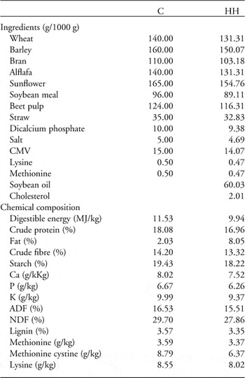
In humans, birth weight is commonly used as a proxy indicator for foetal growth, which can be influenced by maternal nutritional environment.Reference Gluckman and Hanson 3 A few studies have highlighted a relationship between male birth weight and infertilityReference Francois, de Zegher, Spiessens, D’Hooghe and Vanderschueren 4 or hormonal status, such as lower testosterone concentrations in the case of low birth weight.Reference Vanbillemont, Lapauw and Bogaert 5 In sheep, maternal undernutrition during gestation has been linked to altered gonadal development,Reference Alejandro, Perez and Pedrana 6 leading to a decrease in sertoli cell number and a reduction in the diameter of the seminiferous tubulesReference Kotsampasi, Balaskas, Papadomichelakis and Chadio 7 in adulthood; however, effects vary according to the timing of the nutritional stress.Reference Rae, Kyle and Miller 8 Maternal overnutrition in sheep has also been associated with alterations in reproductive functions of male offspring, such as a delay in the onset of puberty associated with lower plasma testosterone concentrations and reduced testicular volume.Reference Da Silva, Aitken, Rhind, Racey and Wallace 9 In the rat, a very recent paper showed that maternal obesity did not affect testicular weight and histology at birth, but the testicular weight of offspring from obese dams was 50% lower at D4.5 post-partum with a high variability in testosterone and oestradiol concentrations, which the authors think as impaired gonocyte development. At D7.5, there was no difference between the groups again.Reference Christante, Taboga, Pinto-Fochi and Goes 10 These studies used different species (sheep, rats) and the dams were overweight, whereas they have increased body fat in the present study. Moreover, the rabbit is a more appropriate model for high-fat nutrition and to assess the effect of developmental programming on reproductive functions.Reference Fischer, Chavatte-Palmer, Viebahn, Navarrete Santos and Duranthon 11 In our laboratory, a model of rabbits with dietary-induced hyperlipidaemia, hypercholesterolaemia and atherosclerosis has been developed.Reference Cordier, Leveille and Dupont 12 , Reference Picone, Laigre and Fortun-Lamothe 13 The hyperlipidic–hypercholesterolaemic (HH) diet was previously designed to obtain hypercholesterolaemia without inducing foetal losses during gestation and was inspired from diets used in the previous literature.Reference Montoudis, Simoneau and Lafond 14 – Reference Marseille-Tremblay, Gravel, Lafond and Mounier 16 The addition of soybean oil as a source of fatty acids was initially meant to have a relatively imbalanced N3/N6 ratio (N3/N6=0.15). Briefly, the control (C) diet (Table 1) was based on a constant composition of vegetal feedstuff with high-fibre contents adapted to rabbits. The experimental HH diet (C diet supplemented with 6% soybean oil and 0.2% cholesterol) contained quantitatively more fatty acids than the C diet from each fatty-acid class, mainly n-6 polyunsaturated fatty acids, and provided 16% more energy than the C diet. Rabbit does fed the HH diet reduce their food intake so that the ingested calories are similar between the two groups.Reference Picone, Laigre and Fortun-Lamothe 13 Recommended daily intake was monitored and validated by a nutritionist, ensuring that the rabbits were not submitted to nutritional deficiency. It has been observed that feeding mothers with the HH diet leads to intrauterine growth retardation.Reference Picone, Laigre and Fortun-Lamothe 13 As in humans, the rabbit male gonadal development begins in utero. In rabbits, the gonads begin to differentiate on the 16th day after fertilization (term=31 days). Primitive seminiferous tubules are formed and contain sertoli cells and germline before birth.Reference Gondos and Conner 17 , Reference Jost, Perlman and Magre 18 The objective of the present study was to assess the impact of maternal HH diet on male offspring reproductive functions at adulthood.
Ingredients and chemical composition of control and HH diets

C, offspring from control dams; HH, offspring from dams fed the hyperlipidic–hypercholesterolaemic diet; ADF, acid-detergent fibre; NDF, neutral detergent fibre.
Percentage of macronutrients refers to weight/weight.
Materials and methods
Animals
New Zealand rabbits (strain INRA 1077) were used. Females (n=8) were fed at libitum with either a control or an experimental hypercholesterolaemic (0.2%)–hyperlipidic (7.7%) dietReference Christante, Taboga, Pinto-Fochi and Goes 10 , Reference Fischer, Chavatte-Palmer, Viebahn, Navarrete Santos and Duranthon 11 from 10 weeks of age, throughout gestation and until offspring’s weaning, as described previously.Reference Christante, Taboga, Pinto-Fochi and Goes 10 , Reference Fischer, Chavatte-Palmer, Viebahn, Navarrete Santos and Duranthon 11 Briefly, females were mated with control males at 18 weeks of age. Pups were weaned at 5 weeks of age and were fed a C diet thereafter. Eight male pups born from four different does fed the HH diet and seven male pups born from four control does were used. Pups were chosen by random selection in each mother. Pups were not identified at birth, and therefore birth weight was not recorded.
Weight and body composition
Body composition was assessed at 18 weeks of age using a total body electrical conductivity technique (TOBEC). This method is valuable to predict energy and lipid body contentReference Fortun-Lamothe, Lamboley-Gaüzère and Bannelier 19 and provides information about total body lipid reserves. All rabbits were weighed before euthanasia at 37 weeks of age.
Arterial blood pressure and heart rate
Systolic, diastolic and mean blood pressures were measured from the ear artery at 37 weeks of age, a few days before euthanasia, as described previously.Reference Picone, Laigre and Fortun-Lamothe 13 An ear artery catheter connected to a pressure transducer (BP sensor; Phymep, Paris, France) was inserted. Data were monitored using a Powerlab system (ADInstruments, Oxfordshire, UK) coupled with computer software (Chart 5; ADInstruments).
Sperm and tissue collection
At 24 weeks of age, semen samples were collected using an artificial vagina. Sperm concentration and sperm-membrane composition were assessed.
At 37 weeks of age, all rabbits were fasted overnight and euthanized. Immediately after euthanasia, the kidneys, liver, heart and perirenal adipose tissues were weighed. The testes and epididymes were collected and weighed. The testes were fixed in 10% buffered formalin for 48 h and stored in phosphate buffered saline until analysis. The caudal part of the epididymes was removed with sharp scissors and placed in 1 ml of PBS to recover spermatozoa in the solution, as described previously.Reference Kumar, Sood, Arora, Singh and Beena 20 Collected spermatozoa were used for sperm DNA fragmentation analysis.
Sperm analysis
Sperm concentration in ejaculates was determined with a haemocytometer counting chamber. Sperm-membrane composition was evaluated. Sperm cholesterol and phospholipids were extracted by the method of Folch et al. adapted for spermatozoa, as described previously.Reference Grizard, Sion, Bauchart and Boucher 21 Lipid extracts were applied on the HPTLC plate and separated by using sequential development system: development to half final distance to resolve polar lipids followed by full development to resolve the non-polar lipids. The quantification was performed after staining. Lipids were quantified against standards by densitometry (Sigma Scan Pro, Sigma-Aldrich).
Sperm DNA fragmentation was assessed in spermatozoa collected from the epididymes with the TUNEL (Terminal Uridine Nick End Labeling) technique using an In Situ Cell Death Detection Kit (In Situ Cell Death Detection Kit, Fluorescein, Roche Applied Science).Reference Dupont, Faure and Sermondade 22 Labelled spermatozoa were measured on a FACScan flow cytometer (BD Biosciences; Becton-Dickinson). Each analysis included 10,000 spermatozoa.
Blood analysis
At euthanasia, a drop of blood was used to measure glycaemia using a glucose strip test (Optium Xceed Medisense, Abbott). Free testosterone concentrations were assayed in one single assay, to avoid inter-assay variability, using radioimmunoassay (DIAsource ImmunoAssays).
Histological analysis
Testis histological analysis was performed for in four HH males and five control males (one testis per animal).
Testes fixed in formalin were dehydrated and embedded in paraffin. Six micrometre sections were stained with haematoxylin and eosin and morphological observations were carried out using Nanozoomer Digital Pathology software (Hamamatsu, Japan).
The seminiferous epithelium height was measured from the basal membrane to the luminal edge. Thirty measures in three different cross-sections were carried for each testis using the public domain image-processing program ImageJ (National Institutes of Health, Bethesda, MD, USA).
Statistical analyses
Non-parametric statistical analyses were used. The Mann–Whitney test was conducted using R software (The R Foundation for Statistical Computing, http://www.R-project.org).
Data are expressed as the median, the first quartile and third quartile (Table 1). P⩽0.05 was considered statistically significant.
Results
Weight, body composition, metabolic status and cardiovascular evaluation
Weight and body lipid composition (measured by TOBEC) were not significantly different between the two groups at 18 weeks of age (P=1 and 0.35, respectively).
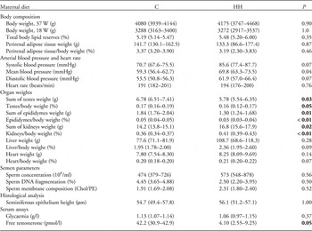
At 37 weeks, body weight and perirenal adipose-tissue weight were not significantly different between the two groups. Glycaemia was similar in both the groups. Systolic and diastolic blood pressure tended to be increased in rabbits born to HH dams (+21.1 and +15.7%, respectively, P=0.067) and mean arterial pressure was significantly increased (+17.7%, P=0.03). The heart rate was not affected by the maternal diet (Table 2).
Physiological and morphological observations in male offspring according to maternal nutrition

Bold values indicates statistically significant results (P<0.05).
C, offspring from control dams; HH, offspring from dams fed the hyperlipidic–hypercholesterolaemic diet.
Median (25th–75th percentile).
Organ weights
Kidney weight was significantly heavier in HH compared with C males (+18.3%, P=0.02; Table 2). Considering the relative weight, the difference remained significant (+14.0%, P=0.037). Absolute and relative weights of the liver and heart did not differ statistically between the two groups (Table 2).
Reproductive functions
Rabbits born from HH dams had significantly lighter testes [5.78 (5.54–6.35) g v. 6.78 (6.51–7.41) g, −14.7%, P=0.03] and epididymes [1.30 (1.24–1.68) g v. 1.84 (1.76–2.04) g, −29.3%, P=0.015] compared with rabbits born from C dams. Considering the relative weight, the difference remained significant (−4.1%, P=0.05 and −26.1%, P=0.009, respectively). The seminiferous epithelium height was comparable between the two groups. No significant differences in sperm concentration, sperm DNA integrity and sperm membrane composition were observed. Interestingly, plasma-free testosterone concentrations were decreased in males born from HH dams [42.2 (30.9–42.9) g v. 4.10 (2.55–9.25) g, −90.0%, P=0.05] at 37 weeks of age (Table 2).
Discussion
The present study indicates that maternal HH diet is associated with reduced testicular and epididymal weight in male offspring. Serum-free testosterone concentrations were decreased in HH males, possibly as a sign of modifications of the hypothalamo–pituitary gonadal axis. No alteration of the seminiferous epithelium structure was observed.
Male gonadal development starts in utero, and thus nutritional stress during this period may challenge testicular and epididymal development. As the testes develop slowly from birth to 5 weeks of age, the maternal diet before weaning (weaning at 5 weeks), that is, lactation period may also have affected the pups’ gonadal development, maybe through a change in the milk contents, as it was previously shown that obese rabbits had a disturbed mammary gland development during gestation.Reference Hue-Beauvais, Chavatte-Palmer and Aujean 23 Indeed, milk contains several hormones, such as leptin, that may be unbalanced by maternal nutrition and are required for the formation of hypothalamic pathways.Reference Sun, Purcell and Terrillion 24 Hypothalamic maturation is essential for subsequent gonadal development.
Sertoli cells play a central role in testis development and their number is correlated with testicular volume and sperm production. Sertoli cell proliferation requires testosteroneReference Sharpe, McKinnell, Kivlin and Fisher 25 that may be challenged by maternal hyperlipidaemia. The alteration of the maternal reproductive hormonal status previously observed may affect the foetal testis development through alterations of the hypothalamic–pituitary axis. Indeed, a decrease in LH levels and hyperinsulinaemia are observed in 18-week-old does fed with the HH diet since puberty,Reference Cordier, Leveille and Dupont 12 , Reference Picone, Laigre and Fortun-Lamothe 13 but no observations are available on their offspring. Moreover, increased aromatase activity in the adipose tissue and reduced production of testosterone by the Leydig cells may contribute to the decrease in plasma testosterone, as described in obese men.Reference Chavarro, Toth, Wright, Meeker and Hauser 26
Besides endocrine disruption, other mechanisms such as oxidative stress and epigenetic changes may be involved in foetal programming and also impair reproductive functions in offspring. Nutritional changes are known to affect oxidative balance.Reference Furukawa, Fujita and Shimabukuro 27 Reactive oxygen species (ROS) control many metabolic pathways by adapting intracellular signalling and ROS excess leads to DNA damages that can be transmitted to offspring. It was observed that obesity or high-fat diet leads to sperm DNA damages in humansReference Dupont, Faure and Sermondade 22 and in rodents.Reference Bakos, Mitchell, Setchell and Lane 28 However, no effect was observed on sperm quality or on sperm DNA fragmentation; this maybe because of the continuous process of spermatogenesis in the male and the fact that offspring were not submitted to the HH diet. Indeed, sperm DNA fragmentation occurs during spermatogenesis or during the transport of spermatozoa through the reproductive tract.Reference Sakkas and Alvarez 29 This does not preclude, however, the possibility of epigenetic effects that may affect fertility.
Finally, consequences of maternal nutrition may not be limited to offspring phenotypes but may also impact subsequent generations’ health. A sex-specific paternal transmission of metabolic disorders and impaired reproductive functions has recently been demonstrated in obese mice over several generations.Reference Fullston, Ohlsson Teague and Palmer 30 Alteration of epigenetic marks in the sperm may contribute to the future embryo development and therefore play a critical role in the long-term health.Reference Jenkins and Carrell 31
Conclusion
Environmental exposures during foetal and neonatal development are known to affect long-term health. In this study, we confirm the importance of maternal nutrition for reproductive organs development and the possible impact on fertility in adulthood. Although gonadal weight was affected, in the present study on a limited number of animals, no effects were observed on sperm-quality parameters. Further studies are needed to explore effects on fertility, the potential apposition of epigenetic marks and any transgenerational effect.
Acknowledgements
The authors thank Y. Lalatonne for testosterone assays, M.C. Aubrière and M. Guinot for technical assistance, B. Dondi for help with flow cytometry and the staff of INRA UCEA unit for the animal care.
Conflicts of Interest
None
Financial Support
This research received no specific grant from any funding agency, commercial or not-for-profit sectors. It was financed by INRA through internal support from the ‘Animal Physiology and Breeding Systems’ and ‘Human Nutrition’ Departments.
Ethical Standards
The experiment was conducted in accordance with the International Guiding Principles for Biomedical Research involving Animals as promulgated by the Society for the Study of Reproduction and in accordance with the European Convention on Animal experimentation. The whole experiment was approved by the local scientific committee for animal use (CSU UCEA) and received ethical approval from the local ethics committee (COMETHA, No. 45 in the National Registry), under protocol number 12/005.