Introduction
Dissociation between caloric and head impulse test results in patients with unilateral Ménière's disease has been well documented in the literature.Reference McGarvie, Curthoys, MacDougall and Halmagyi1–Reference McCaslin, Jacobson, Bennett, Gruenwald and Green5 Specifically, caloric testing indicates impairment of vestibulo-ocular reflex on the horizontal semicircular canal plane; however, video head impulse testing shows near-normal, normal or even increased vestibulo-ocular reflex gain on the horizontal semicircular canal plane.
In 2015, McGarvie et al. proposed a novel theoretical model to explain this dissociation.Reference McGarvie, Curthoys, MacDougall and Halmagyi6 In agreement with work by Valli et al.,Reference Valli, Buizza, Botta, Zucca, Ghezzi and Valli7 they conclude that the hydrostatic model, as proposed by Gentine et al.,Reference Gentine, Eichhorn, Kopp and Conraux8 is the only viable mechanism to explain caloric responses.
The hydrostatic model suggests that during caloric testing, the temperature variation across the temporal bone results in a difference in density, and therefore buoyancy, in the endolymph, between the medial and lateral arms of the horizontal semicircular canal. This produces a pressure differential across the cupula, which causes it to deflect.
Based on this model, McGarvie et al. proposed that in patients with Ménière's disease, the hydropic expansion of the horizontal semicircular canal membranous labyrinth allows for local convective flow and a mixing of the lower- and higher-density endolymph.Reference McGarvie, Curthoys, MacDougall and Halmagyi6 This dissipates the buoyancy that produces the pressure differential across the cupula, and therefore results in an impaired or absent response. Regarding the effect of hydropic expansion on angular acceleration testing (i.e. head impulse testing), they explain that as the membranous duct lies along the outer radius of the bony semicircular canal, there will not be a change in the overall diameter of the duct, rather only an increase in the cross-sectional area of the duct.Reference McGarvie, Curthoys, MacDougall and Halmagyi6 They reference modelling by Grieser et al.,Reference Grieser, McGarvie, Kleiser, Manzari, Obrist and Curthoys9 who hypothesise that the increase in cross-sectional area reduces the duct's natural hydrodynamic flow resistance. This allows greater endolymph flow velocities with an angular rotation, and therefore a larger cupula deflection, resulting in a small increase in vestibulo-ocular reflex gain. This increase in vestibulo-ocular reflex gain is indeed seen in some patients with Ménière's disease.Reference McGarvie, Curthoys, MacDougall and Halmagyi6
Case reports
We present two patients with specific congenital abnormalities of the vestibule and horizontal semicircular canal in whom there is dissociation between caloric and head impulse test results.
Case one
A 51-year-old female presented with a 6-week history of vertigo and mild left-sided tinnitus following a severe upper respiratory tract infection. She had no past medical history of note, and denied any previous episodes of vertigo or balance dysfunction.
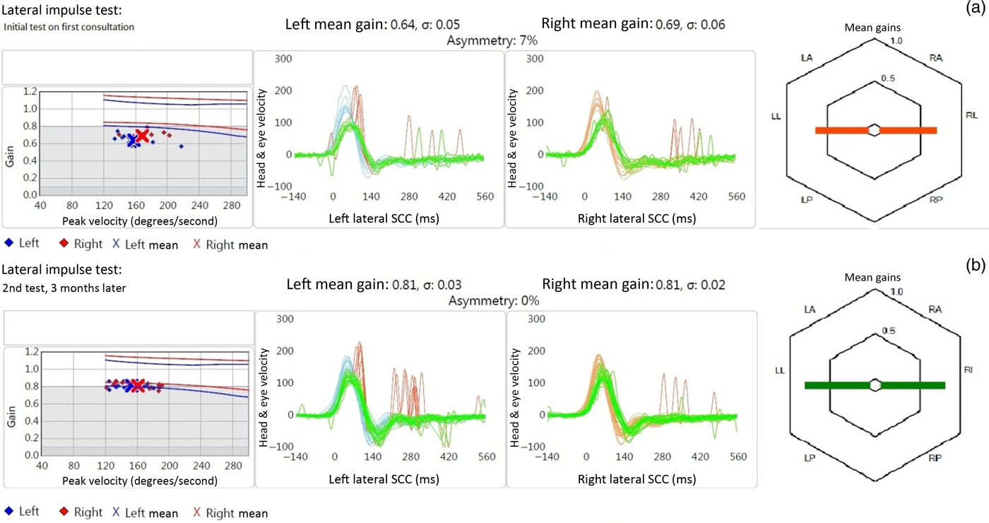
Clinical examination findings were unremarkable. A pure tone audiogram showed a bilateral mild high frequency sensorineural hearing loss, the pattern of which suggested presbycusis. Video head impulse testing on the horizontal semicircular canal plane showed reduced vestibulo-ocular reflex gains bilaterally (0.64 on the left and 0.69 on the right), with some covert catch-up saccades on the left side. The initial impression was a resolving left-sided vestibular neuritis with residual symptoms due to continuous use of vestibular sedatives and lack of vestibular rehabilitation exercises.
The patient was advised to stop taking the vestibular sedatives and to carry out gaze stabilisation exercises following detailed counselling. The repeat video head impulse testing conducted three months later showed improvement in vestibulo-ocular reflex gain (0.81 on both sides), into the normal range, but with persistent covert saccades on the left side (Figure 1). The patient reported significant improvements in her symptoms, although she did not feel there had been a return to her normal self (pre-viral illness state).
Video head impulse test data for case one: vestibulo-ocular reflex gain values showed improvement at the second visit (b), conducted three months after the first visit (a). Despite malformation of the vestibule and semicircular canal on the right side, the vestibulo-ocular reflex gain was similar (normal or near-normal) to the structurally normal left labyrinth. The left side, however, showed persistent covert saccades, indicating some dysfunction. SCC = semicircular canals; LA = left anterior; LL = left lateral; LP = left posterior; RA = right anterior; RL = right lateral; RP = right posterior

At this point, it was felt that the patient was recovering well, and she was discharged with an optional 12-month follow up. She was advised to continue gaze stabilisation exercises at home, with the expectation of further improvement.
The patient returned 12 months later and there had been no further improvement. The repeat video head impulse test results showed a slight reduction in vestibulo-ocular reflex gain (0.74 on the left and 0.79 on the right), with persistent covert saccades on the left side. Although the patient admitted non-compliance with gaze stabilisation exercises, it was felt that further investigations were necessary because of the persistent unilateral symptoms. Caloric testing and imaging were arranged.
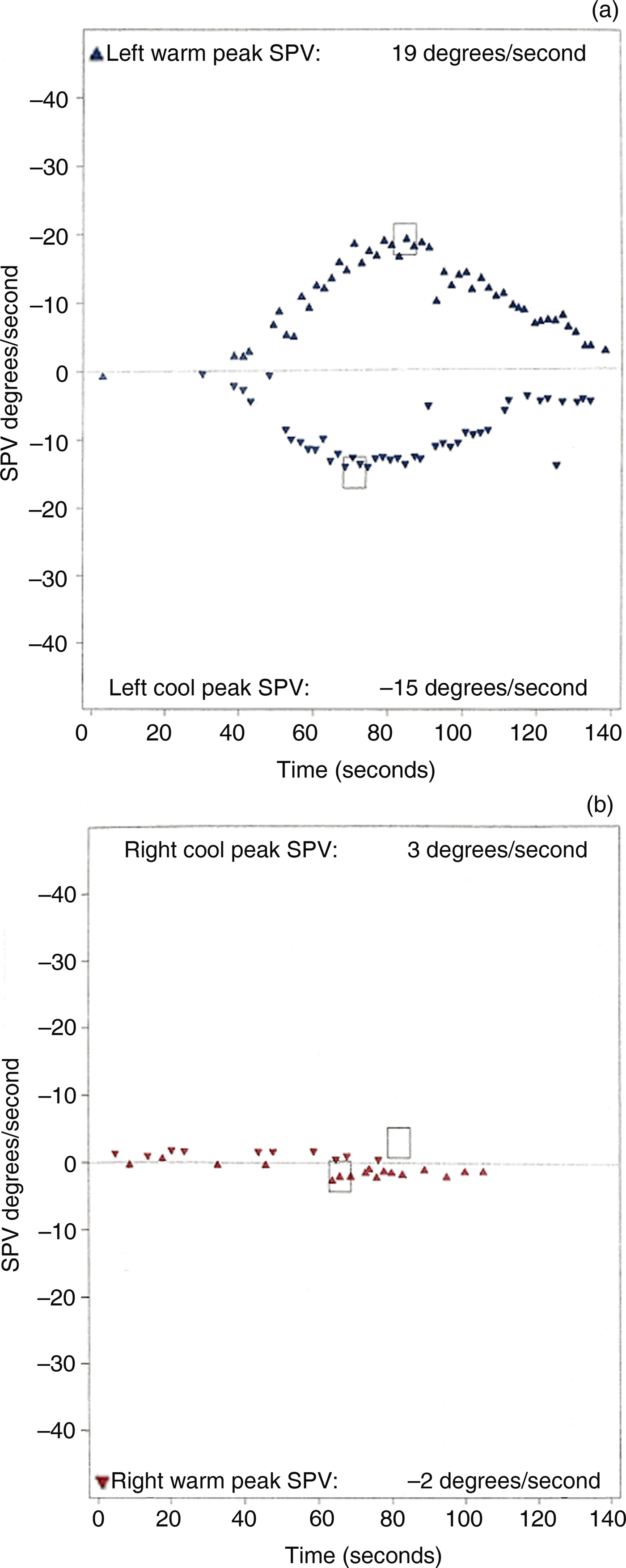
The caloric test showed a normal response on the left side but no response on the right side (Figure 2). Magnetic resonance imaging (MRI) and computed tomography (CT) revealed a developmental malformation of the right vestibule; there was a common cavity and an absence of a horizontal semicircular canal bone island (Figure 3).
Caloric test results for case one, showing a normal response on the left side (b) and an absent response on the right side (a). (Boxes represent peak slow-phase velocity (‘SPV’) for each condition, i.e. right cool, right warm, left warm and left cool.)

Computed tomography scan of temporal bones (axial view), showing a normal appearance on the left (‘L’) side (b) and a common cavity on the right (‘R’) side (a), with the loss of lateral (horizontal) semicircular canal bony island.

Our final diagnosis was vestibular neuritis on the left side, on the background of a congenital abnormality of the right horizontal semicircular canal and vestibule.
Case two
A 75-year-old male presented with left-sided pulsatile tinnitus and an asymmetrical, severe, sloping sensorineural hearing loss (worse on the left side).
Imaging was requested to exclude a vestibular schwannoma. No schwannoma was seen; however, MRI did show a large globular left horizontal semicircular canal, without evidence of a canal bone island, which formed a common cavity with a dilated vestibule. On the right side, there was a small semicircular canal bone island and mild prominence of the vestibule.
Video head impulse testing showed normal vestibulo-ocular reflex gains bilaterally on the horizontal semicircular canal plane, with catch-up saccades on the left side. Caloric testing showed a significant left canal paresis (42 per cent).
With regard to the pulsatile tinnitus, a CT scan showed a subtle erosion of the bony margins of the left jugular fossa, possibly an early sign of a glomus jugulare tumour. The patient was referred to a skull base unit for further opinion.
Discussion
Application of the theoretical dissipation model by McGarvie et al.Reference McGarvie, Curthoys, MacDougall and Halmagyi6 to our cases can explain the dissociation between the caloric and video head impulse test results. In both cases, the horizontal semicircular canal bone island was absent, and there was a common cavity formed by the vestibule and semicircular canal. This anatomical abnormality could also result in local endolymph circulation within the membranous labyrinth, thereby dissipating the hydrostatic pressure differential across the cupula (Figure 4), and resulting in an absent or impaired caloric response (Figure 2). In contrast, on rotational testing such as with video head impulse testing, inertia may still cause deflection of the cupula, resulting in the normal or near-normal responses shown above.
Schematic diagrams, showing: (a) the hydrostatic drive (solid curved arrow) on caloric testing in a patient with a normal lateral (horizontal) semicircular canal, and (b) the local convective flow (solid spiralling arrow) and therefore dissipation of hydrostatic pressure that might occur in patients with a common cavity. (Wavy arrows represent water flow (cold/warm).) Modified from an original drawing in McGarvie et al.Reference McGarvie, Curthoys, MacDougall and Halmagyi6 with permission.

• Dissociation between caloric and head impulse test results is well-known; a dissipation model has been proposed to explain it
• This paper reports similar dissociation in two patients with congenital abnormalities of the vestibule and horizontal semicircular canal
• The findings provide further support for the dissipation model
• The paper raises questions regarding the functional relevance of canal paresis values derived from caloric testing
The McGarvie et al.Reference McGarvie, Curthoys, MacDougall and Halmagyi6 model is supported by a recent study by Choi et al.Reference Choi, Kim, Cho, Lee, Park and Yoon10 They found that, in Ménière's disease patients with normal video head impulse test results, the reduced caloric function was related to vestibular hydrops severity (measured by MRI imaging).Reference Choi, Kim, Cho, Lee, Park and Yoon10
In both the cases reported, the symptoms that prompted referral for specialist assessment do not appear to directly relate to the pre-existing developmental anomaly. In the first case, the patient's symptoms started following a severe viral infection that affected the opposite side to the congenital malformation. In the second case, the pulsatile tinnitus that prompted the referral is likely due to erosion of the bony margin of the jugular fossa. Both patients were asymptomatic despite their anatomical malformations, presumably as they had adequate physiological vestibulo-ocular reflex function required for daily activities.
Although caloric testing provided information on the response of the end organ in our cases, the results did not predict the actual function of the vestibulo-ocular reflex system. Had diagnosis been based on caloric testing alone, the results could have been misleading, even after imaging revealed the anomalies. In both cases, the clinical history, together with the video head impulse test results and imaging (for the second case), rightly identified the new onset pathological process that triggered the symptomatology.
With regard to the management of these patients, it is important to give correct diagnoses, with adequate counselling and reassurance. Management based solely on the caloric test results would have been insufficient, and could even have been detrimental to their rehabilitation because of false beliefs and secondary psychological sequelae (e.g. anxiety, depression).
This report highlights the benefit of using both video head impulse testing and caloric testing in the management of patients with suspected vestibular disorders. It also raises doubts as to the functional significance of canal paresis values based on caloric testing and its relevance to patients’ symptomatology. We suggest that video head impulse testing be the first objective screening test to follow the initial clinical assessment for patients referred with suspected vestibular disorders. Additional tests, including caloric testing, should be used as and when required, based on clinical symptomatology.
Acknowledgements
We thank Professor Ian Curthoys for his valuable comments on the initial draft.
Competing interests
None declared