

Introduction
Understanding the structure and function of the insula has been a terrain filled with obstacles since first named by the anatomist Johann Christian Reil in 1809.Reference Reil and Autenrieth1 This is explained by the location of the structure; the insula lies deep in the brain in the depth of the Sylvian fissure, hidden by parts of the frontal, parietal and temporal lobes – the opercula – and hence is difficult to access for studying. Owing to the development of modern investigative techniques, the insula became nevertheless the subject of extensive anatomic, physiological, and functional studies and reviews, demonstrating a remarkable heterogeneity in architecture,Reference Clark2–Reference Mesulam, Mufson, Peters and Jones4 circuitry,Reference Augustine5–Reference Ghaziri, Tuchloka and Girard7 physiology,Reference Shelley and Trimble8, Reference Kurth, Zilles, Fox, Laird and Eickhoff9 and roles. The insula is now known to be involved in a variety of functions and networks including tasteReference Von Bechterew10, speech,Reference Rose11 sensory processing, motor control, autonomic regulation, higher cognitive functions, and behavioral and social interactions.Reference Mesulam, Mufson, Peters and Jones4, Reference Nieuwenhuys, Hofman and Falk6, Reference Shelley and Trimble8, Reference Gogolla12–Reference Namkung, Kim and Sawa14
Knowledge of this structure first came from the earlier classical morphological, developmental, and comparative studies done in human and primates’ insula and other mammalian models.Reference Clark2–Reference Augustine5 The study of functional neuronal microcircuits in animal models using modern technology in parallel with the increasing use of sophisticated functional imaging methods in humanReference Ghaziri, Tuchloka and Girard7, Reference Morel15, Reference Faillenot, Heckemann, Frot and Hammers16 and human observations during intraoperative functional explorations and brain stimulation studiesReference Penfield and Faulk17–Reference Oppenheimer and Cechetto22 eventually provided a better insight into the complex functional aspects of the insula. The structure is no longer seen as an isolated island or hidden treasureReference Gogolla12, Reference Nguyen23 but rather part of a sophisticated functional system involved in a wide range of neural processes.
These advances explain the re-emerging role attributed to the insula in several pathological contexts in psychiatry and neurology, including epilepsy.Reference Shelley and Trimble8, Reference Gogolla12, Reference Namkung, Kim and Sawa14 The insula contains multiple regions, each characterized by distinct circuits and connections involved in dynamic interactions with the rest of the brain and subserving a variety of functions. This explains the complexity and heterogeneity of the insular epileptic semiology.Reference Geshwind and Picard24 When it became recognized that removal of a portion of the cerebral cortex was a reasonable, and often successful, way to treat patients with severe focal epilepsies, epilepsy surgery was rapidly seen as another opportunity to advance scientific knowledge. Physiologists, neurologists, and neurosurgeons of the late nineteenth and early twentieth centuries understood this, and with the right questions, gaps of knowledge progressively became filled using more direct and sophisticated tools and sound observations.
Understanding the Role of the Insula in Humans and in Temporal Lobe Epilepsy
The Montreal Procedure: A Step Toward a Better Understanding on the Role and Function of the Insula
Between 1945 and 1953 in Montreal, Wilder Penfield and colleagues undertook a series of studies involving insular electrical stimulation performed on awake patients undergoing surgery for focal epilepsy under local anesthesia.Reference Penfield and Faulk17 Scrutinized analysis of a large series of epileptic patients with “patterns of attack … referable to various areas in the temporal and sylvian regions,” provided, decades ago, the very first direct observations on the roles of the insula in the human.
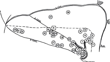
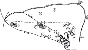
In these classical experiments derived from direct cortical electrical stimulation and later removal of the insula in epileptic subjects, Penfield and colleagues established a visceral sensory and motor response chart of the human insula (Figure 1.1, from Penfield and Faulk, 1955, with case records of these phenomena described in Table 1.1). They showed that electrical stimulations can evoke a variety of gastrointestinal sensations and responses (similar to observations made in mammals) and suggested that the insular cortex exerts motor control over the stomach (positive responses were obtained from the stimulation of a total of 82 different points on the insular cortex of 36 patients). The responses were divided into gastric sensory (e.g., “something funny in the abdomen, gurgling, pain, nausea, scratching”) and gastric motor (e.g., borborygmus, belching, or vomiting), and distributed mostly in the inferior portion of the insula. The authors recognized that the distribution of these responses was influenced by the fact that the inferior surface of the insula was easily accessible, which was not the case of the upper portion of the insula, “for fear of producing severe neurological damage.” Because of these limitations, they were not able to explore the entire region participating in alimentary function and hence to reproduce the sequence of alimentary motor events (“the feeding pattern”) described in dogs.Reference Geshwind and Picard25 In some patients, they could observe a sequence of chewing, swallowing, and salivation attributed to the involvement of the upper bank of the Sylvian fissure and extending “down into it”; they also described a “sensation of mouth and pharynx found on the opercula, and taste with a representation in a zone where the upper Sylvian bank joins the insula.” These early observations were the typical description of the now better understood operculo-insular epilepsy pattern. It also became clear that bilateral sensation in the upper alimentary tract and alimentary movements (e.g., swallowing and eating) were controlled or “represented” by the fronto-central operculum while movements of the stomach were controlled by the insula itself.
Visceral sensory and motor response chart – insula. Positive stimulation points from right and left sides have been summarized on this representation of the right insula. The area above and to the right of the broken line is covered by the parietal and frontal opercula during most operations. The area below is exposed regularly when the temporal operculum is removed in routine partial temporal lobectomy for temporal lobe seizures. Circles represent visceral responses and squares pyloric motor responses. When recorded motor responses were accompanied by visceral sensation, the circle is squared. The numbers correspond with detailed descriptions of the responses for each patient (32 responses in 15 patients), as shown in Table 1.1.
| Patient | Site of stimulation | Semiology described by patient |
|---|---|---|
| SM | 1 | Rising gastric sensation and salivation beginning in epigastrium and moving toward throat (aura). |
| JOL | 2 | Rolling in epigastrium and taste (aura). |
| NK | 3 | Like an attack. A pang of fear in the chest (aura). |
| 3 | Repeated. Feeling as if she were going to be sick. | |
| 3 | Further stimulation at this point repeatedly reproduced the aura. | |
| AKi | 4 | Swallowing, “I smell something funny,” in stomach (left leg numb, “felt like I had to move it”). |
| 5 | Something funny in mouth and stomach. Said he swallowed to get rid of bad taste in his mouth. | |
| 5 | Repeated. “My stomach is upset and I feel something like medicine, a sickly smell.” | |
| 6 | Sensation in abdomen. | |
| 7 | “A sickish taste in my throat coming up into my mouth.” | |
| 8 | Sudden flow of fluid from stomach through tube. | |
| 8 | Repeated. Recorded inhibition of gastric motility and tone. (Aura, feeling in stomach moving to throat where it was a taste.) | |
| JG | 9 | “Almost sick to my stomach.” |
| NH | 10 | Depression of respiration and abdominal sensation. |
| PM | 11 | “My stomach went up and down like you vomit.” |
| JMcC | 12 | “I feel like I cannot swallow.” |
| 12 | Repeated. “I feel like I want to vomit.” | |
| YS | 13 | Sensation in stomach. |
| 14 | Sensation in abdomen. | |
| 15 | Sensation in abdomen like aura. | |
| 16 | “Scratches in stomach.” | |
| 17 | Sensation in abdomen. | |
| 18 | Points to stomach, “Yes.” Like an attack? “Yes.” | |
| 19 | Like before an attack. Aura. Epigastric burning. | |
| JC | 20 | Attack going on to nausea. |
| 21 | Feeling about umbilicus. | |
| 22 | “This is it.” “Going to bring up some gas.” “Scared to death.” | |
| 22 | Repeated. Nausea and “far away” feeling, “scared.” (Aura. Peri-umbilical pain.) | |
| GB | 23 | Sensation at costal margin, left. |
| 24 | Same. | |
| 25 | Bad sensation in stomach (pointed to left costal margin). | |
| 26 | Sensation in epigastrium. | |
| 27 | Disagreeable taste and gastric sensation. | |
| 27 | Repeated twice. Same. | |
| DC | 28 | Sinking feeling in abdomen. |
| 28 | Repeated. Same. | |
| 28 | Repeated. Sinking feeling in abdomen. | |
| JW | 29 | Nausea followed by eructation – vomited 4 min later. |
| WG | 30 | Motor response (12 per min contractions). |
| 31 | Motor response. | |
| AH | 32 | Motor response. Inhibition of tone (relaxation). |
| 32 | Repeated. Inhibition of tone and desynchronization of regular 3 per min activity. |
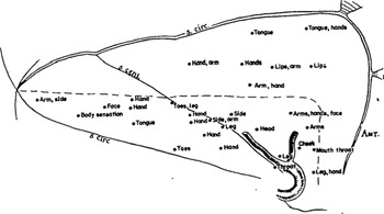
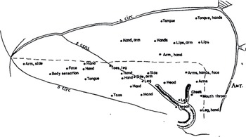
From these studies, a map of somatic sensory responses was also defined (Figure 1.2, somatic responses, from Penfield and Faulk 1955).Reference Penfield and Faulk17 To their great surprise, they found that “somatic sensation is elicited from the insular cortex almost as frequently as visceral sensation.” Their surprise came from the fact that contrary to visceral sensations, somatic sensations are noted infrequently in the “complex of psychical, motor or sensory seizures.” Stimulations of the insula evoked sensations (tingling, numbness, warmth, tightness, vibration, shock, impression of movement, etc.) not much different from those produced by stimulating the Rolandic sensory area (the primary somatosensory cortex SI or Brodmann areas 1, 2, and 3) or the superior bank of the Sylvian fissure in the second-sensory area (the secondary somatosensory cortex SII or Brodmann areas 40 and 43). Similar to the map of visceral responses, a pattern in the distribution of somatic sensory responses appeared: sensatanchorions from stimulations of the posterior and inferior portion of the sulcus centralis were contralateral and involved primarily the upper extremity and hand, while stimulations anterior to the central sulcus of the insula evoked mainly sensations in the lips, tongue, mouth, and throat, sometimes the upper extremities, and with a bilateral distribution. Lower extremity sensations were infrequent (possibly due to difficulty accessing the portion of the insula located posterior and superior to the sulcus centralis as evoked earlier), obviating adequate interpretation and localization.
Somatic sensory responses – insula. Points of stimulation that produced somatic responses on the right and left sides are transferred to this map of the right insula. The anterior end is to the right, posterior to the left. S. cent. = central sulcus, S. circ. = circular sulcus. The broken line separates the portion of the insula covered by the parietal and frontal opercula, above and in front, from the portion covered by the temporal operculum below. The upper extremity responses, below and posterior to the central sulcus, were all contralateral. Above the central sulcus and anterior, the responses were chiefly midline, and when there was spread, it included both arms.
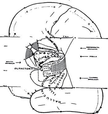
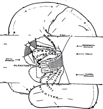
In the same sets of experiments, Penfield and his group made other important observations that are now considered fundamental to understanding insular and other peri-Sylvian focal epilepsies. First, the studies showed (not surprisingly according to Penfield) “that the results of stimulation of the insula are confusingly varied since it is surrounded by such remarkable variety of functional areas”: the anterior and posterior temporal operculum, including the auditory-sensory and vestibular-sensory posterior temporal region, the uncus and amygdaloid nucleus, the orbital operculum, the superior bank of the Sylvian fissure devoted to gustatory sensation, sensation in the throat and mouth and including SII, and, posteriorly, the post-central gyrus and parietal lobe (Figure 1.3, from Penfield and Faulk, 1955).Reference Penfield and Faulk17 They cleverly stated: “It is to be expected, therefore, that local epileptic discharges might cause the patient to experience sensations due to activation of one after another of these functional areas … [and] it follows too that local epileptic discharges often produce a great variety of seizure patterns.”
Functional areas in insular and circuminsular regions, as proposed by Penfield and Faulk in 1955Reference Penfield and Faulk17
In addition, they suggested that old atrophic lesions found in many of their patients, by modifying the normal functional organization of the insula, could contribute to a shift of the functional areas and add to the difficulties in interpreting correctly the electrical responses and the seizure semiology. They finally raised the hypothesis that a cortex subjected to years of epileptic discharges is likely more apt to respond to an electrical stimulation compared to a normal non-epileptogenic cortex, a so-called epileptic facilitation – a phenomenon resulting in a response of one area of the cortex to the stimulation applied to a neighboring “higher threshold” area. This could also contribute to difficulties in interpreting the results of electrical stimulation in the insula.
The difficulty of recording seizures during per-operative electrocorticogram (ECoG) due to an intrinsic time constraint represented an inescapable limitation for these studies, a problem shared during those years by all the groups interested in epilepsy surgery. Still, electrical stimulation studies of the insula cortex and peri-Sylvian cortical areas shed light on the functions of the insula, on its role in what was then believed to be temporal lobe epilepsy (TLE), and on the distinct contribution of the insular cortex in epilepsy. The pioneering work at the Montreal Neurological Institute (MNI) opened new avenues for reflection eventually leading to significant scientific and clinical advances in our understanding of focal epilepsy, TLE, and later insular cortex epilepsy more specifically.
The Beginning of Surgery in Insular Cortex Epilepsy
The first case of insular surgery for epilepsy was reported by Penfield at the Ferrier Lecture of the Royal Society given in 1946. The lecture was entitled “Some Observations of the Cerebral Cortex of Man” and was published in the Proceeding of the Royal Society in 1947.Reference Penfield26 He described the case in this way: “Case 1, J.C. … each attack was ushered in by an epigastric aura which she described as a strange feeling in the abdomen. This was followed in turn by a feeling of panic and evidence of intestinal peristalsis. At operation, stimulation of the pre- and post-Rolandic cortex and first temporal convolution evoked positive responses, motor, somatosensory or auditory (but none of her habitual seizure manifestations).” After the removal of the anterior and inferior portion of the temporal lobe, “stimulation of the insula caused her to feel her familiar abdominal aura, referred to umbilicus, and also to evoked an event ‘This is it’ … explaining after a pause that she had thought it was an attack and was ‘scared to death.’” In the Brain article of 1955, Penfield added: “The partial removal of the insula in addition to the partial temporal lobectomy resulted in a persistent seizure-free condition with both aura and seizures completely abolished, and … ‘the alimentary system functioning normally.’” Several reports of insular surgery were published during the following decade, by Guillaume and Mazars;Reference Guillaume and Mazars27, Reference Guillaume and Mazars28 Guillaume, Mazars, and Mazars;Reference Guillaume, Mazars and Mazars29 Penfield and Flanigin;Reference Penfield and Flanigin30 Penfield and Faulk;Reference Penfield and Faulk17 Ajmone Marsan and Baldwin;Reference Ajmone Marsan, Baldwin, Baldwin and Bailey31 and Silfvenius, Gloor, and Rasmussen.Reference Silfvenius, Gloor and Rasmussen32
The first report of Jean Marie Guillaume and Gabriel Mazars published in La Revue de Neurologie in 1949 concerned five cases of non-tumoral insular epilepsy diagnosed pre-operatively and confirmed by ECoG. The patients underwent a partial anterior temporal resection with a complete resection of the insula in three cases and the anterior ablation of the insula cortex in the remaining two. They did not report the seizure outcome. The authors acknowledged that their interest in insular epilepsy was triggered by the work of Penfield and Jasper and the expertise Gabriel Mazars acquired in Montreal during his visit to the MNI in 1946 or 1947.Reference Penfield and Faulk17, Reference Guillaume and Mazars27 In their second publication, this time concerning seven cases, the two neurosurgeons of La Pitié-Salpêtrière described in some details the techniques and approaches they used for the resection of the insula in insular epilepsies. They suggested that the morphological appearance of the insula is less important than the electrographic findings to determine the extension of the removal. They also emphasized the risk of this surgery due to the presence of numerous and large vessels, and the difficulty to appreciate the thickness of the insular cortex when operating. In the 1953 publication, the group from Paris presented a series of 24 patients operated for insular epilepsy. Most of them (23/24 patients) underwent a partial temporal resection (T1 and T2, or T1, T2, and T3), with a total insulectomy in 17, or a limited anterior insular removal – limen and short gyri – in 6 cases. Only one case had only an insulectomy. This time they reported good follow up results in 16/24 (or 67%), with 5 (21%) patients presenting a post-operative hemiplegia partly regressive in 2 or a transitory paresis in 3. They demonstrated that a variety of epilepsies can be defined as TLE, including the insular subtype, and in which adapted surgical approaches can result in the majority of cases in good outcomes. They also found that with experience and careful surgical planning, neurological sequelae in insular surgery are evitable. The planets appeared then aligned: a better understanding of the role of the insula in TLE and better indications for insular resections opened new perspectives for the hidden lobe, at least for a time. It seemed that the insula had finally emerged from the shadow and insular epilepsy was finally recognized as a distinct type of focal epilepsy.
In 1958, Ajmone Marsan and Baldwin from the National Institutes of Health (NIH) in Bethesda advocated that the removal of an epileptogenic insula may not be necessary in patients who undergo temporal lobe surgery.Reference Ajmone Marsan, Baldwin, Baldwin and Bailey31 In a series of 44 patients, they compared the incidence of insular epileptiform discharges in the final post-excision ECoG with the surgical outcome. They found equally good or satisfactory surgical results in the two groups of patients with (18 patients) or without (25 patients) residual epileptiform anomalies in the insula during the ECoG acquired after TL resection, and suggested that “the excision of an abnormally firing insula does not appear to be necessary.”
After reviewing the 16 years of experience at the MNI (from 1946 to 1962), Silfvenius, Gloor, and Rasmussen also concluded that ablation of the insula in the surgical treatment of TLE patients may not be necessary to achieve successful results – particularly in those patients where the insula appeared morphologically normal, even if electrically abnormal.Reference Silfvenius, Gloor and Rasmussen32 The study included 106 patients, all TLE cases treated by temporal resection, 58 with an additional partial or complete removal of the insula and 48 with no insular ablation despite insular epileptiform abnormalities on the post-TL excision ECoG. The follow-up showed that ablation of the insular cortex in these patients, as a whole, did not increase the number of successful outcomes, and that the presence or absence of residual epileptic activity in the post-excision ECoG did not affect those results. In addition, insular surgery was associated with frequent and serious surgical complications. In the group of patients undergoing a partial or complete insular resection, hemiparesis occurred in 21% of the patients (12/58, either complete, 6/21, partial, 4/31, or minor, 2/6) and speech disturbances occurred in 14% (8/58, either complete, 1/21, partial, 5/31, or minor, 2/6). In comparison, the group of patients with only TL resection leaving the insula intact, hemiparesis occurred in only 2% (1/48) and speech disturbances in 25% (12/48). Interestingly, they underlined that in some cases insular ablation might have favorably influence the surgical outcomes: patients with a higher rate (spike frequency per minute) of residual epileptiform abnormality in the post-excision insular ECoG tended to correlate with a higher incidence of unsuccessful surgical outcome. Moreover, patients who previously had had a TL surgery and were re-operated this time in the insula obtained better results compared to those who were re-operated but without an additional insulectomy. Therefore, and in spite of some clues for an independent role of the insula in certain forms of focal epilepsy presenting with a “temporal lobe flavor,” the conclusion was again that the presence of insular epileptiform abnormalities in the post-excision ECoG did not necessarily represent an indication for removal of the insular cortex. The enthusiasm in the epilepsy world dampened, and the insula was hidden again for three decades.
On the Necessity of Recording Seizures: How the Direct and Chronic Exploration of the Insula Has Changed the Paradigm
After an extended hibernation, the role of the insula in epilepsy received renewed attention. It appeared that the questions raised by the insula would remain unanswered unless the structure could be adequately monitored with chronic EEG recordings. In 1993, the group from UCLAReference Roper, Lévesque, Sutherling and Jr33 studied a young boy with refractory autonomic and motor seizures explained by a low-grade pilocytic astrocytoma located in the right insula. Seizures were of short duration and daily, consisting at onset of a feeling of “butterflies in his throat” followed by hyperkinetic movements with preserved consciousness. Subdural grid electrodes for chronic EEG recording were placed over the fronto-temporo-parietal convexity, and an eight-contact strip in the Sylvian fissure to record directly from the insular cortex. For the first time, the insula was recorded chronically allowing the documentation of the ictal phenomena and confirming in this case the role of the insula in the genesis of the seizures.
Jean Isnard and colleagues at the Neurological Hospital in Lyon then reported in a large series of patients with drug-refractory TLE, interictal and ictal clinical data recorded directly from the temporal and insular lobes using stereo-electroencephalography (SEEG).Reference Isnard, Guénot, Ostrowsky, Sindou and Mauguière34, Reference Isnard, Guénot, Sindou and Mauguière35 They confirmed that semiology and scalp EEG patterns explained by ictal insular involvement could not be clearly differentiated from those generated in the mesial TL. The fact that TL seizures were always associated with propagation into the insula enlightened the role of the insula in the genesis of symptoms that were classically associated with TL seizures. Eight of their patients showed insular spiking, but in all of them, an ictal insular propagation (19/21 patients) or genesis (2/21) was seen consisting either of a recruiting rhythmic activity or a low voltage fast activity. The propagation from the TL to the insula occurred in the majority only after a hippocampal relay and in a few from the temporal neocortex. These observations were novel since data so far had suggested that TL seizures preferentially propagated toward the cingulate gyrus and orbitofrontal cortex. They also correlated semiology with neurophysiology analyzing spontaneous ictal symptoms or evoked behavioral responses to electrical stimulation, and eventually were able to describe a sequence of manifestations reliable enough to characterize insular lobe seizures: “the occurrence in full consciousness of a symptomatic sequence associating a laryngeal discomfort with thoracic oppression or dyspnea, unpleasant paresthesia, or warmth sensation focused on the perioral region or extending to a large somatic territory and dysarthric or dysphonic speech reflects a propagation (or primary involvement) of the discharge to the insular cortex.” They added that “when this sequence is observed at the very onset of the seizure and is followed by focal somatomotor manifestations, it should be considered suggestive of insular epileptic seizures (or an insular generator should be suspected).” Although most of their patients did well after TL surgeries sparing the insula, some rather benefited from insulectomy. It became clear that temporal lobe-like seizures could indeed be generated in the insula itself rather than in the temporal structures, providing an explanation for some of the failures of TL surgeries. Isnard and colleagues finally provided an answer to an issue raised more than 35 years ago: a subgroup of the patients operated on at the MNI could have indeed benefited from an insular ablation.
A major step was undertaken, and following the results of this work published in 2000, they and others presented data that largely contributed to reactivating the interest in insular epilepsy in the adult and pediatric populations.Reference Ostrowsky, Magnin, Ryvlin, Isnard, Guénot and Mauguière18, Reference Isnard, Guénot, Sindou and Mauguière35–Reference Dylgjeri, Taussig and Chipaux43 The increasing use of invasive EEG recordings (in particular, SEEG) in patients with pharmacoresistant focal epilepsies permitted to reach safely and with great spatial and temporal resolution structures, like the insula, considered otherwise unattainable by non-invasive techniques. It allowed in refractory TLE patients to systematically study the ictal clinical and EEG patterns, to disentangle the temporal lobe and insular cortex seizures, and also to characterize patterns of propagation involving other networks than the temporo-insular system. The highly connected insula is now known to be associated with a variety of mesial or orbital frontal-insular, operculo-insular, peri-Sylvian-insular, or parieto-insular epilepsy networks. As with acute per-operative ECoG studies decades ago, chronic intracerebral electrodes studies has provided in the last 20 years a new momentum to study insular functions, understand the localization specificity of insular ictal semiology,Reference Isnard, Guénot, Sindou and Mauguière35, Reference Mazzola, Mauguière and Isnard44, Reference Isnard, Hagiwara and Montavont45 and refine surgical indications.Reference Duffau, Capelle and Lopes46–Reference Barba, Rheims and Minotti49
After More Than a Century Studying the Temporal Lobe, It Sounds Fair to Look at New Horizons
The history of insular epilepsy continues to be written as groups around the world attempt to increase our knowledge of this type of focal epilepsy. The prevalence and incidence of insular seizures and epilepsy remain an unresolved question; insular seizures are probably more frequent than what has been reported so far, but here data are lacking. The insula cortex is at the center of the network concept of epilepsy; this is relatively easy to understand in view of its strategic location and of the extensive connectivity the insula shares with most of the other cortical (temporal, frontal, and parietal) and subcortical areas of the brain. Invasive and direct neurophysiology remains, at this moment, the gold standard: interictal and ictal patterns are detected with a fair accuracy and can be correlated with semiology; and insular function and seizure semiology can be replicated by electrical stimulation. On a larger scale, however, this invasive method is not practical. With its remarkable heterogeneity in circuitry and physiology, a variety of functions, and the ability to generate complex and multiple pathological manifestations, the insula is a perfect field, “a gold mine,” to explore. Hence, the question arises: “How can we study best and understand the variety of insular epileptic networks, and how can we treat this disorder?”
The chapters that follow will review important knowledge gained over the last decade since the early works of Penfield and others. This book finality is to shine the light on this enigmatic structure of the brain, to propose strategies to further understand the neuronal mechanisms of insular function and insular cortex epilepsy, and to make sure that the insula will not anymore be a mysterious island.