Introduction
In vitro culture (IVC) systems used for embryo production still do not reach the level of effectiveness obtained in vivo (Gutiérrez-Adán et al., Reference Gutiérrez-Adán, Lonergan, Rizos, Ward, Boland, Pintado and De La Fuente2001). In several studies involving in vitro production (IVP) of cattle embryos, better results have been obtained when undefined or semi-defined macromolecules of animal origin are used (Rizos et al., Reference Rizos, Gutierrez-Adan, Perez-Garnelo, De La Fuente, Boland and Lonergan2003). Among these, the most common supplements are bovine serum albumin (BSA) and fetal calf serum (FCS). These elements supply the medium with growth factors, hormones, amino acids and proteins, which are beneficial to embryonic development (Gómez et al., Reference Gómez, Rodríguez, Muñoz, Caamaño, Hidalgo, Morán, Facal and Díez2008). Conversely, the use of these macromolecules has been associated with several abnormalities in oocyte and embryo physiology, such as changes in cell organelles, altered patterns of gene expression (Lonergan et al., Reference Lonergan, Rizos, Gutierrez-Adan, Farin and Boland2003), changes in cellular metabolism (Reis et al., Reference Reis, Maccallum and Mcevoy2005; Rizos et al., Reference Rizos, Clemente, Bermejo-Alvarez, De La Fuente, Lonergan and Gutiérrez-Adán2008) and changes in fluidity and function of the plasma membrane (Abe et al., Reference Abe, Yamashita, Satoh and Hoshi2002; Rizos et al., Reference Rizos, Gutierrez-Adan, Perez-Garnelo, De La Fuente, Boland and Lonergan2003). In addition, bovine embryos cultured in serum-containing media tend to present an excessive accumulation of cytoplasmic lipid droplets and fatty acid content when compared with those cultured in serum-free media (Abe et al., Reference Abe, Yamashita, Satoh and Hoshi2002; Reis et al., Reference Reis, Rooke, MacCallum, Staines, Ewen, Lomax and McEvoy2003; Pereira et al., Reference Pereira, Carvalhais, Pimenta, Baptista, Vasques, Horta, Santos, Marques, Reis, Silva Pereira and Marques2008; Sturmey et al., Reference Sturmey, Reis, Leese and Mcevoy2009).
The excessive lipid content in in vitro-produced embryos has been associated with their high susceptibility to cryopreservation (Abe et al., Reference Abe, Yamashita, Satoh and Hoshi2002; Rizos et al., Reference Rizos, Fair, Papadopoulos, Boland and Lonergan2002, Reference Rizos, Gutierrez-Adan, Perez-Garnelo, De La Fuente, Boland and Lonergan2003; Sudano et al., Reference Sudano, Paschoal, Rascado, Magalhães, Crocomo, Lima-Neto and Landim-Alvarenga2011). Although the mechanisms that lead to this lipid accumulation remain unclear, it occurs as a result of FCS supplementation in culture media or abnormalities in energy metabolism. In order to avoid the detrimental effects of embryonic lipids accumulation, some strategies have been proposed, such as the full withdrawal of serum from in vitro culture media (Lim et al., Reference Lim, Jang, Ko, Lee, Park, Kim, Lee, Hwang, Lee and Kang2007), the use of inhibitors of fatty acid synthesis (Sudano et al., Reference Sudano, Paschoal, Rascado, Magalhães, Crocomo, Lima-Neto and Landim-Alvarenga2011), and the addition of antioxidants (Gupta et al., Reference Gupta, Uhm and Lee2010) or molecules capable of modulating the molecular mechanisms of lipid uptake. These strategies are an attempt to change the embryos themselves to make them more cryotolerant (Seidel Jr., Reference Seidel2006), which can be considered the best approach to increase the success of cryopreservation.
One of the biologically active substances capable of modulating the molecular mechanisms of lipid uptake is linoleic acid (LA), which is a polyunsaturated fatty acid (PUFA) of the omega-6 family and is so-called essential because it cannot be synthesized by organisms (Schmitz & Ecker, Reference Schmitz and Ecker2008). Most studies on cryosurvival of bovine blastocysts have focused on the conjugated fatty acids of LA (CLAs), that comprise a group of positional and geometric isomers of LA in which the double bonds are conjugated in either the cis or trans configuration at positions 7,9-, 8,10-, 9,11-, 10,12- or 11,13- of the carbon chain (Sehat et al., Reference Sehat, Kramer, Mossoba, Yurawecz, Roach, Eulitz, Morehouse and Ku1998). The trans-10 cis-12 octadecadienoic CLA (10t,12c CLA) affects the lipid metabolism of adipocytes, reducing the uptake of free fatty acids (FA) without increasing lipolysis (Pariza et al., Reference Pariza, Park and Cook2001) and, due to its ability to modulate lipid uptake, Pereira et al. (Reference Pereira, Baptista, Vasques, Horta, Portugal, Bessa, Chagas, Silva Pereira and Marques2007) demonstrated that supplementation of serum-containing media for embryonic culture with 10t,12c CLA increased the blastocyst cryosurvival rate at 24 h of post-warming culture by reducing the embryonic lipid content.
Although little attention has been given to the effects of the non-conjugated form of LA on embryo development and cryoresistence, a previous study demonstrated that human embryos which developed beyond the 4-cell stage had significantly higher concentrations of unsaturated FAs, particularly LA and oleic acid, explained by the embryo's uptake of individual FA from the medium (Haggarty et al., Reference Haggarty, Wood, Ferguson, Hoad, Srikantharajah, Milne, Hamilton and Bhattacharya2006). As suggested by Hochi et al. (Reference Hochi, Kimura and Hanada1999), embryos presenting higher amounts of LA may be more resistant to cryopreservation as the supplementation of culture medium with unsaturated FA, such as linoleic acid-albumin, may increase membrane fluidity due to the direct incorporation of LA in embryonic cell membranes. However, this hypothesis still needs to be proven.
Besides their anti-lipogenic actions, PUFAs present various biological activities, being reported as anti-atherosclerotic, anti-carcinogenic, hypotensive, immunoenhancing and anti-oxidative (Koba et al., Reference Koba, Belury and Sugano2007; Andreoli et al., Reference Andreoli, Gonzalez, Martinelli, Mocchiutti and Bernal2009). However, contradictory research results indicate that the effects of CLAs on oxidoreduction cell equilibrium have yet to be fully elucidated. Fagali & Catalá (Reference Fagali and Catalá2008) compared the free radical scavenging properties of CLAs, LA and methyl linoleate (LAME) and their results indicate that CLAs would provide protection against free radicals, but LA and LAME would not. Additionally, further studies revealed that LA induces alterations in mitochondrial distribution and activity associated with increased reactive oxygen species (ROS) levels (Marei et al., Reference Marei, Wathes and Fouladi-Nashta2012). In contrast, Rybicka et al. (Reference Rybicka, Stachowska, Gutowska, Parczewski, Baśkiewicz, Machaliński, Boroń-Kaczmarska and Chlubek2011) observed that LA did not exert as strong an effect on ROS production as CLAs in macrophages, particularly 10t,12c CLA.
ROS, including hydrogen peroxide (H2O2), hydroxyl radical (HO·), and peroxyl radical (ROO·), are naturally produced by the cells as byproducts of the cellular respiration process that occurs in the mitochondria. Physiological concentrations of ROS, especially H2O2, are known to signal various cellular functions, such as energy production, cellular proliferation during early embryonic development, cellular differentiation and gene expression correlated with changes in the oxidative response (Harvey, Reference Harvey2007). The oxidative stress occurs when the generation of ROS is higher than the efficiency of the endogenous protective mechanisms (Guérin et al., Reference Guérin, Mouatassin and Ménézo2001; Feugang et al., Reference Feugang, Van Langendonckt, Sayoud, Rees, Pampfer, Moens, Dessy and Donnay2003). As a consequence, high amounts of ROS will adversely affect the development of in vitro-produced mammalian embryos (Corrêa et al., Reference Corrêa, Rumpf, Mundim, Franco and Dode2008), leading to metabolic disorders, lipid peroxidation and spatial modifications of plasma membrane structures (Guérin et al., Reference Guérin, Mouatassin and Ménézo2001; Nedambale et al., Reference Nedambale, Du, Yang and Tian2006; Corrêa et al., Reference Corrêa, Rumpf, Mundim, Franco and Dode2008), culminating in cell death (Halliwell & Gutteridge, Reference Halliwell and Gutteridge1999; Ufer et al., Reference Ufer, Wang, Borchert, Heydeck and Kuh2010). The use of low oxygen tension and/or antioxidant supplementation has been applied in an attempt to reduce the generation of excessive ROS caused by oxidative stress during IVP (Goto et al., Reference Goto, Noda, Mori and Nakano1993).
The aims of the present study were to investigate whether LA supplementation in serum-free culture medium under low oxygen tension can avoid the generation of intracellular ROS in the in vitro-produced embryos and to evaluate the impact of LA in lipid accumulation and embryo survival after cryopreservation.
Material and methods
Reagents and media
Chemicals were purchased from Sigma Chemical Co. (St. Louis, MO, USA), unless otherwise stated. Linoleic acid was diluted in dimethylsulfoxide (DMSO), which final concentration in culture media was < 0.03%) to obtain a stock solution of 300 mM, and was aliquoted and stored at −20°C. At the moment of use, the LA stock solution was diluted in the culture medium (SOF) to a final concentration of 100 μM.
The medium for in vitro maturation (IVM) consisted of tissue culture medium (TCM)-199 (Gibco®, Invitrogen Co., Grand Island, NY, USA) supplemented with 10% (v/v) FCS (Gibco®, Invitrogen Co.), 0.2 mM sodium pyruvate, 25 mM sodium bicarbonate, 50 μg/ml amikacin, 0.5 μg/ml FSH (Follitropin-V; Bioniche Animal Health, Ontario, Canada), and 100 IU/ml hCG (Vetecor®, Hertape Calier, Juatuba, MG, Brasil). IVF medium consisted of Tyrode's albumin lactate pyruvate (TALP) previously described by Parrish et al. (Reference Parrish, Susko-Parrish, Winer and First1988), containing 0.2 mM Na-pyruvate, 6 mg/ml fatty acid-free BSA, 25 mM sodium bicarbonate, 13 mM Na-lactate, 50 μg/ml amikacin, 40 μl/ml PHE solution (1 mM hypotaurine, 2 mM penicillamine and 250 μM epinephrine) and 10 μg/ml heparin. Embryo culture (IVC) medium consisted of modified synthetic oviductal fluid (SOF) previously described by Vajta et al. (Reference Vajta, Rindom, Peura, Holm, Greve and Callesen1999), supplemented with 50 μg/ml amikacin and 8 mg/ml fraction fatty acid-free BSA.
Oocyte collection and maturation
Bovine oocytes from slaughtered cows were obtained in a local abattoir and transported to the laboratory. Intact cumulus–oocyte complexes were aspirated from antral follicles (2–8 mm in diameter). Oocytes with at least four layers of cumulus cells were selected for the experiments. Oocytes were washed and cultured in 500 μl of IVM medium (50 oocytes per well) in a 4-well dish (Nunc®, Nunclon Delta Treated 4-Well IVF Dish, Denmark), without mineral oil covering. Culture was carried out for 22 h, at 38.5ºC under an atmosphere of 5% CO2 in air with maximum humidity.
Sperm separation and in vitro fertilization
Motile spermatozoa were obtained by centrifugation of frozen–thawed semen on a Percoll (GE Healthcare, Uppsala, Uppsala Country, Sweden) discontinuous density gradient (250 μl of 45% Percoll over 250 μl of 90% Percoll, in a 1.5 ml microtube) for 5 min at 2500 g at room temperature. Sperm cells were added to the fertilization droplet at 2 × 10⁶ cells/ml. Oocytes (25 per 90-μl droplet overlaid with mineral oil) and sperm were co-incubated for 18 h, under the same temperature and atmosphere conditions used for IVM. The day of fertilization was defined as day 0.
Embryo culture
Following fertilization, the presumptive zygotes were stripped from cumulus cells via vortex and randomly assigned to the different culture systems. They were transferred to 500 μl of SOF medium (50 oocytes per well) in a 4-well dish (Nunc®, Nunclon Delta Treated 4-Well IVF Dish, Denmark), without mineral oil covering. Embryos were cultured inside a plastic bag (low-density polyethylene, LDPE) at 38.5ºC and gaseous mixture of 5% CO2, 7% O2 and 88% N2, in a humidified atmosphere. Humidification in the plastic bag was achieved by filling a 10-cm plastic plate with 5 ml of sterile water that was introduced inside the bag. The gazing procedure was performed by filling the plastic bag with the gas mixture and then by closing the system with heat sealing. All culture systems were kept inside the water-jacketed CO2 incubator during the entire culture period, except for cleavage assessment. After cleavage assessment, the plastic bag was again filled with the gas mixture, sealed and kept in the incubator until the end of the culture.
Zygotes were incubated up to 72 h post-insemination (hpi) for assessment of cleavage rates. Blastocyst development rates were recorded at day 7 (168 hpi) and at day 8 of IVC (192 hpi).
Quantification of intracellular reactive oxygen species by dichlorofluorescein assay
Intracellular ROS levels in day 7 blastocysts were quantified using the fluorescent probe 6-carboxy-2′,7′-dichlorodihydrofluoresceine diacetate (H2DCFDA; Molecular Probes, Invitrogen, Oregon, USA). The H2DCFDA probe is oxidized directly by H2O2, its derivatives and other peroxides, and is also oxidized indirectly by the superoxide anion, thus providing a reliable tool to assess the intracellular ROS production (Gomes et al., Reference Gomes, Fernandes and Lima2006). A stock solution of H2DCFDA dissolved in DMSO was diluted in phosphate-buffered saline (PBS) to a working concentration of 5 μM (Bain et al., Reference Bain, Madan and Betts2011). Embryos were washed twice in PBS and placed into a 24-well cell culture cluster (Costar®) containing 500 μl of 5 μM H2DCFDA. Embryos were incubated at 38.5ºC in a dark, humidified 5% CO2 atmosphere for 30 min. Stained embryos were washed twice with fresh PBS and imaged immediately using an Olympus, IX51 inverted microscope running image Q-Capture Pro Image software (Media Cybernetics, Inc., Version 5.0.1.26), at excitation wavelength of 495 nm and emission wavelength of 520 nm to quantify the area and fluorescence intensity (pixels) from embryos (arbitrary units of fluorescence). The background signal intensity was subtracted from the measured values of the treatment micrographs.
Quantification of intracellular lipid content by Nile Red staining
The intracellular lipid content of single day 7 blastocysts was quantified using the fluorescent probe Nile Red. Staining was performed according to Genicot et al. (Reference Genicot, Leroy, Van Soom and Donnay2005), with some modifications. Briefly, embryos were fixed for 1 h in 1 ml 3.7% paraformaldehyde and 10% Triton X-100 solution. Then, they were transferred to plates containing 1 ml of a 10 μg/ml Nile Red solution dissolved in physiological saline (0.9% NaCl) with 1 mg/ml polyvinylpyrrolidone. Embryos were stained for 3 h in the dark and at room temperature. Stained embryos were washed twice with fresh PBS and imaged immediately using an Olympus, IX51 inverted microscope running image Q-Capture Pro Image software (Media Cybernetics, Inc., Version 5.0.1.26) to quantify the area and fluorescence intensity (pixels) from embryos (arbitrary units of fluorescence). The background signal intensity was subtracted from the measured values of the treatment micrographs.
Terminal deoxynucleotidyl transferase dUTP nick-end labelling
The terminal deoxynucleotidyl transferase dUTP nick-end labelling (TUNEL) assay was performed using the In Situ Cell Death Detection Kit (Fluorescein; Roche Diagnostics, Indianapolis, IN, USA), according to the recommendations of the manufacturer. Briefly, day 7 blastocysts were washed three times in 100-ml drops of PBS with 1 mg/ml polyvinylpyrrolidone (PBS-PVP) and fixed in 100-ml drops of 4% (w/v) paraformaldehyde solution for 1 h at room temperature. Afterwards, embryos were incubated in a permeabilization solution (0.5% v/v Triton X-100 and 0.1% w/v sodium citrate) for 30 min at room temperature. After washing three times in PBS–PVP, the positive control was incubated in a 50-μl drop of DNase I (50 IU/ml), whereas embryos from the experimental groups were incubated in 100-μl drops of PBS-PVP in the dark in a humidified chamber for 1 h at 37ºC. Subsequently, embryos were washed twice in PBS–PVP and incubated in 15 μl TUNEL reaction mixture (proportion of 1:9, enzymatic and label solution) for 1 h in a humid chamber at 37ºC in the dark. The negative control was incubated in 15 μl containing labelled solution only. Following the TUNEL reaction, embryos were washed twice in PBS–PVP and stained with Hoechst 33342 (1 μg/ml) for 30 min at room temperature. After washing with PBS–PVP, embryos were transferred to glass slides with mounting glycerol and observed under an epifluorescence inverted microscope (IX51; Olympus) at excitation and emission wavelengths of 510 and 590 nm, respectively. Nuclei with green–yellow fluorescence were considered TUNEL positive (fragmented DNA), whereas blue florescence (Hoechst 33342) indicated the total number of cells. To avoid considerable variability in the number of cells, only early blastocysts and blastocysts were stained for TUNEL.
Vitrification, warming and subsequent culture of embryos
All the materials used for vitrification/thawing procedures were supplied from Ingámed Ltda (Perobal, PR, Brazil), including the vitrification solutions (VI-I and VI-II, which contain increasing concentrations of ethylene glycol and DMSO), the thawing solutions (DV-I, DV-II and DV-III, that were sucrose-based medium), the vitrification strips (Vitri-Ingá) and their plastic sheaths. A two-step technique was used for vitrification of blastocysts. Briefly, expanded blastocysts harvested at day 7 of IVC (grades 1 and 2) were washed twice in holding medium (SOF supplemented with 250 mM of HEPES and 20% of FCS) and transferred to VI-I for 5 min at 37.0°C. Then, embryos were transferred to a 10-μl drop of VI-II for 60 s at room temperature. Afterwards, embryos were placed into the hole in the tip of a Vitri-Ingá strip with a minimum amount of vitrification solution, and then directly immersed in liquid nitrogen (N2). The plastic sheaths, which had been previously cooled in N2 liquid vapor for 1 min, were vertically immersed into N2 liquid. The Vitri-Ingá strip was then inserted into plastic sheaths for safe storage.
For thawing procedures, Vitri-Ingá strips were withdrawn from the liquid N2 and transferred to thawing solution DV-I for 1 min, at 37.0°C. After that, embryos were transferred to DV-II for 3 min at room temperature and washed twice in DV-III, for 5 min each. Then, embryos were washed twice and cultured in SOF supplemented with 2.5% of FCS and 5 mg/ml of BSA in saturated humidity and an atmosphere of 5% CO2 in air, up to 48 h. The re-expansion rates were assessed immediately after thawing and after 24 h of culture, and hatching was recorded after 48 h.
Experimental design
Cumulus–oocyte complexes (n = 3100, in 30 replicates) were matured in TCM-199 supplemented with 10% (v/v) FCS. Following IVF, the presumptive zygotes were transferred to a semi-defined IVC medium (supplemented with 8 mg/ml fraction fatty acid-free BSA) that lacked LA supplementation (Control group) or that had been supplemented with 100 μM of LA. The embryos were then cultured until day 7, when their development to the blastocyst stage was evaluated. The intracellular ROS levels of the blastocysts and early blastocysts were quantified using the H2DCFDA assay and the intracellular lipid content was quantified using the Nile Red fluorescent probe. The TUNEL assay was performed in early blastocysts to evaluate the rates of apoptosis. The expanded blastocysts were vitrified and, after warming, the re-expansion rates and hatching were evaluated following 24 h and 48 h of IVC, respectively.
Statistical analysis
Within each replicate, one well of a cell culture plate with 50 oocytes was considered the experimental unit for each group. Treatments were repeated in at least five replicates, on different days. Blastocyst production rates were estimated considering the number of developed blastocysts per oocyte inseminated.
Data were analysed with analysis of variance (ANOVA), using the general linear model (GLM) procedure with the SAS statistical software package 9.0 (SAS Inst. Inc., Cary, NC, USA. If the ANOVA was significant, the means were compared by a Tukey's test. The comparative study between binomial variables was evaluated by chi-squared test. The level of statistical significance was set at P < 0.05. A P-value between 0.051 and 0.1 was considered as tendency.
Results
The effects of supplementation with LA on the cleavage and blastocyst rates, and on the total number of cells and the percentage of apoptotic cells are given in Table 1. There was no effect (P > 0.05) of LA supplementation on cleavage rates (72.7% versus 74.1%, respectively for Control and LA groups). However, embryonic development to the blastocyst stage was higher (P < 0.0001) in the Control group (25.8% and 30.7%, respectively on day 7 and day 8) compared with the LA group (18.5% and 22.4%, respectively on day 7 and day 8). There was no significant difference (P > 0.05) among groups with respect to the total number of cells in day 7 blastocysts (114.9 ± 5.5 and 107.5 ± 5.3, respectively for Control and LA groups) or the percentage of apoptotic cells (1.8 ± 0.2 and 2.0 ± 0.3, respectively for Control and LA groups).
Effects of linoleic acid supplementation during in vitro culture of bovine embryos on cleavage rates and embryonic development to the blastocyst stage, and on the total number of cells and the percentage of apoptotic cells in day 7 blastocysts

Within columns, values with different superscript letters differ significantly (P < 0.05). Cleavage and embryonic developmental rates were evaluated by chi-squared test and the total number of cells and the percentage of apoptotic cells in day 7 blastocysts were evaluated by Tukey's test. Embryos were produced in vitro in medium supplemented with linoleic acid (LA; 100 μM) or without supplementation (Control).
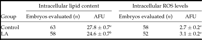
Regarding the results of the mean fluorescence intensity of bovine embryos after Nile Red staining (Table 2), we found a significant reduction (P = 0.0050) in intracellular lipid content in the LA group (27.8 ± 0.7) compared with the control group (24.6 ± 0.7). Intracellular ROS levels (Table 2), also expressed as arbitrary units of fluorescence, were unaffected (P > 0.05) in the LA group (3.1± 0.2) compared with the control group (2.7 ± 0.2).
Effects of linoleic acid supplementation during in vitro culture of bovine embryos on intracellular lipid content and on intracellular reactive oxygen species (ROS) levels [expressed as Arbitrary Fluorescence Units (AFU)] in day 7 blastocysts

Data are the mean ± standard error of the mean (SEM). Within columns, values with different superscript letters differ significantly (P < 0.05) according to Tukey's test. Embryos were in vitro cultured in medium supplemented with linoleic acid (LA; 100 μM) or without supplementation (Control). Treatments were repeated in five replicates.
The re-expansion rates of blastocysts after warming are shown in Table 3. There were no difference (P > 0.05) among groups immediately after warming (59.5% and 63.0%, respectively for Control and LA groups). However, supplementation with LA during IVC resulted in a higher (P = 0.0490) rate of in vitro embryo survival (71.7%) compared with the Control (50.0%) after 24 h of culture post-warming. The hatching rate after 48 h of culture was similar among groups (35.7% and 56.5%, respectively for Control and LA groups), but there was a tendency to obtain a higher hatching rate for LA group (P = 0.0577).
Effects of linoleic acid supplementation during in vitro culture of bovine embryos on the re-expansion rates of vitrified/thawed expanded blastocysts

Data are the mean ± standard error of the mean (SEM). Within columns, values with different superscript letters differ significantly (P < 0.05). *Considered as tendency (P = 0.0577). Embryos were in vitro cultured in medium supplemented with linoleic acid (LA; 100 μM) or without supplementation (Control). Treatments were repeated in five replicates.
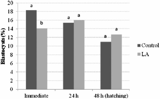
Aiming to evaluate the efficiency of the proposed system of IVP embryo culture for commercial purposes, we estimated the success rate by calculating the expected number of surviving (re-expanded) embryos after warming based on the total number of oocytes matured for each treatment (n = 1504 and n = 1596, respectively for the Control and LA groups, as demonstrated in Table 1). The estimated data were analysed by chi-squared test and results are presented in Fig. 1. Based on this estimation, the success rate (% of expected viable blastocysts after warming) immediately after warming was higher (P = 0.0018) in Control group (18.3%) than in LA group (14.1%), but was similar (P > 0.05) between groups after 24 h of culture post-warming (15.4% and 16.0%, respectively for Control and LA groups). Similarly, the estimated success rate of hatching expected after 48 h of culture post-warming was similar (P > 0.05) between groups (11.0% and 12.7%, respectively for Control and LA groups).
Estimated success rate (% of expected viable blastocysts after warming) calculated based on the total number of oocytes matured in vitro for each treatment. The data represent the means. The total numbers of oocytes used for each treatment group were as follows: Control: n = 1504 [based on results presented in Tables 1 and 3, blastocyst rate at day 8 was 30.7%; re-expansion rates of vitrified/warmed expanded blastocysts was 59.5% (immediately) and 50.0% (after 24 h of culture); and hatching rate after 48 h of culture was 35.7%], and LA: n = 1596 [blastocyst rate at day 8 was 22.4%; re-expansion rates of vitrified/warmed expanded blastocysts was 63.0% (immediately) and 71.5% (after 24 h of culture); and hatching rate after 48 h of culture was 56.5%]. Thirty replicates were performed. a,bDifferent letters above the bars indicate significantly different means (P < 0.05) according to the chi-squared test.

Discussion
The excessive accumulation of cytoplasmic lipid droplets and fatty acid content in in vitro-produced embryos increases their sensitivity to oxidative stress and cryopreservation (Abe et al., Reference Abe, Yamashita, Satoh and Hoshi2002; Rizos et al., Reference Rizos, Fair, Papadopoulos, Boland and Lonergan2002, Reference Rizos, Gutierrez-Adan, Perez-Garnelo, De La Fuente, Boland and Lonergan2003; Pereira et al., Reference Pereira, Carvalhais, Pimenta, Baptista, Vasques, Horta, Santos, Marques, Reis, Silva Pereira and Marques2008; Al Darwich et al., Reference Al Darwich, Perreau, Petit, Papillier, Dupont, Guillaume, Mermillod and Guignot2010; Sudano et al., Reference Sudano, Paschoal, Rascado, Magalhães, Crocomo, Lima-Neto and Landim-Alvarenga2011). The mechanisms that lead to this lipid accumulation remain unclear, but evidence indicates that it is related to the use of undefined components in the culture medium, or is a result of abnormalities in the embryonic energy metabolism caused by the IVC conditions, which also affect the properties and stability of cell membranes (Dinnyes & Nedambal, Reference Dinnyes and Nedambal2009; Leão et al., Reference Leão, Rocha-Frigoni, Cabral, Coelho, Ferreira, Eberlin, Accorsi, Nogueira and Mingoti2015). Results of the present study have shown that although LA supplementation of serum-free culture medium significantly reduced the rates of blastocysts produced under low oxygen tension, this proposed IVC system improved the quality of embryos by reducing their lipid content, thus leading to an increase in their resistance to cryopreservation.
A possible explanation for the decreased developmental capacity of embryos cultured with LA in semi-defined medium could be a putative excessive or unbalanced reduction of its intracellular lipid content. This effect was expected, as PUFAs, (especially CLAs, which are the most studied PUFAs on this subject), can decrease the intracellular lipid content in different cell types by reducing the uptake and synthesis of FA (Loor & Herbein, Reference Loor and Herbein1998; Baumgard et al., Reference Baumgard, Corl, Dwyer and Bauman2000; Brown et al., Reference Brown, Halvorsen, Lea-Currie, Geigerman and Mcintosh2001; Pariza et al., Reference Pariza, Park and Cook2001). Previous studies suggested that the molecular mechanisms involved in the reduction of lipid synthesis involve the coordinated reduction in the mRNA expression of important lipogenic enzymes associated with fat synthesis (Baumgard et al., Reference Baumgard, Corl, Dwyer and Bauman2002; Gervais et al., Reference Gervais, McFadden, Lengi, Corl and Chouinard2009). Although previous reports demonstrated that the addition of CLA to serum-containing media reduces embryo lipid accumulation during in vitro culture and improves cryopreservation survival (Pereira et al., Reference Pereira, Baptista, Vasques, Horta, Portugal, Bessa, Chagas, Silva Pereira and Marques2007, Reference Pereira, Carvalhais, Pimenta, Baptista, Vasques, Horta, Santos, Marques, Reis, Silva Pereira and Marques2008), it is unclear what would be a reasonable decrease in cytoplasmic lipid content of embryos produced in vitro, and which lipids should be decreased or incorporated into the cytoplasmic membrane. Thus, if we assume that LA presents the same anti-lipogenic effects than CLAs, it can be supposed that supplementation with LA in medium without FCS may have led to a marked or unbalanced reduction in lipid content or even have modified the embryo metabolism, which may have affected their developmental capacity.
Although it is well reported that the excessive accumulation of intracellular lipid droplets during in vitro embryonic development impairs the quality of IVP embryos by increasing their sensitivity to oxidative stress and cryopreservation (Pereira et al., Reference Pereira, Carvalhais, Pimenta, Baptista, Vasques, Horta, Santos, Marques, Reis, Silva Pereira and Marques2008; Al Darwich et al., Reference Al Darwich, Perreau, Petit, Papillier, Dupont, Guillaume, Mermillod and Guignot2010), it is important to mention that the lipids have an important role in cell physiology. Lipids are known to play key roles in energy metabolism during oocyte maturation, fertilization and early embryonic development because they provide energy for early preimplantation development before the activation of the embryo’s own genome (Paula-Lopes & Hansen Reference Paula-Lopes and Hansen2002; Ferreira et al., Reference Ferreira, Eberlin, Hallett and Cooks2012). Lipids also determine the structure and the fine-tuning of the physical properties and functions of biological membranes. Additionally, they have potent signaling effects during cell-cell interactions, cell proliferation and intra-cellular and inter-cellular transport (Kim et al., Reference Kim, Kinoshita, Ohnishi and Fukui2001). Thus, there appears to be required a delicate balance between the processes of synthesis, storage and utilization of intracellular lipids that can maintain the cellular activities in appropriated physiological levels.
Regarding the effects of PUFAs on embryo metabolism, it was previously reported that the addition of LA to maturation medium decreased the inner membrane mitochondrial potential in bovine oocytes, indicating mitochondrial dysfunction (Marei et al., Reference Marei, Wathes and Fouladi-Nashta2012). Although LA supplementation was included in the IVC medium in the present study, such possible detrimental effect on mitochondrial function of embryos should be considered and could also be an explanation for the decreased developmental capacity of embryos cultured with LA. The magnitude of the mitochondrial inner membrane potential has been suggested to specify levels of oxidative metabolism and determines levels of energy (ATP) generation, and is strongly correlated with embryo development (Wilding et al., Reference Wilding, Dale, Marino, Matteo, Alviggi, Pisaturo, Lombardi and De Placido2001). In addition, mitochondria are also responsible for the production of ROS and mitochondrial dysfunction could generate supraphysiological levels of ROS, leading to cell death. Thus, for two reasons it was important to assess the ROS production in the present work: to investigate a possible overproduction due to a putative mitochondrial dysfunction, and also because the medium was supplemented with a fatty acid (LA), which could increase lipid peroxidation in the culture system. As an attempt to attenuate or prevent this undesired effect, the embryos were cultured under low oxygen tension, which prevents the formation of free radicals that lead to lipid peroxidation in embryos, thus avoiding cell damage (Nagao et al., Reference Nagao, Lijima and Saeki2008). We found that LA supplementation in serum-free medium did not affect embryo quality, evaluated by intracellular ROS production, and by the total number of blastomeres and the proportion of TUNEL-positive cells (cells undergoing apoptosis). Although apoptosis is a physiological phenomenon that occurs, for example, during embryogenesis, with the purpose of eliminating damaged and unviable cells that could compromise embryo development (Feugang et al., Reference Feugang, De Roover, Moens, Leonard, Dessy and Donnay2004), excessive production of ROS can lead to cell death by apoptosis, resulting in embryonic development failure (Matwee et al., Reference Matwee, Betts and King2000). Thus, the results of this study demonstrate that the proposed strategy of LA supplementation in semi-defined medium under low oxygen tension was effective in preventing the formation of intracellular ROS and therefore prevent increased rates of cell death by apoptosis.
The strategy used in this study, serum-free semi-defined culture medium with LA, reduced embryo lipid content and improved cryosurvival. The positive effect of PUFAs on embryo freezability was first explained by Hochi et al. (Reference Hochi, Kimura and Hanada1999) who suggested that supplementation of embryonic cultures with LA bound to albumin (LAA) could result in an increase in the membrane fluidity (lipid unsaturation level) due to the direct incorporation of LA into the lipid bilayer. In fact, it was recently demonstrated that addition of CLA to serum-containing media significantly improved cryopreservation survival of bovine embryos due to a change in the composition of their membrane lipids, mainly by enriching the contents of sphingomyelin and phosphatidylcholine lipids containing a CLA fatty acyl residue (Leão et al., Reference Leão, Rocha-Frigoni, Cabral, Coelho, Ferreira, Eberlin, Accorsi, Nogueira and Mingoti2015). Authors suggested that a direct incorporation of CLA into the membrane during culture might contribute to an increase in the membrane fluidity (unsaturation level) and so improving embryo cryotolerance. In fact, CLA is responsible for inducing modifications in fatty acid profile and composition of tissues namely by reducing triacylglycerols content or interfering in saturated and unsaturated FA levels (Park et al., Reference Park, Storkson, Albright, Liu and Pariza1999; Alasnier et al., Reference Alasnier, Berdeaux, Chardigny and Sebedio2002; Al Darwich et al., Reference Al Darwich, Perreau, Petit, Papillier, Dupont, Guillaume, Mermillod and Guignot2010). Thus, if LA acts in the same way as CLA, its benefits on embryo resistance to cryopreservation will be both related to a decrease in the cytoplasmic lipid droplet content, as previously reported for CLAs (Pereira et al., Reference Pereira, Baptista, Vasques, Horta, Portugal, Bessa, Chagas, Silva Pereira and Marques2007), as well as to a change in the composition of the membrane lipids, as suggested by Hochi et al. (Reference Hochi, Kimura and Hanada1999) and demonstrated by Leão et al. (Reference Leão, Rocha-Frigoni, Cabral, Coelho, Ferreira, Eberlin, Accorsi, Nogueira and Mingoti2015). However, these mechanisms remain to be investigated.
Finally, the positive effects of LA on embryonic survival after freezing and thawing procedures can be via modulating the embryos’ arachidonic acid content. In a previous study, Kim et al. (Reference Kim, Kinoshita, Ohnishi and Fukui2001) demonstrated that degenerated frozen–thawed oocytes showed significantly lower concentrations of arachidonic acid than those that survived. According to authors, these lower concentrations may be due to the oxidative damage of oocyte membrane lipids during and after freezing and thawing. Arachidonic acid can be synthesized from linoleic acid, and oocytes may benefit by storing the more versatile precursor preferentially, which is at lower risk of free radical damage (McEvoy et al., 2000). However, arachidonic acid was not quantified in the present work and further studies are needed to fully understand the role of LA-mediated protection of in vitro-produced bovine embryos cryoinjury. Typification and monitoring of some lipid changes during the developmental stages and/or culture conditions should contribute to improve cryopreservation and in vitro culture conditions of mammalian embryos.
In summary, we demonstrated that culturing in vitro-produced embryos in serum-free medium supplemented with LA and under low oxygen tension leads to a reduction in the rate of embryo development to the blastocyst stage. Conversely, the proposed system was effective in promoting the decrease in the intracellular lipid content, consequently increasing the cryopreservation survival. Thus, aiming to evaluate the efficiency of the proposed system of IVP embryo culture for commercial purposes, we estimated the success rate by calculating the expected number of surviving (re-expanded) embryos after warming based on the total number of oocytes matured for each treatment. Based on this estimation, the success rate, i.e. the percentage of expected viable embryos for transfer after warming was similar between Control and LA groups after 24 h of culture post-warming, as well as the success rate of hatching expected after 48 h of culture post-warming. Then, it seems obvious that although the proposed system has provided minor conditions to allow the full embryonic development, the embryos presented higher qualities that enabled them to withstand the freezing stress. The present results question the commonly used criteria to evaluate the efficiency of an IVP system specifically with relation to the maximum number of blastocyst produced and suggest that might be more appropriate to establish a suitable culture system that could improve the quality of embryos generated according to the specific purpose of the in vitro embryo production, commercial or scientific.
In conclusion, despite supplementation with LA to the serum-free culture medium have impaired embryonic development, the cryopreservation survival was improved and this finding was related to a reduction in embryo's lipid accumulation.
Acknowledgements
The present study was supported by Fundação de Amparo à Pesquisa do Estado de São Paulo (FAPESP), Brazil. MFA was supported by a PhD scholarship from Conselho Nacional de Desenvolvimento Científico e Tecnológico (CNPq), Brazil. The authors wish to thank Alta Genetics Brazil Ltda for the donation of the straws of bull semen.