Introduction
Cognitive impairment is common in late-life depression (LLD). The impairment could be present in almost all cognitive domains, including processing speed and visuospatial and executive function (Butters et al., Reference Butters, Whyte, Nebes, Begley, Dew, Mulsant and Becker2004). Certain impairment may carry a prognostic value. For instance, executive dysfunction is associated with poor antidepressant treatment outcomes in both adult (Li, Su, Wang, Tu, & Hsieh, Reference Li, Su, Wang, Tu and Hsieh2015; Li et al., Reference Li, Lin, Chou, Chen, Hsieh, Wu and Su2010) and elderly patients (Morimoto, Kanellopoulos, Manning, & Alexopoulos, Reference Morimoto, Kanellopoulos, Manning and Alexopoulos2015) with major depressive disorder (MDD). Some cognitive impairments in MDD are strongly associated with depressive symptoms (i.e. ‘hot’ cognition), and some cognitive abnormalities do not disappear completely, even after remission of depression (Roiser & Sahakian, Reference Roiser and Sahakian2013). Patients with treatment-resistant depression (TRD), which refers to a history of poor responses to adequate antidepressant treatment, have prominent cognitive impairment (Li et al., Reference Li, Su, Wang, Tu and Hsieh2015; Rao et al., Reference Rao, Xu, Lu, Liang, Lin and Tang2019).
Dementia is a neurodegenerative illness characterized by prominent cognitive impairment, which considerably affects daily functioning of an individual. LLD is associated with a two- to five-fold increased dementia risk (Byers & Yaffe, Reference Byers and Yaffe2011). A population-based twin study in Sweden revealed that individuals with a recent diagnosis of LLD were 3.9 times more likely to have dementia than those without a depression history (Brommelhoff et al., Reference Brommelhoff, Gatz, Johansson, McArdle, Fratiglioni and Pedersen2009). The mean for the first depression episode was 11.6 years before dementia onset for the probands in patients with LLD (Brommelhoff et al., Reference Brommelhoff, Gatz, Johansson, McArdle, Fratiglioni and Pedersen2009). However, studies have shown inconsistent results (Becker et al., Reference Becker, Chang, Lopez, Dew, Sweet, Barnes and Reynolds2009; Byers & Yaffe, Reference Byers and Yaffe2011; Lindsay et al., Reference Lindsay, Laurin, Verreault, Hebert, Helliwell, Hill and McDowell2002). For example, a community-based cohort in the USA followed 288 participants up to 9 years, reporting no association between baseline depressed mood and incident dementia (Becker et al., Reference Becker, Chang, Lopez, Dew, Sweet, Barnes and Reynolds2009). Another nationwide population-based study in Canada included 194 patients with Alzheimer disease (AD) and 3894 cognitively normal controls but reported no significant association between a history of depression and development of dementia (Lindsay et al., Reference Lindsay, Laurin, Verreault, Hebert, Helliwell, Hill and McDowell2002). The inconsistent results echo a common clinical finding that cognitive impairment in MDD does not usually progress to dementia, and dementia may occur only in a subgroup of patients with depression.
Because AD has no cure to date, early prediction and intervention in individuals with a high risk of the disease is an essential but unsolved clinical requirement. A positive amyloid-PET is considered a surrogate marker of brain fibrillary amyloid pathology, which is one of the characteristic pathologies for AD (Dubois et al., Reference Dubois, Feldman, Jacova, Hampel, Molinuevo, Blennow and Cummings2014). The diagnosis of AD could be facilitated through β-amyloid (Aβ)-PET by using a validated radiotracer, such as Pittsburgh compound-B (11C-PIB) (Butters et al., Reference Butters, Klunk, Mathis, Price, Ziolko, Hoge and Meltzer2008). Nonetheless, accumulating evidence indicates that the accumulation of brain amyloid deposits may serve a protective function (Treusch, Cyr, & Lindquist, Reference Treusch, Cyr and Lindquist2009; Wolfe & Cyr, Reference Wolfe and Cyr2011). Therefore, our primary objective was to investigate whether LLD increases the AD neuropathology of the brain. We hypothesized that patients with LLD may have increased Aβ deposits in the brain regions correlated with impaired neurocognitive functioning. It is plausible because pilot research revealed that increased depressive symptoms in patients with LLD were associated with greater amyloid burden in the anterior cingulate cortex (Eyre et al., Reference Eyre, Siddarth, van Dyk, St Cyr, Baune, Barrio and Lavretsky2017).
A systemic review of case–control and cohort studies revealed that greater frequency and severity of depressive episodes apparently increase dementia risk (da Silva, Goncalves-Pereira, Xavier, & Mukaetova-Ladinska, Reference da Silva, Goncalves-Pereira, Xavier and Mukaetova-Ladinska2013). Further research indicated that dementia risk could not be captured through a single assessment of depressive symptoms but is associated with the depression course (Mirza et al., Reference Mirza, Wolters, Swanson, Koudstaal, Hofman, Tiemeier and Ikram2016). The high risk of dementia was observed only in patients with increased depressive symptoms (Mirza et al., Reference Mirza, Wolters, Swanson, Koudstaal, Hofman, Tiemeier and Ikram2016). A recent review indicated that patients with AD tend to respond poorly to standard antidepressants (Lozupone et al., Reference Lozupone, La Montagna, D'Urso, Piccininni, Sardone, Dibello and Panza2018). Therefore, our secondary objective was to compare brain Aβ deposits between patients with TRD and non-TRD (nTRD). We hypothesized that compared with patients with nTRD, patients with TRD have decreased cognitive functioning and increased Aβ deposits in the brain regions responsible for depression control. Given that the approved medications (e.g. donepezil, galantamine, and memantine) for AD provide only symptomatic relief and do not reverse or stop the disease progression (Hameed et al., Reference Hameed, Fuh, Senanarong, Ebenezer, Looi, Dominguez and Simon2020), the results of the present study may provide valuable information for identifying high-risk individuals and developing disease-modifying therapies.
Methods and materials
Participants
Eligible participants were adult patients aged >50 years and diagnosed as having recurrent MDD on the basis of the Diagnostic and Statistical Manual of Mental Disorders, Fourth Edition (DSM-IV) criteria. Diagnoses were established after thoroughly examining the medical history and conducting a semistructured interview through the administration of the Mini International Neuropsychiatric Interview published by American Psychiatric Association in 1994. Patients in the TRD group included those who failed to respond to at least two adequate antidepressant trials in their current episode (e.g. failed to achieve 50% improvement of depression to an equivalent daily dose of 10–20 mg of escitalopram for at least 8 weeks), whereas patients in the nTRD group included those who responded to an adequate antidepressant treatment in their current episode. Finally, a group of age-, sex-, and education-matched healthy controls (HCs) was recruited.
Participants were excluded if they had a psychiatric history of schizophrenia, psychotic disorders, bipolar disorder, or organic mental disorder, including substance abuse and dependence (on the basis of the DSM-IV criteria). People with a medical history of major systemic illness or neurological disorders (e.g. stroke, seizure, traumatic brain injury, post-brain-surgery disorder, and any type of dementia), brain implants (neurostimulators), and cardiac pacemakers and pregnant women were excluded. The study was performed in accordance with the Declaration of Helsinki and was approved by the local ethics review committee. All participants had provided written informed consent.
Depression and cognition assessments with apolipoprotein E (APOE) gene
We collected clinical depression variables including duration of illness and length of the current episode. Symptomatic rating was assessed using the Hamilton Depression Rating Scale (HDRS-17) (Hamilton, Reference Hamilton1967), since it is a validated and widely-used scale used in patients with MDD and TRD (Li et al., Reference Li, Chen, Juan, Huang, Chen, Hsieh and Su2014, Reference Li, Chen, Lin, Hong, Yang, Liu and Su2016a, Reference Li, Cheng, Chen, Juan, Tu, Bai and Su2020). Mini-Mental State Examination (MMSE) (Folstein, Folstein, & McHugh, Reference Folstein, Folstein and McHugh1975) and Montreal Cognitive Assessment (MoCA) (Nasreddine et al., Reference Nasreddine, Phillips, Bedirian, Charbonneau, Whitehead, Collin and Chertkow2005) were used to detect global cognitive functions. MoCA is more sensitive than MMSE in detecting mild cognitive impairment (Nasreddine et al., Reference Nasreddine, Phillips, Bedirian, Charbonneau, Whitehead, Collin and Chertkow2005). Memory function was measured using a Word List Test (Wechsler, Reference Wechsler1997). Immediate recall (i.e. score on the first word list task) and delayed recall (score on the second word list task) were used as outcome measures. Higher scores indicated better memory and recall. The Wisconsin Card-Sorting Test (WCST) was used to evaluate executive function (Heaton, Chelune, Talley, Kay, & Gurtiss, Reference Heaton, Chelune, Talley, Kay and Gurtiss1993). Two WCST indices were used for analysis: (1) percent (%) conceptual level responses, and (2) categories completed. More conceptual responses or categories completed indicated better executive function. The APOE gene is the most common and important genetic risk factor for late-onset sporadic AD (Coon et al., Reference Coon, Myers, Craig, Webster, Pearson, Lince and Stephan2007) and was also analyzed using the same procedures previously validated (Yang et al., Reference Yang, Huang, Liu, Liou, Hong, Lo and Tsai2014).
Imaging studies
Structural MRI
Magnetic resonance images were acquired using a 3.0 GE Discovery 750 whole-body high-speed imaging device for all participants. High-resolution, structural T1-weighted images were acquired for improving the co-registration of PET images in the sagittal plane using a high-resolution sequence (repetition time, 2530 ms; echo spacing, 7.25 ms; echo time, 3 ms; and flip angle, 7°) with isotropic 1 mm voxels and FOV = 256 × 256 mm2.
PET
PET data acquisition: 11C-PIB PET scans of Aβ depositions and fluorodeoxyglucose (18F-FDG) PET scans of glucose use at rest were acquired using a PET/CT scanner (Discovery VCT; GE Healthcare, USA) or an integrated PET/MRI scanner (SIGNA; GE Healthcare) for patients who agreed to receive simultaneous PET scans. The distribution of PET machines (PET/CT v. PET/MRI) was consistent among HC (8 v. 6), nTRD (9 v. 5), and TRD (9 v. 5) groups (χ2 = 0.202, p = 0.904). All participants fasted for at least 8 h before the PET examination. 11C-PIB PET images were acquired and averaged over frames corresponding to 40–70 min after an intravenous injection of approximately 555 MBq per 70 kg of 11C-PIB (Yamane et al., Reference Yamane, Ishii, Sakata, Ikari, Nishio, Ishii and Group2017). After the 11C-PIB PET acquisition, an intravenous injection of approximately 370 MBq per 70 kg of 18F-FDG was applied (Li et al., Reference Li, Hsieh, Wang, Yang, Bai, Lin and Su2012). 18F-FDG PET images were then acquired and averaged over frames corresponding to 45–60 min postinjection intervals. Other details of PET/CT acquisitions were identical to those of our previous study (Li et al., Reference Li, Hsieh, Wang, Yang, Bai, Lin and Su2012). All the acquired three-dimensional (3D) PET scan images in time-of-flight mode data were reconstructed into a 256 × 256 mm2 matrix and 3D ordered subset expectation maximization algorithm with five iterations, 32 subsets, and a 3 mm Gaussian filter. As for the PET/MRI scanner, scatter correction, MRI-based attenuation correction, point spread function correction, dead time correction, and decay correction were performed.
PET analysis: PET data were analyzed using Statistical Parametric Mapping [version 8 (SPM8); Wellcome Department of Cognitive Neurology, Institute of Neurology, University College London, London, England] implemented in MATLAB (version 7.1; The Mathworks Inc., Sherborn, MA, USA). First, all PIB images were visually inspected and classified as PIB negative or positive on the basis of the criteria validated previously (Akamatsu et al., Reference Akamatsu, Ikari, Ohnishi, Nishida, Aita, Sasaki and Senda2016). To improve the normalization accuracy, two group-specific MRI-aided 11C-PIB templates were created, one for PIB-negative participants and the other for PIB-positive participants (online Supplementary Fig. S1), and used separately to normalize each participant's PIB PET images. Furthermore, a group-specific MRI-aided 18F-FDG template was created (Gispert et al., Reference Gispert, Pascau, Reig, Martinez-Lazaro, Molina, Garcia-Barreno and Desco2003; Signorini et al., Reference Signorini, Paulesu, Friston, Perani, Colleluori, Lucignani and Fazio1999) and used to normalize each participant's 18F-FDG PET images. Parametric whole-brain standardized uptake value image was then calculated by dividing the radiotracer activity concentration in the normalized PET image (MBq/kg) by the radiotracer dose (MBq) divided by the body weight (kg) (Li et al., Reference Li, Chen, Lin, Hong, Yang, Liu and Su2016a). For PIB images, all voxel values were further normalized on the basis of the mean value of the cerebellar cortex (‘Cerebellum_Crus’ in the AAL-ROI) to generate standardized uptake value ratio (SUVR)-scaled images (Akamatsu et al., Reference Akamatsu, Ikari, Ohnishi, Nishida, Aita, Sasaki and Senda2016) (online Supplementary Fig. S1). Cerebellum is an ideal reference region for parametric 11C-PIB PET analysis (Tolboom et al., Reference Tolboom, Yaqub, Boellaard, Luurtsema, Windhorst, Scheltens and van Berckel2009). Finally, all normalizations were followed by smoothing with a 3D Gaussian kernel (FWHM = 8 mm).
To assess potential group differences in normalized and smoothed images, analysis of covariance models with age, gender, education, PET machines, HDRS-17 scores, and global uptake values as covariates of no interest were used for between-group comparisons (Li et al., Reference Li, Chen, Lin, Hong, Yang, Liu and Su2016a). The significance thresholds for between-group comparisons were set at a voxel-level p < 0.05 [family-wise error (FWE)-corrected for multiple comparisons]. If failing to pass the strictest criteria, we reported potential group differences by setting the significance thresholds at voxel-level uncorrected p < 0.001 and cluster-level FWE-corrected p < 0.05. Using a voxel-level corrected p < 0.05 could be too conservative for a priori brain regions involved in dementia-related cognitive impairment with 11C-PIB PET (e.g. the prefrontal, parietal, and posterior-cingulate cortices and precuneus) (Devanand et al., Reference Devanand, Mikhno, Pelton, Cuasay, Pradhaban, Dileep Kumar and Parsey2010).
Statistical analysis for other variables
For analyzing demographic data and clinical variables, SPSS 16.0 software (SPSS Inc., Chicago, IL, USA) was used. One-way ANOVA (or Student's t test) and the χ2 test (or Fisher's exact test, when the expected value in a cell was <5) were used to compare continuous and categorical variables among groups, respectively. Before the analysis for continuous variables, we used Levene's test for homogeneity of variances to ensure that the homogeneity assumption has not been violated. If the results from Levene's test were significant (p < 0.05), non-parametric analysis would be applied. All variables, but the duration of illness, passed the Levene's test. Mann–Whitney U test was used for the two-group comparisons of the duration of illness. Sample size was predetermined on the basis of high PIB deposits in normal elderly with prominent depressive symptoms (Yasuno et al., Reference Yasuno, Kazui, Morita, Kajimoto, Ihara, Taguchi and Nagatsuka2016) and calculated using the Power and Sample Size Program (version 3.0.43) (Dupont & Plummer, Reference Dupont and Plummer1990). The estimated parameters used to reject the null hypothesis included the population means of the experimental and control groups being equal with a probability (power) of 0.8, and the type I error probability associated with this test's null hypothesis was 0.05. Pearson correlation was used to investigate relationships between general cognitive function (i.e. MMSE and MoCA) and the brain region identified to be the most significantly differed in the between-group comparison (i.e. prefrontal cortex). We then used multiple regression analyses to examine the effects of PIB deposits in the prefrontal cortex on general cognitive function after adjusting for age, sex, educational level, and HDRS-17 scores. A p value of <0.05 was considered statistically significant.
Results
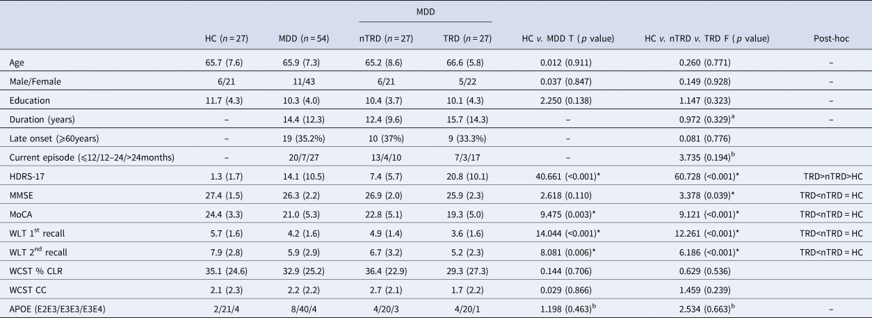
We screened 106 subjects and eventually recruited 81 subjects (27 TRD, 27 nTRD, and 27 HCs) for the present study. Demographic data did not differ between MDD and HC groups (Table 1). MDD patients had significantly higher HDRS-17 (p < 0.001), lower MMSE (p = 0.039) and MoCA (p < 0.001), and lower WLT 1st recall (p < 0.001) and 2nd recall (p = 0.006) than HC (Table 1). TRD patients had significantly higher HDRS-17 (TRD > nTRD > HC, p < 0.001), lower MoCA (TRD < nTRD = HC, p < 0.001), and lower (WLT 1st and 2nd recall; TRD < nTRD = HC, p < 0.001). No significant differences between TRD and nTRD were found for total illness duration, duration of the current episode, and ratios of late-onset depression. No between-group differences for the APOE gene were found (Table 1). Late-onset patients performed worse in global cognition and WCST than non-late-onset patients. For other details please refer to the online Supplementary material.
Demographic data and clinical variables

MDD, major depressive disorder; HC, healthy control subjects; TRD, treatment-resistant depression; nTRD, non-TRD; HDRS-17, 17-item Hamilton Depression Rating Scale; MMSE, Mini-Mental Status Examination; MoCA, Montreal Cognitive Assessment; WLT, Word List Test; WCST, Wisconsin Card Sorting Test; CLR, conceptual level responses; CC, categories completed; APOE, apolipoprotein E gene. *p < 0.05.
a P = 0.658 as revealed by Mann–Whitney U analysis.
b Fisher's exact test was applied.
MDD v. HC: PIB SUVR and normalized brain glucose use
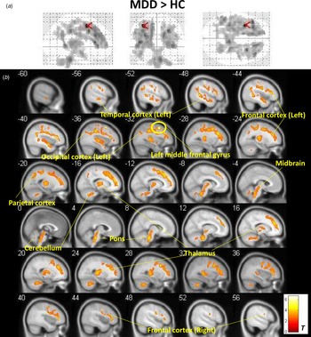
Forty-two subjects (14 TRD, 14 nTRD, and 14 HCs), whose neurocognitive results were similar to those in the 81 subjects (online Supplementary Table S1), completed the PET scans. The voxel-wise comparisons of PIB scans showed that MDD had significantly higher PIB SUVR than HC in many brain regions, including the frontal cortex, parietal cortex, temporal cortex, occipital cortex, thalamus, brainstem, and even cerebellum, after adjusting for age, sex, education levels, PET machines, HDRS-17, and global counts (cluster-level FWE-corrected p < 0.001; Fig. 1). The most significant difference between MDD and HC was in the left middle frontal gyrus (peak MNI coordinate: −34, 10, 52; Fig. 1a: red arrows, Fig. 1b: a yellow circle; voxel-level FWE-corrected p < 0.05). No brain region showed significantly lower Aβ deposits for MDD than HC. If patients were divided into late-onset (onset ⩾60 years old) and non-late-onset groups, no differences were observed between these groups in the frontal, temporal, parietal, or occipital PIB SUVR (for details please refer to online Supplementary material). Furthermore, no differences were noted in the PIB SUVR among three different APOE genotypes. For brain glucose use, no significant difference was found between the two groups under the threshold of either cluster-level or voxel-level FWE-corrected p < 0.05. (For other details please refer to online Supplementary material.)
Glass-brain view (a) and sagittal view (b) demonstrate that patients with MDD had more global PIB deposits in the brains than HC subjects. The most prominent difference was in bilateral prefrontal cortex, especially the left middle frontal gyrus (peak MNI coordinate: −34, 10, 52) as pointed out by red arrows (a) and yellow circle (b).

TRD v. nTRD: PIB SUVR and normalized brain glucose use
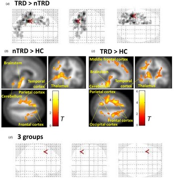
The voxel-wise PIB SUVR results showed that patients with TRD had more PIB deposits in the left-sided brain regions than did patients with nTRD after adjusting for age, sex, education levels, PET machines, HDRS-17, and global counts (cluster-level FWE-corrected p < 0.001; Fig. 2). The most prominent difference between the two groups existed in the left inferior parietal gyrus (peak MNI coordinate: −38, −36, 38; red arrows in Fig. 2a). Additionally, we compared 11C-PIB SUVRs between patients with nTRD and HCs (Fig. 2b) and between patients with TRD and HCs (Fig. 2c), revealing that both patients with TRD and nTRD have abnormally high Aβ deposits in many brain regions, including the frontal cortex, parietal cortex, temporal cortex, occipital cortex, thalamus, and even brainstem (cluster-level FWE-corrected p < 0.001). If we compared 11C-PIB SUVR among the three groups, namely TRD, nTRD, and HC, only the left middle frontal gyrus (peak MNI coordinate: −34, 10, 52; Fig. 2d) survived after correcting for multiple comparisons (voxel-level FWE-corrected p < 0.05; post hoc analysis: TRD < HC, nTRD < HC). For brain glucose use, no significant difference was found between the TRD and nTRD groups or among the three groups under the threshold of either cluster-level or voxel-level FWE-corrected p < 0.05.
Glass-brain view (a) shows that TRD patients had more PIB deposits in the left-sided brain than non-TRD patients (nTRD), in which the red arrows indicate the peak difference in the left inferior parietal gyrus (peak MNI coordinate: −38, −36, 38). Exploratory comparisons of 11C-PIB SUVR between nTRD and HC subjects (b) and between TRD and HC subjects (c), revealing that both TRD and nTRD patients have abnormally high PIB deposits in many brain regions, including frontal cortex, parietal cortex, temporal cortex, occipital cortex, thalamus, and even brainstem. The three-group comparison showed the most significant finding was in the left middle frontal gyrus (peak MNI coordinate: −34, 10, 52) as pointed out by red arrows (d).

Differential correlations between PIB SUVR and neurocognition in HC and MDD
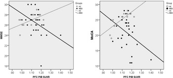
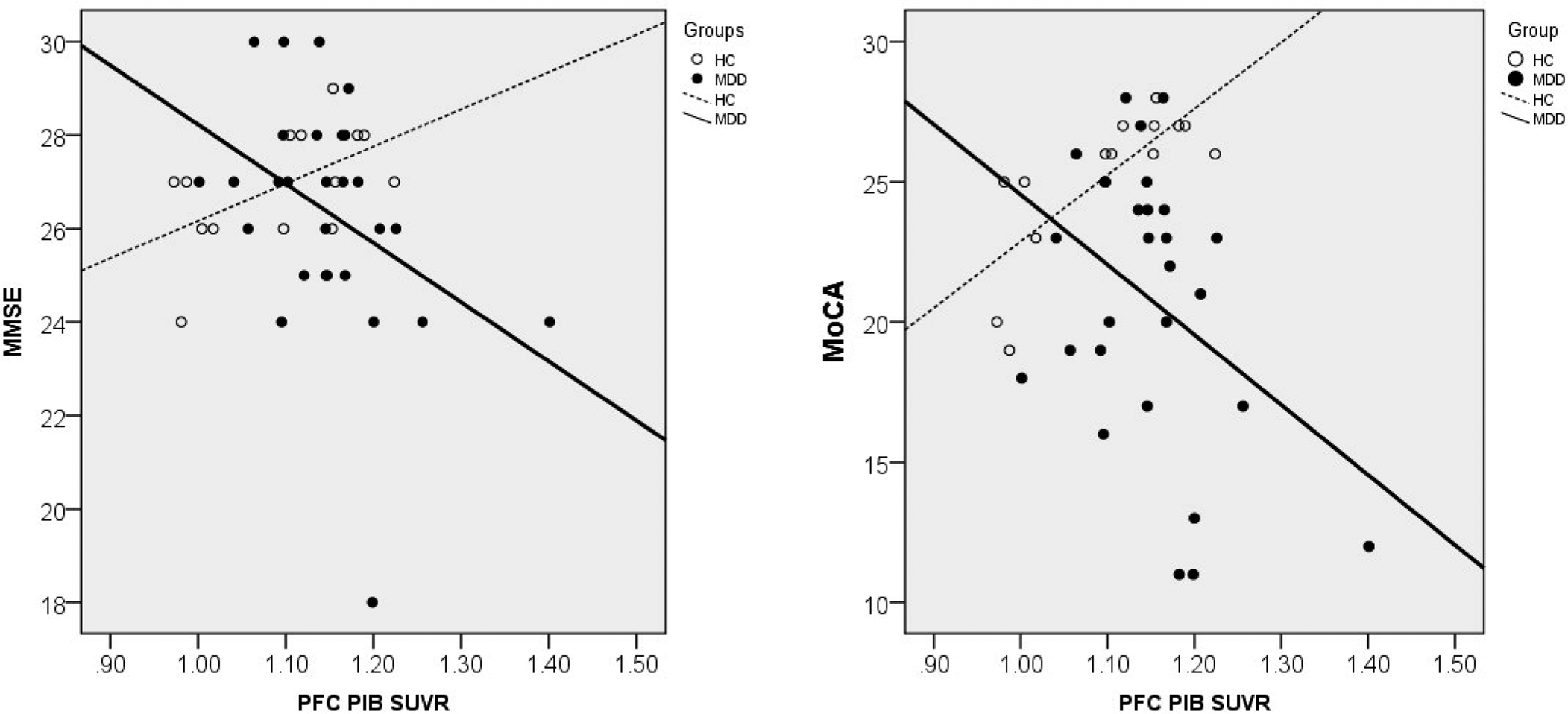
The correlation analysis further showed that better neurocognitive functioning was significantly correlated with higher PIB SUVR in the prefrontal, parietal, and occipital cortex in the HC group (p < 0.05, Table 2), whereas opposite findings were found between neurocognitive functioning and PIB SUVR across all the cortical regions in the MDD group (p < 0.05, Table 2). For example, high global cognitive functioning was significantly correlated with high PIB SUVR in the prefrontal cortex in the HC group (MMSE: r = 0.546, p < 0.05, dotted line in Fig. 3a; MoCA: r = 0.766, p = 0.001, dotted line in Fig. 3b), whereas low functioning was correlated with high PIB SUVR in the prefrontal cortex in the MDD group (MMSE: r = − 0.395, p < 0.05, solid line in Fig. 3a; MoCA: r = − 0.381, p < 0.05, solid line in Fig. 3b). If the TRD and nTRD groups were separately analyzed, all the results were still negative correlations. Such an opposite finding of global cognition and PIB deposits between patients with MDD and HCs also existed for temporal cortex, parietal cortex, and occipital cortex (online Supplementary Fig. S3). Likewise, multiple linear regression analyses also showed that PIB deposits in the prefrontal cortex positively predicted global cognitive function [i.e., MoCA (β = 20.055, T = 2.977, p = 0.018)] in the HC group and negatively predicted global cognition [i.e., MMSE (β = −12.686, T = −2.346, p = 0.028)] in the MDD group.
Correlations between prefrontal PIB deposits and global cognitive function as measured by MMSE (a) and MoCA (b), showing positive correlations between MMSE (r = 0.546, p < 0.05) and MoCA (r = 0.766, p = 0.001) in the HC subjects but negative correlations between MMSE (r = −0.395, p < 0.05) and MoCA (r = −0.381, p < 0.05) in the MDD patients. SUVR, standardized uptake value ratio.
Correlations between neurocognitive functioning and PIB SUVR

MDD, major depressive disorder; HC, healthy control subjects; PFC, prefrontal cortex; TEMP, temporal cortex; PAR, parietal cortex; OCC, occipital cortex; MMSE, Mini-Mental Status Examination; MoCA, Montreal Cognitive Assessment; WLT, Word List Test; WCST, Wisconsin Card Sorting Test; CLR, conceptual level responses; CC, categories completed. *p < 0.05.
Furthermore, we investigated correlations between PIB SUVR and illness duration. None of the brain regions survived after correcting for multiple comparisons (for other details please refer to online Supplementary material). In addition, if illness duration was entered as another covariate of no interest, the result of voxel-wise PIB SUVR analysis did not change, showing that patients with TRD had more PIB deposits in the left-sided brain regions than did patients with nTRD.
Discussion
As hypothesized, we found that patients with LLD, particularly TRD, had significantly impaired neurocognitive functioning (Table 1) and the characteristic feature of AD with abnormally increased brain Aβ deposits (Fig. 1). MoCA has been reported to be more sensitive than MMSE to detect mild cognitive impairment (Nasreddine et al., Reference Nasreddine, Phillips, Bedirian, Charbonneau, Whitehead, Collin and Chertkow2005) and may be more useful clinically in LLD, since no significant between-group difference was found for MMSE (Table 1).
Among the regions, the left prefrontal cortex was the most prominent brain region differentiating MDD and HC groups. Furthermore, patients with TRD had more Aβ deposits in the left-sided brain regions, particularly the left parietal cortex, than did patients with nTRD (Fig. 2). A review research indicated that, most often, dementia following a depressive episode is AD (Bazin & Bratu, Reference Bazin and Bratu2014). Accumulating evidence has suggested that LLD and AD have a common neuropathophysiology (e.g. NMDA-regulated signaling pathways) (Hoeffer & Klann, Reference Hoeffer and Klann2010). Low-dose ketamine as an NMDA antagonist has shown its antidepressant efficacy for treating refractory MDD (Li, Yang, & Lin, Reference Li, Yang and Lin2018; Li et al., Reference Li, Chen, Lin, Hong, Yang, Liu and Su2016a). The present study provided the first direct evidence to support a common feature of LLD and AD involving the Aβ-related neuropathology. Moreover, we found a differential relationship between prefrontal PIB deposits and global cognitive function in patients with MDD and HCs (Fig. 3 and online Supplementary Fig. S3). The supporting evidence was that correlations between MMSE/MoCA and PFC Aβ deposits in patients with MDD were significantly negative, whereas those between MMSE/MoCA and PFC Aβ deposits in HCs were significantly positive (Fig. 3).
The first major finding was that late-life MDD led to abnormally increased Aβ deposits in many cognition-related brain regions, especially in the left PFC. The increased PFC Aβ deposits correlated negatively with global cognitive functioning as measured using MMSE and MoCA. The results were consistent with a well-established finding of negative correlations between global cortical PIB deposits and cognitive function in elderly patients with MDD (Wu et al., Reference Wu, Liu, Chen, Chen, Hsiao, Hsieh and Lin2016) and dementia (Devanand et al., Reference Devanand, Mikhno, Pelton, Cuasay, Pradhaban, Dileep Kumar and Parsey2010). However, cortical PIB deposits may not be linearly associated with cognitive impairment in a specific domain or may not be cognitively harmful while the deposits are still in low doses. One of the supporting facts is the lack of significant negative correlations between PFC Aβ deposits and WCST or WLT in the MDD (Table 2). However, the exact mechanisms need to be further investigated.
In addition, whether MDD is a prodromal or risk factor for AD is still debatable (Brommelhoff et al., Reference Brommelhoff, Gatz, Johansson, McArdle, Fratiglioni and Pedersen2009) (Gao et al., Reference Gao, Huang, Zhao, Ma, Qiu, Zhang and Xiao2013), and the results so far have been inconsistent (da Silva et al., Reference da Silva, Goncalves-Pereira, Xavier and Mukaetova-Ladinska2013). For instance, although some studies have indicated that late-life MDD may be a prodrome of, rather than a risk factor for, AD (Brommelhoff et al., Reference Brommelhoff, Gatz, Johansson, McArdle, Fratiglioni and Pedersen2009), some have suggested it to be a risk factor for dementia (Gao et al., Reference Gao, Huang, Zhao, Ma, Qiu, Zhang and Xiao2013). Our results of increased PIB deposits in patients with MDD and TRD v. HCs imply that LLD and high depression refractoriness could be a risk factor for, but not a prodrome of, AD. One of the reasons for this conclusion was that no significant differences were observed in PFC PIB deposits between late-onset and non-late-onset LLD. In addition, we found that late-onset LLD had decreased global cognitive function and executive function than non-late-onset ones. The finding was in line with previous research showing that patients with late-onset depression have more impaired neurocognitive functioning, particularly the executive function, than patients with early-onset depression (Herrmann, Goodwin, & Ebmeier, Reference Herrmann, Goodwin and Ebmeier2007). Taken together, it is plausible that executive dysfunction observed in the late-onset LLD could be related to mechanisms other than Aβ-related neuropathology.
In addition to the prefrontal cortex, the present study identified widespread neocortical regions (e.g. the parietal, temporal, and occipital cortices) and brain regions frequently identified in MDD pathophysiology (e.g. the thalamus, pons, and brainstem) to have increased Aβ in the MDD group compared with the HC group. The involvement of depression-related brain regions was not surprising because the emotional limbic system has been found to be regularly involved in AD (Hopper & Vogel, Reference Hopper and Vogel1976; Li et al., Reference Li, Wang, Tian, Zhou, Li, Wang and Yu2016b), even as an early target for Aβ (Acosta-Cabronero & Nestor, Reference Acosta-Cabronero and Nestor2014). Thus, a late-life diagnosis of MDD may accelerate the process of Aβ deposition across the brain, or participants with high Aβ deposit levels may be vulnerable to MDD. However, our results supported the former because high MoCA scores were significantly correlated with high PIB SUVR in the prefrontal cortex in the HC group (Fig. 3b).
The present study found a differential relationship between prefrontal PIB deposits and global cognitive function in patients with MDD and HCs. Significant positive correlations between MMSE/MoCA and PFC Aβ deposits in HCs are in contrast to the findings in patients with MDD (Fig. 3). The lack of statistical significance in some multiple regression analyses may have partly resulted from a small sample size in the present study. Although the exact mechanisms for the positive correlation in HCs remain elusive, the positive correlations may explain why brain PIB deposition did not always predict AD in HCs and support the theory that brain amyloid accumulation serves a protective function (e.g. sequestration of neurotoxic misfolded protein in amyloid inclusion bodies and plaques) (Treusch et al., Reference Treusch, Cyr and Lindquist2009). However, relevant longitudinal studies with larger sample sizes are warranted.
Another crucial finding was that TRD had more Aβ deposits in the left-sided brain regions than did patients with nTRD. Left frontal hypoactivity has long been considered a characteristic feature of depression, particularly in patients who responded poorly to treatment (Li et al., Reference Li, Lin, Chou, Chen, Hsieh, Wu and Su2010, Reference Li, Su, Wang, Tu and Hsieh2015). Repetitive transcranial magnetic stimulation frequently targets the left prefrontal cortex to treat refractory depression (Li et al., Reference Li, Chen, Juan, Huang, Chen, Hsieh and Su2014, Reference Li, Cheng, Chen, Juan, Tu, Bai and Su2020). Brain lateralization may be crucial in many cognitive functions. For example, left hemisphere predominance is found for, but not limited to, language function (left frontal, parietal, and temporal cortices), gesture imitation (left parietal cortex) (Muhlau et al., Reference Muhlau, Hermsdorfer, Goldenberg, Wohlschlager, Castrop, Stahl and Boecker2005), and local relative to global visual attention processing (left occipital cortex) (Han et al., Reference Han, Weaver, Murray, Kang, Yund and Woods2002). The finding of left-brain predominance of Aβ deposits in TRD could not only be a consequence of TRD (i.e. TRD increases vulnerability to amyloid deposits specifically in the left brain) but might also be a cause for TRD (i.e. left-brain Aβ deposits lead to refractory depression). This finding is based on the fact that prominent deficits of glucose metabolism were absent but abnormal Aβ deposits were present in our patients with MDD and TRD. However, brain PIB deposits were noted before cerebral metabolic disruption in participants with cognitive impairment (Lowe et al., Reference Lowe, Kemp, Jack, Senjem, Weigand, Shiung and Petersen2009). The exact mechanisms by which Aβ deposits tended to occur in the left-sided brain regions of patients with TRD remain unclear and need to be studied. However, our current findings extend our understanding of late-life TRD from a purely functional depression to an amyloid-related depression.
This study had some limitations. The first limitation is the cross-sectional design. Future studies with a long follow-up period are needed to determine the long-term cognitive outcomes of the patients. However, our recent nationwide study with a 9-year follow-up period has confirmed that late-life TRD is associated with an elevated risk of AD (Chan et al., Reference Chan, Chen, Tsai, Bai, Tsai, Cheng and Li2020). Second, the cerebellum may not be an ideal reference region for depression studies. To minimize potential bias, we considered global PIB uptake as a covariate of no interest in the model. We also followed the standard procedure for parametric 11C-PIB PET imaging analysis (Tolboom et al., Reference Tolboom, Yaqub, Boellaard, Luurtsema, Windhorst, Scheltens and van Berckel2009) and used the cerebellar cortex, which was a more ideal reference region than the whole cerebellum, to obtain the SUVR (Akamatsu et al., Reference Akamatsu, Ikari, Ohnishi, Nishida, Aita, Sasaki and Senda2016). Finally, previous depressive history (e.g. past depressive episodes and chronicity) may have negative impacts on the brain (Gorwood, Corruble, Falissard, & Goodwin, Reference Gorwood, Corruble, Falissard and Goodwin2008), but we did not evaluate past depressive episodes because of the difficulty to correctly retrieve the information in TRD patients who usually had long and non-remitting depression. However, the duration of illness and length of the current episode were not different between nTRD and TRD (Table 1). It warrants further studies to study the effects of previous depressive history on brain functions.
Conclusions
The present study provided evidence for abnormally increased brain Aβ deposits in many cognition-related brain regions in patients with late-life MDD, particularly TRD, a characteristic feature of AD. Here, patients with TRD demonstrated more Aβ deposits in the left-sided brain regions, particularly in the left parietal cortex, than did patients with nTRD. Our findings revealed that Aβ deposits were not negatively associated with global cognition in HCs, whereas a detrimental role of Aβ deposits in global cognition in patients with late-onset or non-late-onset MDD supported the theory that late-life MDD could be a risk factor for AD.
Supplementary material
The supplementary material for this article can be found at https://doi.org/10.1017/S0033291721001070.
Acknowledgements
We would like to thank Robert Innis (from molecular imaging branch, National Institute of Mental Health, USA) for the instructions and comments for the study. We also want to thank all the patients who kindly participated in this study and all the research assistants, physicians, and staff who assisted with the study and imaging procedures.
Financial support
This study was sponsored by grants from Taipei Veterans General Hospital (V108D44-003-MY3-1), and the Ministry of Science and Technology (MOST 106-2314-B-075-034 -MY3; 108-2321-B-075-004-MY2). In addition, this work was supported by the Brain Research Center (108BRC-B502), National Yang-Ming University from The Featured Areas Research Center Program within the framework of the Higher Education Sprout Project by the Ministry of Education (MOE) in Taiwan. Neither of the aforementioned organizations had any role in the study design, data collection, analysis, interpretation of results, writing of the report, or ultimate decision to submit the paper for publication. None of the authors in this study has any conflict of interest to declare.
Conflict of interest
The authors declare that they have no conflicts of interest. All authors have no financial relationships relevant to this article to disclose.