Case report
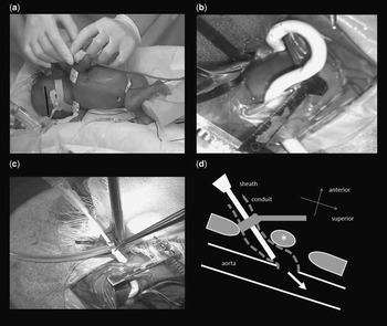
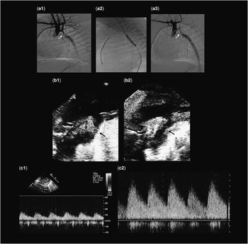
A 530 g female baby was referred to our institute on day 7 of life due to anuria after undergoing ductus arteriosus ligation on day 5. She was one of the monochorionic diamniotic twins and a donor of twin-to-twin transfusion syndrome. We diagnosed the patient with a coarctation of the aorta, which became apparent after the operation. The coarcted segment had a diameter of 1 mm (Fig 2b(1)). Doppler flow of the abdominal aorta showed a very low blood flow rate and pulse pressure (Fig 2c(1)). We initially believed that we could dilate the coarcted segment using intravenous prostaglandin E1; however, this method was ineffective. We, therefore, chose balloon angioplasty using the descending aorta as the access route. On day 10, we performed thoracotomy and approached the descending aorta from the eighth left intercostal space (Fig 1a), then an inosculated vascular graft with a diameter of 3 mm was placed in the descending aorta with a diameter of 3.5 mm (Fig 1b). We snared the 3-Fr sheath inside the vascular graft (Fig 1c). A guide wire was directly inserted into the sheath using an Ironman, and the tip of the guide wire was fixed to the internal thoracic artery across the coarcted segment. To dilate the coarcted segment, a 2.5×13 mm Raiden balloon catheter was utilised and we expanded it with a pressure of 15 atmospheres (Fig 2a(2)). After balloon angioplasty, both angiography and an echocardiogram demonstrated coarcted segment dilation (Fig 2a(3) and b(2)). The pressure gradient decreased from 30 to 10 mmHg between the ascending aorta and the descending aorta. The echocardiogram showed that the maximal flow rate of Doppler imaging of the abdominal aorta improved from 20 to 70 cm/second (Fig 2c(2)). The blood pressure of the lower limbs became pulsatile and anuria improved. There were no complications in the central nerve system. We followed-up the patient for the next 3 months and no re-stenosis was observed.
( a ) In the intervention room, under general anaesthesia, we approached the descending aorta from the left eighth intercostal space.( b ) An inosculated vascular graft with a diameter of 3 mm was placed in the descending aorta with a diameter of 3.5 mm. ( c ) The snared 3-Fr sheath, with the tip remaining inside the graft. ( d ) A schematic illustration of the procedure.

( a ) Images of catheterisation. (1) Before balloon angioplasty. (2) Treated with a balloon catheter with a diameter of 2.5 mm. (3) After balloon angioplasty. The isthmus was dilated. Arrow (→) indicates the coarcted segment. ( b ) Echocardiographic examination before (1) and after (2) balloon angioplasty. Arrow (→) indicates the coarcted segment. ( c ) Doppler ultrasound waveform of the descending aorta before (1) and after (2) balloon angioplasty.

Discussion
The most difficult problem in the intervention for extremely low birth weight infants is the vascular access route.Reference Abraham, Alvaro, William, Juan and Humberto 1 It has been reported that the internal carotid artery and the umbilical artery could be used for the vascular access route in the intervention for extremely low birth weight infants;Reference Schamberger and Lababidi 2 , Reference Dryzek, Goreczny and Kopala 3 however, in the present case, we could not evaluate the internal carotid artery by ultrasound due to severe oedema, and it was not possible to approach the internal carotid artery for dissection. The use of the femoral artery for the vascular access route may interrupt the blood flow to the lower half of the body. Furthermore, intervention through the internal carotid artery or the umbilical artery may infarct brain vessels or cause thrombosis of an abdominal artery, respectively. After intervention, re-stenosis can invariably occur; however, the peripheral arteries always occlude after intervention. In addition, by using the internal carotid artery and femoral artery for the access route, there are a limited number of opportunities for re-stenosis. Thus, we developed a new access route to prevent these complications using the descending aorta. As the descending aorta is thicker than the peripheral vessels, it is possible to repeatedly use it as an access route, and it reduces the potential of thrombosis. Moreover, we used a vascular graft, which made the fixation of the devices stronger and the operability easier. We expect this method to be used in various interventions for extremely low birth weight infants, such as aortic valve stenosis and pulmonary valve stenosis via the arterial duct.
Acknowledgement
None.
Financial Support
This research received no specific grant from any funding agency, commercial, or not-for-profit sectors.
Conflicts of Interest
None.