The COVID-19 infection started to spread rapidly all over the world since it was first seen in Wuhan, China, in December 2019. In March 2020, World Health Organization declared the COVID-19 pandemic. Reference Dong, Du and Gardner1 It was thought to have a mild course in children compared with adults at the onset of the severe acute respiratory syndrome coronavirus 2. Reference Dong, Mo and Hu2,Reference Lu, Zhang and Du3 But in April 2020, some children with symptoms similar to Kawasaki and toxic shock syndrome were reported in Italy and the United Kingdom. Reference Riphagen, Gomez, Gonzalez-Martinez, Wilkinson and Theocharis4,5 Afterwards, similar cases were also reported in Europe and the United States of America. 6–Reference Chiotos, Bassiri and Behrens12 Thereby, some concerns began to be raised about this hyperinflammatory process in the children associated with severe acute respiratory syndrome coronavirus 2. This new disease was defined by the Royal College of Pediatrics and Child Health as a paediatric inflammatory multisystemic syndrome and as a multisystem inflammatory syndrome in children by the World Health Organization and the Centers for Disease Control and Prevention. 6,7,13
Cardiac involvement has been described in up to 67–80% of children with multisystem inflammatory syndrome in children. Ventricular dysfunction is a common finding of multisystem inflammatory syndrome in children with 33–50% of patients affected. Reference Belhadjer, Méot and Bajolle14–Reference Greene, Saleh, Roseman and Sinert16 The pathophysiology underlying myocardial dysfunction in multisystem inflammatory syndrome in children has not been fully understood yet. Etiologic factors for myocardial involvement in adults with COVID-19 include acute myocarditis, hypoxic injury, ischaemic injury caused by cardiac microvascular damage or coronary artery disease, right heart strain, stress cardiomyopathy (Takotsubo), and systemic inflammatory response syndrome. Reference Sanna, Serrau, Bassareo, Neroni, Fanos and Marcialis17–Reference Tersalvi, Vicenzi, Calabretta, Biasco, Pedrazzini and Winterton21 Coronary artery dilation or aneurysms occurs in 6–24% and arrhythmias in 7–60% of children with multisystem inflammatory syndrome in children. Reference Licciardi, Pruccoli and Denina22–Reference Whittaker, Bamford and Kenny26 Data on multisystem inflammatory syndrome in children are still limited but rapidly increasing in the literature.
In this study, we aimed to evaluate the electrocardiographic and echocardiographic findings, 24-hour Holter recordings of children diagnosed with multisystem inflammatory syndrome in children, and to determine the factors predicting cardiac involvement in multisystem inflammatory syndrome in children.
Materials and method
The data of children hospitalised with the diagnosis of multisystem inflammatory syndrome in children were retrospectively collected. Multisystem inflammatory syndrome in children was diagnosed according to the criteria of World Health Organisation. 6 Patients with known arrhythmia, electrolyte imbalance, hormonal/metabolic disorders and patients using arrhythmogenic drugs were excluded from the study. The study has been approved by the research ethics committee of our institute before the experiment was started and that has been conducted in accordance with the principles outlined in the Helsinki Declaration.
Demographic characteristics, medical data (comorbidities, symptoms, and physical examination findings), laboratory findings (including leukocyte, lymphocyte and neutrophil counts, neutrophil/lymphocyte ratio , B-type natriuretic peptide levels, troponin I levels, C-reactive protein, d-dimer, fibrinogen, ferritin, and sedimentation), electrocardiogram and echocardiographic findings, 24-hour Holter recordings, need for ICU, and extracorporeal membrane oxygenation were analysed. With the beginning of arrhythmias reported in patients with COVID-19, 24-hour Holter monitoring has been started to be performed in all children with multisystem inflammatory syndrome in children in our clinic. If arrhythmia was detected in the Holter recording, 24-hour Holter monitoring was repeated before discharge. All the Holter recordings were evaluated by a paediatric electrophysiologist.
Acute left ventricular systolic dysfunction was defined as left ventricular ejection fraction (EF) ≤%55 in echocardiographic. A coronary artery aneurysm was reported based on echocardiographic findings and z scores (Boston criteria).
Severe acute respiratory syndrome coronavirus 2 was diagnosed by using a reverse transcription-polymerase chain reaction test on the nasopharyngeal and oropharyngeal swab or positive serology (IgM or IgG).
Statistical analysis
In the descriptive statistics of the data, mean, standard deviation, median, minimum, maximum, frequency, and ratio values were used. The distribution of variables was measured with the Kolmogorov–Smirnov test. The Mann–Whitney U-test was used in the analysis of quantitative independent data, the Wilcoxon test for dependent quantitative data, and the Chi-square test for qualitative independent data. Spearman correlation analysis was used in the correlation analysis. Cut-off values were analysed with the Receiver Operating Characteristic (ROC) curve. SPSS 27.0 program was applied in the analysis.
Results
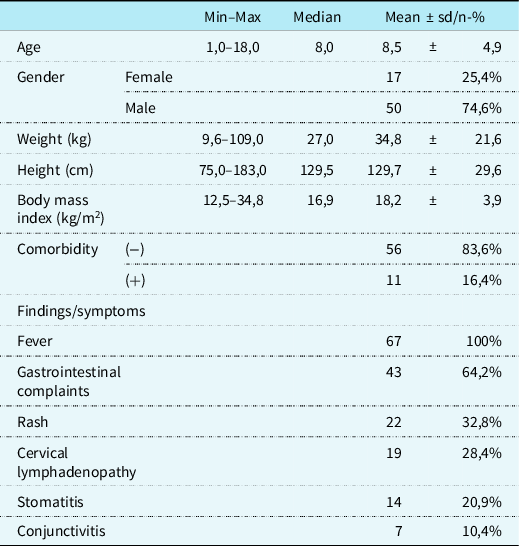
Sixty-seven children were included in the study. A total of 74.6% of the cases were male with a median age of 8 years (8.5 ± 4.9 years). Comorbidities including juvenile idiopathic arthritis (3), familial Mediterranean fever (2), operated Fallot tetralogy (1), operated ventricular septal defect (1), epilepsy (1), autism (1), and obesity (2) were present in 16.4% of patients.
Fever and gastrointestinal system findings were the most common presenting symptoms. Demographic characteristics and symptoms are shown in Table 1. In total, 10.4% of the patients had positive severe acute respiratory syndrome coronavirus 2 reverse transcription-polymerase chain reaction test, 73.1% had positive severe acute respiratory syndrome coronavirus 2 IgG antigen, and 40% had both positive severe acute respiratory syndrome coronavirus 2 IgM and IgG antigen. While 79.1% of patients had a history of contact with a COVID positive patient proven with positive severe acute respiratory syndrome coronavirus 2 antigen and/or polymerase chain reaction tests, 16.4% had only a history of contact.
Demographic characteristics and symptoms

Min = minumum; max = maximum; sd = standart deviation.
Electrocardiographic findings were normal in 77.6% of patients. On admission, electrocardiogram demonstrated sinus tachycardia in nine patients, Qtc prolongation in three patients, sinus bradycardia in one patient, and PR interval prolongation in two patients. However, minor electrocardiogram changes recovered, and all electrocardiograms were normal on discharge. The median heart rate in electrocardiogram was 98/min (99.2 ± 23.6/min), median Qtc was 422 ms (420.4 ± 33.9 ms), and median PR interval was 0.18 ± 0.08 ms. None of our patients received pro-arrhythmogenic therapy such as hydroxychloroquine or azithromycin.
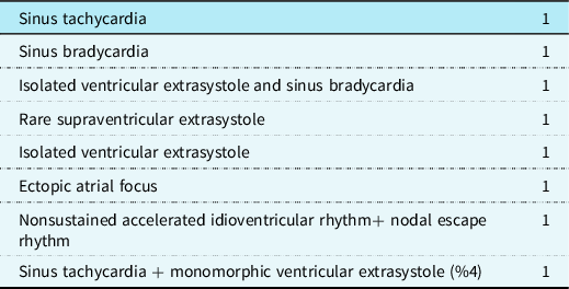
Holter results were recorded in 61.2% of the patients and 49.2% were normal. The mean heart rate on the Holter recording was 78.8 ± 24.4/min. There was no statistically significant difference in EF values between the group with arrhythmia in Holter and the group with normal Holter results (p ≥ 0.05). The Holter findings are summarised in Table 2. All arrhythmias were observed transiently, and none of the patients required antiarrhythmic therapy, pacing, or emergency intervention.
Abnormal Holter findings (n)

On echocardiographic examination, 14.9% of the patients had systolic dysfunction (EF ≤ 55%). While the mean EF was 65.5 ± 9.5% on admission, the mean EF before discharge was 64.1 ± 13.1% (p = 0.036). Also, the mean EF on discharge was statistically higher than on admission (p = 0.003). While 32.8% of patients had mild mitral regurgitation, 3% had moderate mitral regurgitation, and 6% had mild aortic regurgitation. Mitral regurgitation was seen in 17.9% of patients on discharge, while mild mitral regurgitation incidence decreased to 6% at the 1-month follow-up.
All of the aortic regurgitations disappeared on discharge. While minimal pericardial effusion was present in 7.5% of the patients, none of the patients had signs of tamponade. Coronary artery involvement (mild aneurysm) was observed in one patient. This patient met the criteria for Kawasaki disease and had a history of contact with a COVID-positive individual. Coronary artery diameters normalised with IVIG and aspirin therapy.
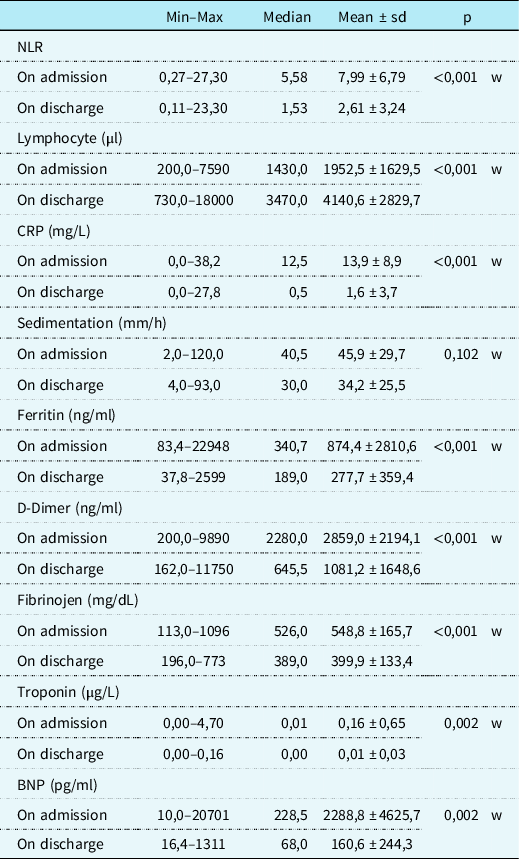
Laboratory findings on admission and discharge are shown in Table 3.
Laboratory findings on admission and discharge

w Wilcoxon test.
BNP = B-type natriuretic peptide; CRP = c-reactive protein; min = minimum; max = maximum; NLR = neutrophil/lymphocyte ratio; sd = standard deviation.
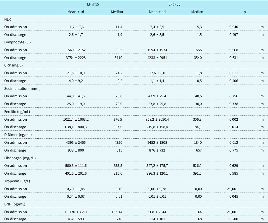
The COVID polymerase chain reaction, IgG, and IgM positivity rates did not differ significantly in the group with EF ≤ 55 and EF > 55 (p > 0.05). Also, the COVID antibody titer did not differ in the group with EF ≤ 55 and EF > 55 (p > 0.05). Laboratory findings of patients with EF ≤ 55 and EF > 55 are shown in Table 4.
Comparison of the laboratory findings with EF ≤ 55 and EF > 55

m Mann–Whitney U test.
BNP = B-type natriuretic peptide; CRP = c-reactive protein; EF = jejection fraction; NLR = neutrophil/lymphocyte ratio; sd = standart deviation.
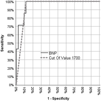
Significant effectivity of the BNP value was observed in the differentiation of those with EF ≤ and > 55%. The cut-off value of 1700 pg/ml for the BNP was found to be significantly effective in this distinction. For this value, the sensitivity was 100%, the specificity was 88.4%, the positive and negative predictive value was 58.3 and 100%, respectively (p < 0.01, 95% CI: 0.0879–1.000) (Fig 1).
ROC curve for BNP.

A significant positive correlation was observed between troponin and B-type natriuretic peptide , neutrophil/lymphocyte ratio, C-reactive protein, ferritin, d-dimer. B-type natriuretic peptide was positively correlated with C-reactive protein, ferritin, and fibrinogen. Lymphocyte value was negatively correlated with both B-type natriuretic peptide and troponin (Table 5).
Correlation BNP and troponin with laboratory values

Spearman Correlation.
BNP = B-type natriuretic peptide; CRP = C-reactive protein; NLR = neutrophil/lymphocyte ratio..
The average hospital stay of the patients was 10.5 ± 6.0 days. Totally, 29.9% of patients (n = 20) were hospitalised in the ICU. In the group with EF ≤ 55, the length of hospital stay and the rate of admission to the ICU were significantly higher than the group with EF > 55 (p < 0,001). The most common indication for admission to the ICU was hypotension, and the second most common reason was altered consciousness. The mean systolic blood pressure of the patients hospitalised in the ICU was 78.4 mmHg, and the diastolic blood pressure was 44.7 mmHg.
In total, 24.6% of patients needed inotropic support, and extracorporeal membrane oxygenation support was needed for three (4.5%) patients. One of the three patients with extracorporeal membrane oxygenation died, one was successfully weaned off extracorporeal membrane oxygenation , one was implanted with a ventricular assist device under extracorporeal membrane oxygenation, and is waiting for heart transplantation. One patient who died had systemic juvenile idiopathic arthritis.
Discussion
Multisystem inflammatory syndrome in children is a novel syndrome that is serious and potentially life threatening. Although a mild course was observed in children at the beginning of the COVID-19 pandemic, concern for children increased with reported multisystem inflammatory syndrome in children cases later on. Reference Dong, Mo and Hu2,Reference Riphagen, Gomez, Gonzalez-Martinez, Wilkinson and Theocharis4,7,Reference Verdoni, Mazza and Gervasoni9–Reference Chiotos, Bassiri and Behrens12 Cardiac manifestations are common, and cardiac involvement is one of the most important factors determining prognosis in multisystem inflammatory syndrome in children. Reference Kabeerdoss, Pilania, Karkhele, Kumar, Danda and Singh27
In our single-centre study, we reported the laboratory, clinical findings and cardiac involvement of 67 patients with multisystem inflammatory syndrome in children. Totally, 61.2% of patients had a Holter recording, and it was normal in 49.3%. Similar to previous studies, the median age of the patients in our study was 8 years, and there was a male predominance. Reference Riphagen, Gomez, Gonzalez-Martinez, Wilkinson and Theocharis4,Reference Verdoni, Mazza and Gervasoni9,Reference Toubiana, Poirault and Corsia10 Unlike Kawasaki disease, multisystem inflammatory syndrome in children is usually seen in children over 5 years of age. Reference Kabeerdoss, Pilania, Karkhele, Kumar, Danda and Singh27 Fever and gastrointestinal involvement were the most common findings compatible with the literature. Reference Feldstein, Rose and Horwitz28,Reference Valverde, Singh and Sanchez-de-Toledo29
While there was 10.4% reverse transcription-polymerase chain reaction COVID positivity in our study, IgG positivity was 73.1% and the majority of patients had contact with a COVID-19-positive individual. This suggests that multisystem inflammatory syndrome in children is triggered by an adaptive immune response rather than an acute infection with COVID-19. Reference Kabeerdoss, Pilania, Karkhele, Kumar, Danda and Singh27
Valverde et al. reported the incidence of arrhythmia as 35% in their study in which they examined 286 patients with multisystem inflammatory syndrome in children. Reference Valverde, Singh and Sanchez-de-Toledo29 Also, Whittaker et al. reported four patients (6%) developed arrhythmia: one patient had a first-degree atrioventricular block with frequent supraventricular ectopic beats; one had intractable broad complex tachycardia, associated with low cardiac output, necessitating extracorporeal membrane oxygenation; one had atrial fibrillation managed with amiodarone; and one had second-degree heart block, which resolved without treatment. Reference Whittaker, Bamford and Kenny26 Premature ventricular contractions, Reference Kim, Kim, Kim and Han30 monomorphic ventricular tachycardia, Reference Peigh, Leya and Baman31 diffuse ST-segment changes, T wave inversions in lateral leads, low-voltage QRS, atrioventricular conduction block, and sinus node dysfunction have also been reported. Reference Azarkish, Laleh far, Eslami and Mollazadeh32–Reference Paul, Charles, Richaud, Caussin and Diakov36 In our study, electrocardiogram demonstrated sinus tachycardia in nine patients, Qtc prolongation in three patients, sinus bradycardia in one patient, and PR interval prolongation in two patients. Qtc prolongation in multisystem inflammatory syndrome in children patients may be due to inflammation and changes in ion channel activity. Reference Merino, Martínez‐Cossiani and Iniesta37 We performed 24-hour Holter recording on 61.2% of patients, and arrhythmia was observed in 50.7% and did not require any antiarrhythmic drug therapy or emergency intervention. Although findings other than nonsustained accelerated idioventricular rhythm detected in the Holter recordings can also be seen in the healthy population, the Holter recordings completely normalised without any anti-arrhythmogenic treatment before discharge. While there are studies of Holter recordings in adults with COVID-19 infection, data in children are very limited. Reference Laleh Far, Mehrakizadeh and Eslami38 Only one study evaluated the 24-hour Holter monitoring in the short-term follow-up of multisystem inflammatory syndrome in children patients, and the Holter records of all patients were normal. Reference Vukomanovic, Krasic and Prijic39 However, in our study, Holter recordings were performed in the acute period and their relationship with the systolic function was also evaluated. Since Holter monitoring is not routinely performed, the incidence of arrhythmia in multisystem inflammatory syndrome in children may be underestimated. Because of no active infection in multisystem inflammatory syndrome in children, there is no contamination risk of staff and Holter devices so it should be used more.
On echocardiography, systolic dysfunction was determined in 14.9% of 67 children diagnosed with multisystem inflammatory syndrome in children. The most frequently affected valves were the mitral and aortic valves, respectively. Coronary artery dilation or aneurysms have been described in 6–24% of patients in multisystem inflammatory syndrome in children. Reference Sperotto, Friedman, Son, VanderPluym, Newburger and Dionne40 In this study, coronary artery involvement was seen in one (1.5%) patient, and this patient fulfilled the criteria for Kawasaki disease and had contact with COVID-19-positive individuals. As we know, multisystem inflammatory syndrome in children overlaps with Kawasaki and Kawasaki-like disease. Reference Kabeerdoss, Pilania, Karkhele, Kumar, Danda and Singh27,Reference Lee, Day-Lewis and Henderson41 A systematic review and meta-analysis demonstrated that myocardial dysfunction is a more common cardiovascular complication in multisystem inflammatory syndrome in children than coronary artery dilation or aneurysms, unlike in Kawasaki disease. Reference Yasuhara, Watanabe, Takagi, Sumitomo and Kuno42 Our study also supports this finding.
Feldstein et al. have shown that 80% of patients required intensive care and 4% received extracorporeal membrane oxygenation. Reference Feldstein, Rose and Horwitz28 Extracorporeal membrane oxygenation support was 0.3% in Valverde et al.‘s study and one death occurred. Reference Valverde, Singh and Sanchez-de-Toledo29 In the study by Belhadjer et al., the need for extracorporeal membrane oxygenation was in 28% of patients and no death was observed. Reference Belhadjer, Méot and Bajolle14 In our study, 29.9% of the patients were admitted to the ICU, and 4.5% of the patients needed extracorporeal membrane oxygenation due to persistent hypotension. Of patients who underwent extracorporeal membrane oxygenation, one patient with systemic juvenile idiopathic arthritis died, one was successfully weaned off extracorporeal membrane oxygenation, and one was implanted with a ventricular assist device under extracorporeal membrane oxygenation and is waiting for heart transplantation.
Some laboratory findings support that multisystem inflammatory syndrome in children is an Macrophage Activation Syndrome (MAS)-like cytokine storm. Reference Lee, Day-Lewis and Henderson41 Pouletty et al. have compared patients with severe and non-severe forms of multisystem inflammatory syndrome in children, and they reported that higher age and a serum ferritin >1400 μg/L were the best indicators for severe disease. Reference Pouletty, Borocco and Ouldali43 Overall, C-reactive protein, procalcitonin, and erythrocyte sedimentation rate are highly elevated, as well as ferritin and interleukin-6. Reference Sperotto, Friedman, Son, VanderPluym, Newburger and Dionne40 B-type natriuretic peptide and troponin values are important markers in terms of cardiac involvement and were found to be high in most multisystem inflammatory syndrome in children patients. Reference Sperotto, Friedman, Son, VanderPluym, Newburger and Dionne40 Many biomarkers, including C-reactive protein, ferritin, troponin, and NT-proBNP levels may help predict the progression and severity of the disease. Salido et al. compared children with multisystem inflammatory syndrome in children and COVID-19 infection and found that the multisystem inflammatory syndrome in children patients presented severe inflammation, severe lymphopenia, with a high neutrophil/lymphocyte ratio . Reference García-Salido, de Carlos Vicente and Belda Hofheinz44 In adults, neutrophil/lymphocyte ratio has been observed to distinguish mild from severe cases of COVID-19. Reference Wang, Hu and Hu45–Reference Chen, Wu and Guo47 In our study, supporting the literature, a significant positive correlation was observed between troponin and B-type natriuretic peptide, neutrophil/lymphocyte ratio, C-reactive protein, ferritin, d-dimer. B-type natriuretic peptide was positively correlated with C-reactive protein, ferritin, and fibrinogen. Lymphocyte value is negatively correlated with both B-type natriuretic peptide and troponin. Also, neutrophil/lymphocyte ratio, C-reactive protein, d-dimer, ferritin, troponin, and B-type natriuretic peptide values were found to be significantly higher in patients with systolic dysfunction. The cut-off value of 1700 pg/ml for the B-type natriuretic peptide was found to be significantly effective in predicting systolic function. Therefore, these factors can be used to determine the severity of the disease.
There are some limitations of our study. First, this is a retrospective study. We did not perform cardiac CT or MRI for cardiac evaluation, and Holter recordings of some patients were absent.
Due to the limited data on this subject, our study is important because it presents Holter data of multisystem inflammatory syndrome in children. No serious arrhythmia was observed in electrocardiogram and Holter recordings. On echocardiographic examination, 14.9% of the patients had systolic dysfunction and three patients needed extracorporeal membrane oxygenation support one of whom died. One patient is waiting for a heart transplant with a ventricular assist device. Neutrophil/lymphocyte ratio, C-reactive protein, d-dimer, ferritin, troponin, and B-type natriuretic peptide values were found to be significantly higher in patients with systolic dysfunction. Also, the cut-off value of 1700 pg/ml for B-type natriuretic peptide was significantly effective. These parameters can be used to indicate the severity of the disease.
Financial support
This research received no specific grant from any funding agency, commercial, or not-for-profit sectors
Conflicts of interest
None.