INTRODUCTION
Sulphide, which usually refers to three forms of hydrogen sulphide—hydrogen sulphide gas (H2S), the bisulphide anion (HS−) and the sulphide anion (S2−) (Millero et al., Reference Millero, Plese and Fernandez1988)—is a well-known toxin that inhibits many enzymes involved in aerobic metabolism, such as cytochrome c oxidase (CCO), a mitochondrial terminal oxidase. The CCO causes harm to organisms, including sulphhaemoglobin formation (Affonso et al., Reference Affonso, Polez, Correa, Mazon, Araujo, Moraes and Rantin2004; Bailly & Vinogradov, Reference Bailly and Vinogradov2005), mitochondrial depolarization (Eghbal et al., Reference Eghbal, Pennefather and O'Brien2004) and increased free radical production by inducing reactive oxygen species (ROS) leading to oxidative stress (Affonso & Rantin, Reference Affonso and Rantin2005; Attene-Ramos et al., Reference Attene-Ramos, Wagner, Gaskins and Plewa2007). Nevertheless, a variety of marine invertebrates living in habitats such as hydrothermal vents, hydrocarbon seeps, coastal mudflats and marshes that periodically encounter or are continuously exposed to sulphide can survive (Bagarinao, Reference Bagarinao1992; Goffredi et al., Reference Goffredi, Childress, Desaulniers and Lallier1997; Julian et al., Reference Julian, Wieting, Seto, Bogan and Arp1999; Buchner et al., Reference Buchner, Abele and Pörtner2001; Joyner-Matos et al., Reference Joyner-Matos, Predmore, Stein, Leeuwenburgh and Julian2010).
The capability of animals to tolerate sulphide is thought to result from specialized sulphide oxidation detoxification mechanisms, such as mitochondrial sulphide oxidation, which reduces the toxicity of thiosulphate (Grieshaber & Völkel, Reference Grieshaber and Völkel1998). Some invertebrates such as the gutless clam Solemya reidi (Powell & Somero, Reference Powell and Somero1986; Kraus et al., Reference Kraus, Doeller and Powell1996) and the lugworm Arenicola marina (Hildebrandt & Grieshaber, Reference Hildebrandt and Grieshaber2008a; Theissen & Martin, Reference Theissen and Martin2008) can oxidize sulphide in their mitochondria. The first step in mitochondrial sulphide oxidation is presumably catalysed by a mitochondrial membrane flavoprotein, sulphide: quinone oxidoreductase (SQR), which oxidizes sulphide to persulphide. The sequential actions of a sulphur dioxygenase and a sulphur transferase (or rhodanese) are proposed to convert SQR-bound persulphide into sulphite and thiosulphate, respectively. Finally, the product of the sulphur dioxygenase reaction, sulphite, can be directly oxidized to sulphate by sulphite oxidase and alternatively converted by sulphur transferase to thiosulphate (Hildebrandt & Grieshaber, Reference Hildebrandt and Grieshaber2008b; Taniguchi et al., Reference Taniguchi, Matsunami, Kimura, Yonezawa, Ishiki, Sekiguchi, Nishikawa, Maeda, Ishikura and Kawabata2009; Tiranti et al., Reference Tiranti, Viscomi, Hildebrandt, Di Meo, Mineri, Tiveron, Levitt, Prelle, Fagiolari, Rimoldi and Zeviani2009; Kabil & Banerjee, Reference Kabil and Banerjee2010; Ma et al., Reference Ma, Zhang, Shao, Kang, Tan and Li2010) (Figure 1).
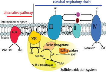
The proposed model of two different pathways for sulphide oxidation in the mitochondria (modification from Kabil & Banerjee, Reference Kabil and Banerjee2010). Electrons from the sulfide oxidation system can be transferred to oxygen either by the alternative pathway or the classical respiratory chain. The enzymes, complexes and respective substrates in relation to the inner mitochondrial membrane are depicted in the schematic drawing. II, complex II (succinate: ubiquinone oxidoreductase); III, complex III (ubiquinol: cytochrome c oxidoreductase); IV, complex IV, (cytochrome c oxidase); AOX, alternative oxidase; SQR, sulphide: quinol oxidoreductase; UQ, ubiquinone pool.

Eukaryotic anaerobes containing the same basic set of genes and enzymes for anaerobic energy metabolism and alternative oxidase (AOX) in mitochondria may help to maintain redox balance (Muller et al., Reference Muller, Mentel, van Hellemond, Henze, Woehle, Gould, Yu, van der Giezen, Tielens and Martin2012). In addition, the same low glycolytic rate was observed in mitochondria or eukaryotic anaerobes during either anoxia or sulphide stress (Oeschger & Storey, Reference Oeschger and Storey1990). Anoxia and sulphide could be inhibited CCO. In marine invertebrates, AOX also maintains respiratory electron flux, substrate oxidation and cellular redox potential when CCO is inhibited (Abele et al., 2007). High concentrations of sulphide can bind reversely at the haeme site of cytochrome aa3 (Nicholls, Reference Nicholls1975), to inhibit the transfer of electrons from sulphide oxidation via CCO. In this case, electrons are transferred to oxygen by another pathway, the alternative mitochondrial respiratory pathway (Figure 1). This pathway branches at the ubiquinone pool and consists of AOX, which presumably resembles the enzyme found in plant mitochondria and has a central role in cyanide-resistant respiration, as well as acting as an alternative terminal oxidase in the electron transport chain (ETC). It catalyses the oxidation of ubiquinol, reducing molecular oxygen to water, without translocation of protons across the inner mitochondrial membrane. The redox energy that is not conserved for ATP synthesis is released as heat (Vanlerberghe & McIntosh, Reference Vanlerberghe and McIntosh1997).
Recently, AOX studies have focused on plants and microbes. The structure of the plant AOX model has been determined and all plant AOX amino acid sequences (in the C-terminal domain) contain two copies of the conserved iron-binding motif (E-X-X-H). Three fully conserved residues (T, E, Y) in the pocket were thought to be potential ligands for ubiquinone (Sluse & Jarmuszkiewicz, Reference Sluse and Jarmuszkiewicz1998). However, limited data have been reported for AOX in animals. Nucleic acid sequences similar to plant AOX have been identified in some invertebrates, such as the chordate Ciona intestinalis Linnaeus and the oyster Crassostrea gigas. The AOX of Ciona intestinalis was first expressed in human kidney cells (Hakkaart et al., Reference Hakkaart, Dassa, Jacobs and Rustin2006) and then Drosophila melanogaster cells (Fernandez-Ayala et al., Reference Fernandez-Ayala, Sanz, Vartiainen, Kemppainen, Babusiak, Mustalahti, Costa, Tuomela, Zeviani, Chung, O'Dell, Rustin and Jacobs2009), based on cDNA sequences from C. intestinalis genomic data. The oyster, Crassostrea gigas, AOX sequence was recently cloned (McDonald et al., Reference McDonald, Vanlerberghe and Staples2009). In permeabilized fibres from the sandworm Nereis (Neanthes) virens, AOX activity could be triggered both by the redox state of the cell and the type of substrates provided to mitochondria (Pichaud et al., Reference Pichaud, Rioux and Blier2012).
Owing to its character, AOX may allow animals to acclimate to stressful conditions, particularly those that inhibit the cytochrome pathway. Biochemical studies in A. marina demonstrated rapid mitochondrial sulphide detoxification via the AOX pathway that was regulated by the redox state of the organism (Hildebrandt & Grieshaber, Reference Hildebrandt and Grieshaber2008a).
In the present study, full-length cDNA of AOX was cloned from the Echiuran worm Urechis unicinctus, mainly present in China, Korea, Russia and Japan, and inhabiting marine coastal sediments. In addition, we measured AOX mRNA expression patterns coupled with the activity of CCO in the body wall and hindgut of the worm during sulphide exposure. Our aim was to determine the AOX expression characteristics during sulphide exposure, and extend our understanding of the regulation of mitochondria sulphide detoxification in animals.
MATERIALS AND METHODS
Animals and experimental design
Urechis unicinctus, collected from a coastal intertidal flat in Yantai, China, were non-damaged and of a homogeneous size (20 ± 2 cm body length). The worms were maintained for one week in an aerated, circulating seawater aquarium (20 ± 1°C, pH 7.8 ± 0.1, salinity 28‰) and fed with microalgae (Chlorella vulgaris and Mtzschia closterium). Feeding was stopped 24 h before the experiment.
The concentration of sulphide treatment was defined as 50 and 150 µM by diluting a stock sulphide solution (10 mM Na2S, pH 8.0). Natural seawater was used as a control. Each group contained three replication tanks. Each tank (60 × 40 × 40 cm) contained 15 worms with 30 l seawater. The continuous sulphide concentration was maintained by adding the stock solution every 2 h based on a previous examination for measuring the change of sulphide concentration in each tank. Two worms were sampled from each tank (i.e. four individuals from each group) at 0, 6, 12, 24, 48 and 72 h after initiation of sulphide exposure. The body wall and hindgut of the worms were excised, frozen in liquid nitrogen, and stored at −80°C for subsequent analysis.
RNA isolation and cloning of full-length cDNA
Total RNA was extracted from the body wall and hindgut tissues using Trizol (Invitrogen), according to the manufacturer's instructions. All RNA isolations exhibited an A260/A280 >1.5. First-strand cDNA was synthesized using a reverse transcription system (Takara).
To obtain an AOX cDNA fragment, nested degenerate primers were designed based on evolutionarily conserved domains from known AOX sequences available at the National Center for Biotechnology Information (NCBI). The primary degenerated primers were: Forward 5′-ARGCNGARAAYGARMGNATGCA-3′ and Reverse: 5′-GCYTCYTCYTCNARRTANCC-3′; pair of nest primers were: N1 5′-GAYYAYGGNTGGATHCAYAC-3′ and N2: 5′- CKRTGRTGIGCYTCRTCNGC-3′. The obtained fragment was sequenced by an ABI PRISM 3730 DNA sequencer. Sequence alignment by Blastx from NCBI was performed using the DNA fragment, and demonstrated 75% identity to Crassostrea virginica AOX sequence. Thus, the fragment was preliminarily confirmed as part of an AOX sequence.
The full-length cDNA of AOX was obtained using 5′- and 3′-rapid amplification of the cDNA ends (RACE) PCR technique performed by the SMARTer™ RACE cDNA Amplification kit (Clontech). The 5′- and 3′-RACE-Ready cDNAs were prepared according to the manufacturer's instructions. Two gene-specific primers (GSP), GSP-5′ 5′-AGGTATCCCACAAAACGATGGCAGAG-3′ and GSP-3′ 5′-AGAACGAGCGTATGCACCTGATGGT-3′ were designed to clone the 5′ and 3′ ends of AOX cDNA, respectively. The RACE PCR reactions were performed by Advantage II Polymerase Mix (Clontech). The 3′- and 5′-RACE products were sequenced and assembled.
Sequence analyses
Sequence similarity search for nucleotides and amino acids used the BLAST program from the NCBI (http://www.ncbi.nlm.nih.gov/BLAST/). The subcellular location of proteins was predicted with TargetP (http://www.cbs.dtu.dk/services/TargetP) and the transmembrane region was determined by TMHMM-2.0 (http://www.cbs.dtu.dk/services/TMHMM-2.0). Multiple sequence alignments of AOX proteins were generated using the Clustal X program. The phylogenetic tree was computed and constructed using MEGA 4.0 with the neighbour-joining method using the Poisson correction amino acid substitution model and the complete deletion gaps option. Bootstrap values from 1000 replicates were calculated and indicated at branch points on the neighbour-joining tree.
Analysis of relative AOX gene expression in tissues
Body wall and hindgut RNA isolation and cDNA synthesis were achieved as described above. The synthesized cDNA was diluted (1:100) for real-time RT-PCR. Relative quantifications of AOX mRNA expression were performed in duplicate, at least, for every sample. Real-time RT-PCR was performed using a fluorescence temperature cycler (7500 Real-Time PCR Systems, Applied Biosystems, Foster City, USA) in the presence of SYBR-green. The optimized reactions of real-time RT-RCR were conducted according to the manufacturer's instructions (Takara SYBR Premix Ex Taq) using U. unicinctus β-actin (GenBank Accession No. GU592178.1) as an internal standard. The primer sequences were as follows: AOX-F 5′-GTAGCCTTGCCTCATCCCATC-3′, AOX-R 5′-TTCATCTTGTCTCCCTCTGCTG-3 giving a product of 165 base pairs (bp); β-actin-F 5′-CACACTGTCCCCATCTACGAGG-3′, β-actin-R 5′-GTCACGGACGATTACACGCTC-3′ generating a product of 153 bp. Data were analysed using the 7500 System Sequence Detection Software (Applied Biosystems) with the 2−ΔΔ C t method.
Isolation of mitochondria and cytochrome c oxidase (CCO) activity assay
The isolation of mitochondria was performed according to the protocol of Schroff (Schroff, Reference Schroff1977). Each sample (400 mg) was homogenized in a glass homogenizer with 9 × volume of pre-cold isolation solution (58.4 mM sucrose, 140.2 mM glycin, 40 mM Tris, 2 mM EGTA, 0.2% bovine serum albumin, pH 7.5). The homogenates were centrifuged (×4000 g, 15 min at 4°C), and the supernatants were kept and the pellets discarded. The supernatants were then centrifuged (×12,000 g, 10 min at 4°C) in isolation buffer and the pellets contained isolated mitochondria, which were suspended for the enzyme assay. Protein levels in the mitochondria extract were determined using the Bradford (1976) method using bovine serum albumin as a standard.
Cytochrome c oxidase activity was evaluated by determining the rate of oxidation of reduced cytochrome c using methods described by Hand & Somero (Reference Hand and Somero1983) with some modification. Briefly, the assay solution contained 0.1 M Tris-HCl (pH 6.0), and 1 ml 0.2% reduced cytochrome c (Sigma from equine heart), in a total volume of 2.0 ml. Then, 10 µl mitochondria extract was added and the reaction was measured by recording the decrease in absorbance at 550 nm. One unit of CCO was defined as 0.001 decrease of OD550 at 1 min per 1 mg protein (U/mg protein/min). Each sample was detected at least in duplicate.
STATISTICAL ANALYSIS
All data were presented as mean ± standard error (SE). Significant differences between treatments were evaluated using one-way analysis of variance (ANOVA) followed by the Duncan method, and between pairs of data by the Student’s t-test, using the SPSS statistical package (Version 18.0). Values of P < 0.05 were considered statistically significant.
RESULTS
Sequence and analysis
The PCR product amplified by the degenerate primers was 165 bp and its amino acid sequence was significantly homologous to other known AOXs, with 75% homologous identity to Crassostrea virginica. Based on the partial sequence, two fragments of 1091 bp and 791 bp were cloned by 5′- and 3′-RACE, respectively, and assembled to a full-length AOX cDNA of 1725 bp (GenBank Accession No. HQ822262).
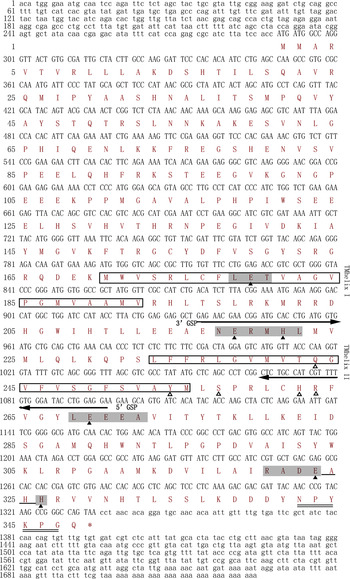
The complete sequence of AOX cDNA consisted of a 5′terminal untranslated region (UTR) of 288 bp, a 3′UTR of 390 bp and an open reading frame (ORF) of 1047 bp, which contained a termination codon (TAA) and encoded a putative protein of 348 amino acids (Figure 2) with a molecular mass of 39.49 kDa and a theoretical isoelectric point (pI) of 8.49. The deduced protein contained conserved LET, NERMHL, LLEEA, RADE_ _H regions and a Q-binding site, which are the main characteristics of AOX. TargetP analysis showed that Urechis unicinctus AOX was located in the mitochondria, with a score of 0.673. Topology prediction for the 348 amino acid protein by TMHMM demonstrated the protein had two transmembrane helices (TMHelix I 170-192 aa and TMHelix II 233-255 aa, Figure 2).
The full-length cDNA sequence and deduced amino acid sequence of Urechis unicinctus AOX. The highly conserved iron-binding motifs are highlighted as shaded regions in which the Glu and His residues are marked by ▴ and the residues for quinol binding are indicated by △. The transmembrane helices (TMhelix) are shown in an open box; RACE gene-specific primers (GSP) are underlined with an arrow; the animal-specific C-terminal motif is demonstrated by double lines.

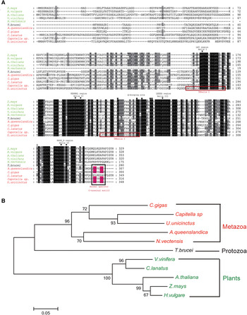
Sequence analysis by the NCBI BLAST program revealed that the putative amino acid sequence of U. unicinctus AOX shared high identity with other AOXs in plants, parasites, green algae, fungi and some metazoans. Alignment of AOX sequences demonstrated they shared well-conserved motifs characteristic of alternative oxidase. Similar to other animal AOXs, U. unicinctus AOX possesses a C-terminal motif N-P-Y-[KE]-P-G that is not present in AOX proteins from other kingdoms (Figure 3A). A phylogenetic tree was constructed based on amino acid alignment using the MEGA program and the result indicated that U. unicinctus AOX was clustered together with Capitella sp., as a sub-cluster of metazoans, while others formed distinct paraphyletic clusters (Figure 3B). Overall, the relationships displayed in the phylogenetic tree were generally in accordance with the taxonomy.
Sequence comparison of protein encoded by AOX from different species: (A) multiple sequence alignment of AOX sequences from different species. Identical and similar residues are highlighted in black and grey, respectively. Residues for iron-binding and quinol binding are indicated by ▾ and ■, respectively. The transmembrane helices (TMhelix) and the animal-specific C-terminal motif are denoted in the red box; (B) the phylogenetic relationships between AOX from different species constructed by MEGA 4.0 using the neighbour-joining method. The Accession numbers are: Amphimedon queenslandica (XP_003385247.1), Arabidopsis thaliana (BAB09852.1), Capitella sp. (JGI genome), Citrullus lanatus (ADD84880.1), Crassostrea gigas (EKC33714.1), Hordeum vulgare subsp. Spontaneum (AEC33278.1), Nematostella vectensis (XP_001635929.1), Trypanosoma brucei brucei (AAB46424), Urechis unicinctus (ADZ36698.1), Vitis vinifera (XP_002274470.1) and Zea mays (AAB97839). Unless otherwise specified, the sequences were obtained from NCBI. Metazoan species are listed in red, protozoa in black and plants in green. See online publication for colour figure, doi:10.1017/S0025315413000696

Expression of AOX mRNA and activity of CCO under different sulphide treatments in Urechis unicinctus body wall and hindgut
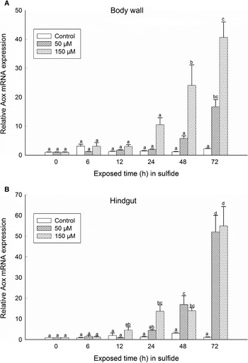
The expression dynamics of U. unicinctus AOX mRNA in the body wall and hindgut of worms exposed to different concentrations of sulphide are shown in Figure 4. Distinct dose- and time-dependent elevations of AOX mRNA expression were present in both tissues from the treatment groups during the experiment. In the body wall of worms exposed to 50 µM sulphide, increased AOX mRNA was not significant(P > 0.05) during the first 72 h of exposure, while a significant increase (P < 0.05) was detected at 72 h and was 16.7-fold higher compared with controls. At 150 µM sulphide, a significant increase (P < 0.05) was observed at 48 h after exposure with a 24.1-fold and 40.6-fold increase at 72 h compared with controls, respectively (Figure 4A). In the hindgut, a similar pattern was observed (Figure 4B). However, the timing of the significant increase occurred earlier in the hindgut than in the body wall. In the 50 µM sulphide group, AOX mRNA increased significantly at 48 h with a 17.2-fold higher increase and reached a maximum (52.1-fold higher) at 72 h. In the 150 µM sulphide group, the expression was higher than that of the 50 µM sulphide group except at 48 h (Figure 4B).
Relative expressions of AOX mRNA in body wall (A) and hindgut (B) of Urechis unicinctus exposed to sulphide. The β-actin gene was chosen as a reference gene, and the relative gene expression was analysed according to the 2−ΔΔCT method. All data are presented as mean ± SE, N = 4 with two replicates. Groups containing the same letters on the bar indicate no significant difference while different letters on the bar indicate significant difference (P < 0.05).

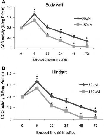
The CCO activity, a terminal electron acceptor of classical mitochondrial ETC, increased in the body wall and hindgut of worms at the beginning and then declined during sulphide exposure (Figure 5). At 6 h of sulphide exposure, the CCO activities of both worm tissues exposed to 50 µM sulphide significantly increased (P < 0.05), while the increase due to 150 µM sulphide was not significant (P > 0.05). The CCO activity decreased significantly (P < 0.05) in both worm tissues exposed to 150 µM sulphide for 24 h, and was inhibited (approximately 0) at 48 h. In the 50 µM group, the timing of the significant decrease occurred later than the 150 µM group, which was at 72 h (Figure 5).
CCO activity in body wall (A) and hindgut (B) of Urechis unicinctus exposed to sulphide. Data represent the mean ± SE, N = 4 with twp replicates. *, indicates significant difference from the 0 h control, P < 0.05.

DISCUSSION
In this study, the full-length cDNA of AOX from the Echiuran worm Urechis unicinctus was cloned, and shown to contain a conserved 4-helix bundle, Q-binding site and an animal-specific C-terminal motif. Furthermore, we experimentally demonstrated the expression dynamics of AOX mRNA for the first time, and suggested that alternative pathways exist in the worm by the expression of U. unicinctus AOX mRNA and CCO activity when the animal is exposed to sulphide.
Sequence and structure analysis of the alternative oxidase from Urechis unicinctus
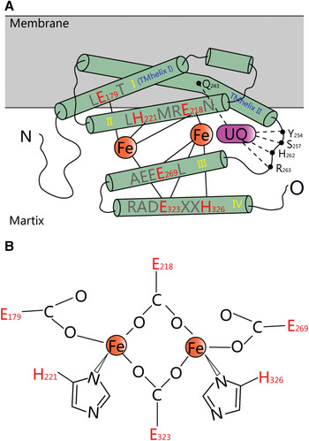
The currently adopted view is that the active site of AOX comprises a non-haeme di-iron centre, in which the metal atoms are ligated by glutamate and histidine residues within a 4-helix bundle (Gomes et al., Reference Gomes, Le Gall, Xavier and Teixeira2001). Based on the di-iron centre and assignments of the proposed Glu and His ligands, AOX was predicted to interact with one leaflet of the membrane bi-layer as an integral interfacial membrane protein, referred to as the Andersson and Nordlund (AN) model (Berthold et al., Reference Berthold, Andersson and Nordlund2000; Moore & Albury, Reference Moore and Albury2008). The model is supported by extensive site-directed mutagenesis studies (Affourtit et al., Reference Affourtit, Albury, Crichton and Moore2002; Albury et al., Reference Albury, Affourtit, Crichton and Moore2002), and the presence of a binuclear iron center was later confirmed by electron paramagnetic resonance (EPR) spectroscopy (Berthold et al., Reference Berthold, Voevodskaya, Stenmark, Graslund and Nordlund2002; Moore et al., Reference Moore, Carre, Affourtit, Albury, Crichton, Kita and Heathcote2008). Using electrochemistry and Fourier transform infrared (FTIR) spectroscopy, the redox properties of recombinant alternative ubiquinol oxidase from the parasite Trypanosoma brucei were studied and interpreted in terms of the possible structure of the active site and mechanism of oxygen reduction to water by AOX. This study confirmed the di-iron carboxylate protein model of alternative oxidase (Maréchal et al., Reference Maréchal, Kido, Kita, Moore and Rich2009). Based on the AN model and bioinformatics, we predicted a schematic structure of U. unicinctus AOX in the present study, with four conserved regions (LE 179 T, NE 218 RMH 221 L, LE 269 EEA, RADE 323 AHH 326 ) involved in the formation of the 4-helix bundle, in which the Glu and His residues of the motifs participate in the formation of the bundle required for iron-binding (Figure 6).
Schematic structure of Urechis unicinctus AOX based on a model proposed by Andersson and Nordlund (modification from Berthold et al., Reference Berthold, Andersson and Nordlund2000): (A) the modified AN model. Cylinders with yellow Roman numerals represent the four-helix bundle in which the red letters represent conserved residues acting as iron-binding ligands (iron atoms are shown as red spheres). Cylinders with blue letters indicate transmembrane helices predicted by TMHMM. Black dots signify residues for quinine binding and UQ in pink represents the ubiquinone; (B) ligands of the di-iron centre in the modified AN model. See online publication for colour figure, doi: 10.1017/S0025315413000696

To fulfil its main function of transferring electrons to oxygen, a binding site for ubiquinol, the reducing substrate of AOX, must be contained. Albury et al. (Reference Albury, Elliott and Moore2010) proposed a model for the ubiquinol-binding site in AOX between helices II and III, leading from a proposed membrane binding domain to the catalytic domain, by bioinformatics analysis and site-directed mutagenesis in plant Sauromatum guttatum AOX expressed in yeast Schizosaccharmyces pombe. It was demonstrated that Gln242, Tyr253, Ser256, His261 and Arg262 were critical for ubiquinol-binding (Albury et al., Reference Albury, Elliott and Moore2010). Multiple sequence alignment of AOX proteins showed that these residues were highly conserved and existed in animals (Figure 3A). Transmembrane prediction of AOX in U. unicinctus by TMHMM located the ubiquinol-binding pocket (composed of Gln243, Tyr254, Ser257, His262 and Arg263) in the end of the transmembrane helix (TMhelixII, Figure 2), thus placing the ubiquinol ring near the membrane interface, similar to cytochrome bo3 ubiquinol oxidase from Escherichia coli (Abramson et al., Reference Abramson, Riistama, Larsson, Jasaitis, Svensson-Ek, Laakkonen, Puustinen, Iwata and Wikstrom2000). Such a structure, a transmembrane helix containing Q-binding site followed by a part of the 4-helix bundle (helix III), moved the reduced quinol as a substrate of AOX to the di-iron centre and promoted its functions.
Proposed role of AOX in mitochondrial sulphide oxidation detoxification
In the classical respiratory chain of mitochondria, CCO (complex IV) acts as a terminal oxidase to accept electrons from cytochrome c and direct them to the four-electron reduction of O2 with the formation of water and the concomitant translocation of protons across the inner membrane coupled with ATP synthesis (Figure 1). Sulphide can inhibit the activity of CCO by reversibly binding at the haeme site of cytochrome aa3 (Cooper & Brown, Reference Cooper and Brown2008). As ATP synthesis is dependent on the flow of electrons down the electrochemical gradient mediated by enzyme cytochrome complexes, inhibition of any complexes of the respiratory chain will disrupt the electron flow, increasing the cellular NADH/NAD+ ratio and placing the cell under reductive stress (Truong et al., Reference Truong, Eghbal, Hindmarsh, Roth and O'Brien2006). In addition, the inhibition of CCO will lead to incomplete oxygen reduction to water. All of these factors can increase the formation of ROS, which has the potential to disturb the homeostasis of the intracellular redox state, which is crucial for the correct functioning of most biological processes.
In the lugworm Arenicola marina, electrons from sulphide oxidation at lower sulphide levels can be passed to oxygen via the classical CCO pathway and coupled to ATP production. However, activity of CCO was blocked when sulphide concentrations were higher than 10 µM. The electrons from sulphide oxidation are thought to be transferred to oxygen via AOX (Völkel & Grieshaber, Reference Völkel and Grieshaber1996). In our study, the activity of CCO decreased significantly at 24 h and was approximately zero at 48 h after 150 µM sulphide exposure, but significantly declined at 72 h after 50 µM sulphide exposure. Our results might imply that CCO of U. unicinctus has a greater tolerance to sulphide than A. marina. The mRNA expression of U. unicinctus AOX increased significantly at 48 h after sulphide exposure suggesting U. unicinctus AOX promoted the transfer of electrons from sulphide oxidation to oxygen, and thus decreased tissue damage in the worm caused by excess ROS production when CCO activity was inhibited. Our data experimentally support the hypothesis that an alternative sulphide oxidation pathway exists in animals and indicates a relevant role for AOX in establishing a protective mechanism against sulphide stress.
FINANCIAL SUPPORT
This work was supported by the Natural Science Foundation of China (NSFC) [40776074 and 31072191].