Introduction
Thelohania solenopsae Knell Allen and Hazard was first discovered in ethanol-fixed collection samples of the red imported fire ant, Solenopsis invicta and was formally described as a new species on the basis of light and electron microscopy of S. invicta collected around the city of Cuiabá, Matto Grosso, Brasil (Knell et al. Reference Knell, Allen and Hazard1977). Later, the same microsporidium was discovered in Argentina, Uruguay, Paraguay, and several southern states of the United States (Briano et al. Reference Briano, Patterson and Cordo1995; Williams et al. Reference Williams, Knue and Becnel1998, Reference Williams, Oi, Porter, Pereira and Briano2003; Cook, Reference Cook2002; Sokolova et al. Reference Sokolova, Bossard, Fuxa, Sanson and Foil2004a). The most prominent feature of this species – the presence of pansporoblasts containing 8 octospores – was the reason this species was placed into the genus Thelohania. The microsporidiums's additional Nosema-like sequence and infection of insect hosts, however, were not consistent with the characteristics of this genus, which exhibits monotypic development and parasitization of marine crustaceans (Hazard and Oldacre, Reference Hazard and Oldacre1975). Furthermore, at least 4 developmental sequences, which produce morphologically distinguishable spore types, have been described for T. solenopsae (Sokolova and Fuxa, Reference Sokolova and Fuxa2001; Shapiro et al. Reference Shapiro, Becnel, Oi and Williams2003; Sokolova et al. Reference Sokolova, McNally, Fuxa and Vinson2004b). Morphological and life-cycle studies together with recently obtained SSU rDNA sequence data for Thelohania butleri (Brown and Adamson, Reference Brown and Adamson2006), a close relative of T. giardi, the type species of the genus Thelohania, as well as all phylogenies based on molecular data (Lom et al. Reference Lom, Nilsen and Dykova2001; Moodie et al. Reference Moodie, Le Jambre and Katz2003a, Reference Moodie, Le Jambre and Katzb; Brown and Adamson, Reference Brown and Adamson2006; Sokolova et al. Reference Sokolova, Kryukova, Glupov and Fuxa2006), indicate that Thelohania solenopsae is not related to the species parasitizing crustaceans and thus must be removed from the genus Thelohania.
T. solenopsae has been considered a promising biocontrol agent because it can be detrimental to host populations over long periods (Williams et al. Reference Williams, Oi and Knue1999; Cook, Reference Cook2002; Cook et al. Reference Cook, Lowery, Frey, Rowe and Lynch2003; Oi et al. Reference Oi, Valles and Pereira2004; Fuxa et al. Reference Fuxa, Sokolova, Milks, Richter, Williams and Oi2005b; Milks et al. Reference Milks, Fuxa and Richter2008). It has been successfully introduced into fire ant field populations in the United States (Fuxa et al. Reference Fuxa, Sokolova, Milks, Richter, Williams and Oi2005b). However, the lack of knowledge of its life-cycle significantly hampers development of methods for use of this microsporidium in biological control. For example, numerous attempts to perorally infect fire ants with formulations of spores have been unsuccessful, because the spore type(s) involved in horizontal transmission of T. solenopsae is unknown. The mode of horizontal transmission of this microsporidium in monogyne (single-queen) colonies also remains enigmatic; the prevalence of natural infections of monogyne colonies was estimated to be as low as 0% in Florida (Oi et al. Reference Oi, Valles and Pereira2004) and 3% in Louisiana (Milks et al. Reference Milks, Fuxa and Richter2007), and experimental introductions into strictly monogyne populations were either not successful (Oi et al. Reference Oi, Valles and Pereira2004; Oi, Reference Oi2006) or not long-lasting (Fuxa et al. Reference Fuxa, Milks, Sokolova and Richter2005a).
The goals of this paper were (1) to present new results on the fine morphology and biology of T. solenopsae collected from Louisiana populations of Solenopsis invicta and to compare these data with the original description of T. solenopsae (Knell et al. Reference Knell, Allen and Hazard1977) based on the Brazilian isolate of the microsporidium, (2) to clarify the microsporidium's life-cycle based on the new data and (3) to establish a new genus, Kneallhazia, for T. solenopsae based on peculiarities of its life-cycle, which are unique among microsporidia as adaptations to the social life-style of the host.
We also present SSUrDNA-sequence divergence data placing T. solenopsae as a separate clade on phylogenetic trees. Additional data are presented in this paper on morphology and life-cycle accumulated during a 6-year study of T. solenopsae in Louisiana pastures in experimental and field conditions, to help understand the parasite's biology, which is of key significance for its successful application in biological control.
Free diplokaryotic spores developing in pupae (Oi et al. Reference Oi, Becnel and Williams2001; Shapiro et al. Reference Shapiro, Becnel, Oi and Williams2003) and larvae 4, previously named ‘juvenile spores’ (Sokolova et al. Reference Sokolova, McNally, Fuxa and Vinson2004b), are referred to in this paper as Type 1 DK spores. Free diplokaryotic spores developing in imagoes, previously named Nosema-like spores (Sokolova and Fuxa, Reference Sokolova and Fuxa2001; Sokolova et al. Reference Sokolova, McNally, Fuxa and Vinson2004b), are referred to in this paper as Type 2 DK spores. Other terms for spore types produced by this microsporidium are the same as in the original descriptions: octospores (Knell et al. Reference Knell, Allen and Hazard1977) and megaspores (Sokolova et al. Reference Sokolova, McNally, Fuxa and Vinson2004b).
Materials and methods
The microsporidium and fire ants
Microsporidia-infected colonies of S. invicta were removed either from naturally infected field populations near Rosepine, Louisiana (Sokolova et al. Reference Sokolova, Bossard, Fuxa, Sanson and Foil2004a) or from sites of experimental releases of the microsporidium near St Joseph and Clinton, Louisiana. Brood for both releases was obtained originally from infected colonies in Florida (Fuxa et al. Reference Fuxa, Sokolova, Milks, Richter, Williams and Oi2005b). There were no morphological differences (Sokolova et al. Reference Sokolova, Bossard, Fuxa, Sanson and Foil2004a) or variations in SSrDNA sequence (Y. Sokolova and J. Fuxa, unpublished results) between the native Louisiana microsporidium from Rosepine and the Florida isolates. Colonies were maintained in the laboratory with standard rearing methods (Banks et al. Reference Banks, Lofgren, Jouvenaz, Stringer, Bishop, Williams, Wojcik and Glancey1981). Larval instars were identified according to O'Neal and Markin (Reference O'Neal and Markin1975). Workers were considered as minors if their head capsules measured less than 550 μm across, and majors if the heads were more than 700 μm (Sokolova et al. Reference Sokolova, Fuxa and Borkhsenious2005a). Queens of polygyne colonies were differentiated from de-alate females by the presence of spermatheca filled with spermatozoids and follicles containing mature eggs at the proximal region of ovarioles (Hermann and Blum, Reference Hermann and Blum1965). The morphology and life-cycle of T. solenopsae were studied during the years 2001–2006.
Overall, 1059 individual insects (including fourth-instar larvae, pupae, and imagoes were examined individually; 378 of them were infected with the microsporidium.
Light microscopy
Infection of the colonies by T. solenopsae was confirmed by modified trichrome staining (Weber et al. Reference Weber, Bryan, Owen, Wilcox, Gorelkin, Visvesvara, Juranek, Addiss, Spencer, Hightower, Stewart, Roberts, Wahlquist, Horsburgh, Castro, Tauxe, Vugia, Glass, Thompson, Schwartz, Kozarsky, Steinberg, Shulman, Dismukes, Dupuis, Nickerson, Rimland, Hogan, Johnson and Elliott1992) and by PCR (Milks et al. Reference Milks, Sokolova, Isakova, Fuxa, Mitchell, Snowden and Vinson2004; Sokolova et al. Reference Sokolova, Sokolov and Fuxa2004c). Infection of individual ants was detected by direct observation of fresh smears under phase-contrast optics or methanol-fixed smears stained with trichrome. Dissection of ants and isolation of specific tissue and organs were performed under a Leica MC125 dissecting microscope. Microsporidian development was characterized, in general, from microscopical observation of Giemsa-stained smears of host tissues at a magnification of 1000X. A working solution of Giemsa stain was prepared by 5-fold dilution of commercial stock solution (Sigma) in phosphate buffer, pH 7·0. Tissue specificity was confirmed by examination of hemi-thin araldite-embedded sections stained with methylene blue (1% methylene blue in 4% sodium borate in water). For visualizing nuclei, fresh or methanol-fixed smears were stained with DAPI (Sigma, 10 μm solution in phosphate-buffered saline). Slides were examined, and stages and spores were measured and photographed under a Nikon Eclipse E-600 Microscope equipped with the fluorescence unit and a Metaview digital camera and software (MetaView. 1998, Meta Imaging Series 4.5. Universal Imaging Corporation, West Chester, PA).
Electron microscopy
Only imagoes – 3 minor workers, 5 major workers, 10 alates of both sexes, and 8 infected queens – were examined by electron microscopy. Ovaries of the latter were isolated and observed separately under EM. Pupae infected with Type 1 DK spores have previously been examined ultrastructurally by Shapiro et al. (Reference Shapiro, Becnel, Oi and Williams2003).
Electron microscopy was performed as previously described (Sokolova et al. Reference Sokolova, Fuxa and Borkhsenious2005a). Briefly, samples were fixed in a mixture of 2% paraformaldehyde and 1·25% glutaraldehyde in sodium cacodylate buffer, pH 7·4, post-fixed in 1% OsO4 in the same buffer, incubated overnight in 0·5% uranyl acetate, dehydrated in a descending ethanol series, infiltrated, and embedded in Epon-araldite resin. All reagents were from EMS Chemicals (Fort Washington, PA). Thin (80 nm) sections were cut on a MT-XL ultratome (RMC Product, Tucson, AZ) or Reichert Ultracut Microtome (Reichert-Jung, Austria), contrasted with lead citrate, and examined with a Zeiss 10 or JEOL JEM 100CX electron microscope at 70–80KV.
Identification of infection in brood by PCR
Approximately 30 mg of eggs or 1–3 instars from the infected or control colony were pooled together, washed in 1% sodium dodecyl sulfate (SDS) and macerated in 150 μl of lysis buffer (50 mm Tris-HCl, pH 8, 4% SDS, and 5% 2-mercaptoethanol) in a 1·5-ml microtube with a disposable pellet pestle (Koates Glass Co., Vineland, NJ). DNA was isolated by phenol-chloroform extraction followed by isopropanol precipitation, and the DNA was PCR-amplified with TsSSU841F and TsSSU1059R primers as previously described (Milks et al. Reference Milks, Sokolova, Isakova, Fuxa, Mitchell, Snowden and Vinson2004).
Statistical and phylogenetic analyses
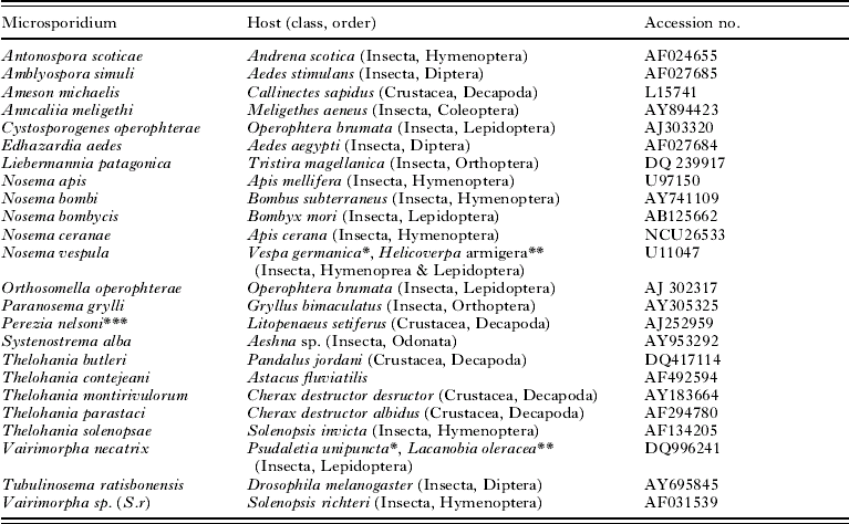
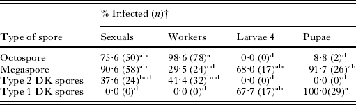
SATATISTICA for Windows software, version 6.0 (Anonymous, 1995) was used for all statistical analyses. Distribution of spore types among ant castes, larvae, and pupae infected with T. solenopsae was analysed by two-way ANOVA with the Tukey HSD test among means. Individuals (n=413) from 3 localities (Rosepine, Clinton, and St Joseph, LA) were checked for T. solenopsae, and 198 appeared to be infected and were split into 3 replicates (63 from St Joseph; 67 from Rosepine, and 68 from Clinton) for analyses (Table 1). The interactions among 2 independent variables, the spore type and the caste or stage of the ant (sexual female or male, major or minor worker, larvae 4 or pupae), and a dependent variable, the percentage of individuals infected with certain type of spores, were estimated. The Arcsine transformation was used for data expressed as percentages; Bonferonni adjustment was applied to multiple comparisons (Zar, Reference Zar1999).
Hosts and GenBank Accession numbers for the SSU rRNA sequences of 24 microsporidian species used in the phylogenetic analysis

* Natural host.
** Experimental host.
*** Cited in GenBank as Pleistophora sp. (LS); identified as Perezia nelsoni by Canning et al. (Reference Canning, Curry and Overstreet2002).
Phylogenetic relationships among microsporidian species were assessed by the neighbour-joining (NJ) algorithm applied to the alignment of 24 SSUrDNA sequences of approximately 1300 bp each, obtained through the GenBank. Sequences chosen for the analysis represent the major groups of terrestrial and polysporous species, representatives of the genus Thelohania spp., and microsporidia parasitizing hymenopterans. Table 1 shows Accession numbers of the sequences, the list of microsporidia and host species, and systematic positions of hosts. An ascomycete, Basilobolus ranarum, was chosen as an outgroup. Sequences were aligned and subjected to NJ and Phylip distance matrix analyses by the Clustal X program (Thompson et al. Reference Thompson, Gibson, Plewniak, Jeanmougin and Haggins1997).
Results
Light microscopy
Spore types and distribution of T. solenopsae infection among castes and instars of fire ants
The life-cycle of T. solenopsae includes 4 regularly reproduced sequences, each resulting in the formation of specific type of spores. The spore sequence occurring exclusively in brood produces the Type 1 diplokaryotic (DK) spores (length 3·9±0·33×width 2·0±0·24 μm, n=10, based on methanol-fixed smears stained with trichrome or Giemsa) with prominent posterior vacuoles, that develop in pupae and occasionally in fourth instars. The Type 2 DK spore sequence produces thin-walled diplokaryotic spores (4·6±0·29×2·3±0·20 μm, n=30) with easily discharged polar filaments, in imagoes of all castes. The ‘megaspore sequence’ produces large spores (6·2±0·41×3·6±0·29 μm, n=26) that can be found in all castes of imagoes as well as in pupae and fourth instars, but mostly in queens (de-alate females laying eggs). The ‘octospore sequence’ produces octospores (3·1±0·38×2·1±0·23 μm, n=148) in sporophorous vesicles in all castes of imagoes, predominantly in workers and alates, but never in brood (Fig. 1; Table 2). No spores or stages could be definitely identified in 1–3 instar larvae by trichrome, Calcofluor, or Giemsa staining. However, PCR with specific T. solenopsae primers amplified microsporidian DNA in eggs and 1–3 instars in 80% (n=15) of infected colonies, clearly suggesting the presence of latent infection in eggs and brood.
Four spore types of Thelohania solenopdsae, phase contrast. (a) Type 1 DK spores with prominent posterior vacuole (arrow). (b) Type 2 DK spores with a few octospores (arrow). (c) Megaspores and individual octospores, indicated by arrow. (d) Octospores; arrow points to a macrospore resulting from the failure of the last post-meiotic division.

Distribution of Thelohania solenopsae spore types among castes and stages of fire ants collected from three infected colonies revealed by trichrome stainingFootnote *

* 413 individuals were examined, and 198 of them were infected, generally with more than one spore type.
† Means throughout the table followed by the same letter did not differ at P<0·05, Tukey HSD test.
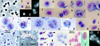
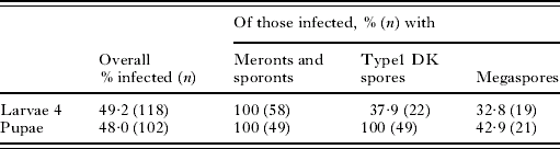
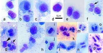
Thelohania solenopsae infection in fourth instars and pupae. Transition to the fourth instar (larvae 4) obviously triggered microsporidian development, because spores and vegetative stages were consistently detected in larvae 4 and in pupae (Fig. 2; Tables 2 and 3). Pre-spore stages were found in 100% of infected larvae 4 and pupae; Type 1 DK spores were detected in 37·9% of larvae 4 and in 100% of pupae. Megaspores were identified in 32·8% of the microsporidium-infected larvae and in 42·9% of pupae (Table 3, Fig. 2). The most peculiar stages were the unicellular round cells of about 1·7–2·5 μm in diameter, observed in unfixed or Giemsa-stained smears only from larvae 4 (Fig. 2a–e). These stages were often seen in pairs. DNA-specific staining by DAPI revealed that 1 cell of the pair occasionally lacked a nucleus, whereas its counterpart might contain 2 separate nuclei (Fig. 2d) or 1 large nucleus resulting from the fusion of 2 nuclei (Fig. 2e, f). The large nucleus of such monokaryotic cells (Fig. 2g, h) divided to form a diplokaryon (Fig. 2i). Monokaryotic cells regularly displayed a characteristic appendage (Fig. 2g). Diplokaryotic cells underwent nuclei dissociation and cytokinesis (Fig. 2j, k), likely followed by mitotic divisions resulting in formation of round ‘early sporonts’ (5–10 μm in diameter) with large diplokaria (Fig. 2l), especially abundant on smears from larvae 4 and pupae. Their diplokarya divided to produce tetranuclear sporonts (Fig. 2m–q), which gave rise to 2 diplokaryotic sporoblasts that matured eventually into the Type 1 DK spores (Fig. 2r–x). These spores, discovered by Oi et al. (2001 ) and described ultrastructurally by Shapiro et al. (Reference Shapiro, Becnel, Oi and Williams2003), could be easily distinguished from other spore types by prominent posterior vacuoles seen both in unfixed and fixed spores (Fig. 1a; Fig. 3u, v, x). ‘Empty’ spores with discharged polar filaments looked evenly greyish under phase contrast (Fig. 2t); their presence on smears suggested that the Type 1 DK spores play a role in autoinfection.
Stages of Thelohania solenopsae developing in larvae 4 and pupae. (a and b) Uninucleate miniature cells, often observed in pairs, phase contrast, a smear from larvae 4. (c) The same cells, stained with Giemsa. (d) The same cells, stained with DAPI. One of 2 cells in a pair lacks nucleus (arrow). (e–g) Mononuclear cell with a large nucleus, presumably a product of karyogamy of 2 haploid nuclei. Size of the nuclei, which exceeds 2 haploid nuclei put together, suggests that DNA replication took place before the cell entered meiosis. Arrow points to characteristic appendix. (h–j) Dikaryotic cell, in which nuclei eventually dissociate. (k) Stage with 2 separated nuclei at the onset of cytokinesis. (l) Cell division resulting in 2 cells with large diplokaria. (m–q) Tetranuclear sporonts with comparatively small nuclei, observed in larvae 4 (m, q) and pupae (n–p). (r and s) Sporoblasts of the Type 1 DK spore sequence from larvae 4 (r) and pupae (s). (t) Discharged spores (arrows) on smears from pupal fat body, suggesting autoinfection; intact spore is indicated by arrowhead. (u) Megaspores (arrow) and Type 1 DK spores (arrowhead) on a smear from pupal fat body. (v) Type 1 DK spores (arrowhead) and sporoblasts (arrows), the most abundant pre-spore stage observed in pupae. (w and x) Mature Type 1 DK spores. (y) Megaspore in pupa. (z) A few octospores revealed by DAPI staining in larvae did not contain nuclei, which might indicate that their sporoplasms had been already discharged. (a, b, t, u) Phase contrast; (d, z) DAPI staining of fresh smears; (e–s, v and w) Giemsa staining of methanol-fixed smears; (x and y) trichrome staining of methanol-fixed smears. Scale bars for a–s, v–z, are as shown on c and x.

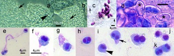
Fresh smear of fat body tissue of Solenopsis invicta minor worker infected with Thelohania solenopsae observed under a dissecting microscope (a) and phase-contrast optics at 400× (b and c). (a) Uninfected regions of fat body consist of loosely packed ovoid adipocytes of uniform diameter (black arrow); the infected cell (asterisk) is hypertrophied, and mature spores are released from the disturbed region (white arrow). (b) A smear from the hypertrophied cell (immature cyst) revealing stages of sporogony: uninuclear cell (arrowhead) presumably resulting from karyogamy of diplokaryon counterparts of a meront/sporont transitional stage undergoes meiosis followed by 3 successive divisions to give rise to 8 sporoblasts transforming eventually into octospores. Arrows (numbers 1–3, respectively) indicate cells produced as a result of the 1st, 2nd, or 3rd meiotic divisions. (c) A smear from a cyst: octets of octospores with refractive spore walls recently liberated from the cyst.

Infection by Thelohania solenopsae revealed by Giemsa stain in larvae 4 and pupae

Some of the early sporonts (Fig. 2l) probably were directly (without nuclear division) transformed into sporoblasts of the megaspore sequence (Fig. 2y), because no other potential precursors of the megaspores were seen in larvae 4, particularly when the potential octoploidy of megaspores was strongly suggested by measurements of nuclear volumes stained with DAPI (Y. Sokolova and J. Fuxa, unpublished data).
Octospores were extremely rare on smears from larvae 4 and pupae and likely derived from the disrupted fourth larval intestines or remnants of pupal meconium (Chen et al. Reference Chen, Snowden, Mitchell, Sokolova, Fuxa and Vinson2004), which normally were removed from smears. Interestingly, DAPI staining of these occasional octospores revealed that many of them lacked a nucleus (Fig. 2z).
Development of the octospore sequence in workers and alates
The most conspicuous type of sporogony in workers consisted of numerous octospores (meiospores), maturing inside sporophorous vesicles (SVs) in sets of 8 (Fig. 1d). SVs containing mature spores were fragile; they readily disintegrated and liberated octospores. Light microscopical detection of microsporidian infection in fire ants has been based on visualization of this particular spore type (Milks et al. Reference Milks, Sokolova, Isakova, Fuxa, Mitchell, Snowden and Vinson2004; Sokolova et al. Reference Sokolova, Bossard, Fuxa, Sanson and Foil2004a). In some imagoes, infection with octospores caused false hypertrophy of fat body tissue which, at the late stages of disease, led either to total substitution of fat body tissue by masses of spores or to depletion of most of the fat body cells except for a few (1–20) that eventually transformed into large ‘sporocytosacs’ (Sokolova et al. Reference Sokolova, Fuxa and Borkhsenious2005a), called also ‘cysts’ (Knell et al. Reference Knell, Allen and Hazard1977) (Fig. 3a–c). More than 90% of all dissected workers contained mature octospores exclusively; this, together with the absence of any noticeable gross pathology, made it difficult to study the microsporidium's development in imagoes. Fortunately, some immature cysts contained a few stages of sporogony (Fig. 3b, Fig. 4) in addition to spores. The probability of observing pre-spore stages of T. solenopsae was greater in infected alates (Fig. 5) than in workers, probably because the alates live longer (Hölldobler and Wilson, Reference Hölldobler and Wilson1990) and thus may host several overlapping generations of the pathogen. In workers and alates the octosporogonic sequence was initiated by fusion of 2 parts of the meront diplokaryon (Fig. 4a, b). The dikaryotic arrangement of the nucleus was restored in the process of the first meiotic division (Fig. 4c), which was followed by nuclei dissociation (Fig. 4d, e) and plasmotomy (Fig. 4e). The resultant uninucleate sporogonial plasmodia (Fig. 4f) underwent 3 rounds of DNA replication and successive binary nuclear fissions (Fig. 4g–n). The boundaries between individual sporonts were noticeable beginning with the 4-nuclear stage (Fig. 4j, l–o). Sporophorous vesicles (SV) normally contained 8 sporoblasts (Fig. 4p), which transformed gradually into octospores (Fig. 4p–r, v). In heavily infected insects, SVs often contained ‘macrospores’ (Knell et al. Reference Knell, Allen and Hazard1977), which are abnormal spores with 2–3 nuclei produced due to a failure of the last division of the sporogonial plasmodium (Fig. 4q–s). Type 2 DK sporoblasts and spores were seen occasionally in smears from infected workers (Fig. 4t, u), but this type of spore appeared en masse only in a few individuals (Fig. 1b). Megaspores (Fig. 4u, v) were less abundant but not uncommon in smears from workers. Megaspores often were concentrated in the subcuticular layer of abdominal muscles of alates (Sokolova et al. Reference Sokolova, McNally, Fuxa and Vinson2004b). Smears from these tissues displayed developmental stages of the megaspore (Fig. 5a–l) and Type 2 DK spore sequences (Fig. 5m–p). Observations suggested that the formation of megaspores as well as of octospores was preceded by fusion of DK counterparts presumably followed by meiosis (Fig. 5a–f). In some smears, cells were observed that contained 2 diplokarya of much smaller diameter than the diplokarya of the stages of the megaspore sequence (Fig. 5m). We assume that these were tetranuclear sporonts of the Type 2 DK spore sequence, which after cellular division produced binucleate sporonts that transformed into sporoblasts and Type 2 DK spores (Fig. 5n–p).
Stages of microsporidian development observed on smears prepared from immature cysts and fat body fragments isolated from workers. (a) Octets of octospores (OO) and a diplokaryotic meront (DK – diplokaryon). (b and c) Meront (M) undergoing fusion of DK counterparts. (d and e) Dissociation of nuclei (1st meiotic division). (f) Uninuclear sporont, the precursor of the sporogonial plasmodium. (g and h) Sporogonial plasmodia with 2 large nuclei (products of the 1st nuclear division) presumably in the process of DNA replication. (i) Sporogonial plasmodium at 3-nuclei stage (undergoing 2nd nuclear division). (j and k) Uninuclear sporont (arrowhead) and 4-nuclei sporogonial plasmodia (arrows) at various stages of separation of nuclei. (l–n) Sporogonial plasmodia in transition to 8-nuclei stage (undergoing 3rd nuclear division) and uninuclear sporont (arrowhead). (o) Octet of sporoblasts in sporophorous vesicle. (p) Eight immature (left) and mature octospores (right) spores in sporophorous vesicles. (q and r) Megaspore (q, left upper corner) and sporophorous vesicles (q and r) containing 7 octospores, one of which is an aberrant macrospore (arrow). (s) Sporophorous vesicle with 4 macrospores, occasionally observed in heavily infected imagoes. (t) A sporoblast and a spore of the Type 2 DK spore sequence. (u) Megaspores and individual octospores. (v) Three spore types as they were observed on trichrome-stained preparations from imagoes: octospores (O); megaspore (M) and Type 2 DK spores (N). (a–s) Giemsa staining; (t) trichrome staining of methanol-fixed smears.

Pre-spore stages of the megaspore and Type 2 DK -spore sequences observed on smears from abdominal muscle tissue of alates. (a) Two diplokaryotic meronts. (b) Diplokaryotic meront undergoing fusion of 2 nuclei and a uninuclear stage. (c and d) Uninucleate stages derived from diplokaryotic meronts after fusion of 2 nuclei, presumably undergoing meiosis. (e) Binucleate stage, product of first meiotic division, in the process of cytokinesis. (f) Two early sporonts, in one of which the diplokaryotic arrangement of the nucleus has been restored (arrow). (g) Three sporonts with various conditions of the nuclei: white arrow points to the sporont with 4 tightly packed nuclei (the diplokaryon in the process of division); black arrow points to sporont with 3 nuclei, from which 2 form a diplokaryon, and the third is not yet divided; arrowhead indicates diplokaryotic sporont. (h) Uninucleate stage and 3 diplokaryotic sporonts. (i and j) Diplokaryotic sporonts of the megaspore sequence. (k) Sporoblasts of the megaspore sequence. (l) Two megaspores. (m) Tetranucleate sporont with 2 diplokarya. (n and o) Two sporoblasts of the Type 2 DK spore sequence derived from a tetranucleate sporont. (p) Mature Type 2 DK spores. Giemsa staining of methanol-fixed smears.

Pattern of infection in queens
In determining the distribution of spore types among castes and stages of ants, we checked 24 queens, of which 12 were infected. In Table 2 queens were pooled with alate males and females, because in light microscopy the queens did not differ in the distribution of spore types compared with alate or de-alate females and males (data not shown). All three types of spores – octospores, Type 2 DK spores, and megaspores – were detected in smears from queens, and the percentage of queens infected with megaspores was significantly higher than workers. Occasionally queens, like workers and alates, contained cysts with octospores in abdominal fat body (Fig. 6 a). Examination of isolated ovaries of infected females revealed conglomerates of megaspores inside muscle fibres, tracheoles, and fat body adjacent to ovaries, as well as in ovarioles themselves (Fig. 6b, c). All the queens examined were inseminated. Observation of fresh and Giemsa-stained smears from the infected ovaries showed numerous empty spores (Fig. 6 d), sporoplasms (Fig.6e–g), and presumably early stages of development, including stages with dissociated nuclei, monokaryotic cells, and diplokaryotic meronts (Fig. 6h–k).
Infection in queens. (a) Smear from the queen fat body: 2 adjacent cells are infected with either octospores (arrow) or megaspores (arrowhead). (b) Ovarioles filled with megaspores (arrow). (c) Group of megaspores surrounded by spermatozoids (arrows) in a smear from the ovary of an inseminated queen. (d) Empty (asterisk) and intact spores. (e) Megaspore and a discharged sporoplasm. (f and g) Sporoplasms with 2 nuclei. (h) Early stage with dissociating nuclei. (i) Stages observed on smears from ovaries of inseminated queens: a monokaryotic cell, arrowhead; a 2-nucleate-stage, arrow; and a cell with 2 nuclei presumably in the process of division. (j) Two monokaryotic cells. (k) Two cells with diplokarya. (a and b) Phase contrast; (c) trichrome staining; (e–k) Giemsa staining.

Electron microscopy
Ultrastructural manifestations of T. solenopsae infection in workers and alates
Octospores were concentrated in cysts in most imagoes (Fig. 7a) (Sokolova et al. Reference Sokolova, Fuxa and Borkhsenious2005a). Octospores also developed occasionally inside the layer of host adipose tissue attached to cuticle. Electron microscopy of this region revealed numerous pre-spore diplokaryotic stages (Fig. 7b) that replaced fat body and occasionally the muscular layer underlying the cuticle. In the majority of the examined ants, the subcuticular space looked empty in thin sections because mature octospores were regularly washed out during fixation and washing; only few intact sporophorous vesicles were seen (Fig. 7c). Megaspores were fixed extremely poorly; they resided in half-destroyed muscle cells of workers and alates (Fig. 7d), as well as in adipocytes and tracheoles surrounding undeveloped ovaries and testes in alates. Diplokaryotic stages (Fig. 7e) were common in sections through abdominal adipose tissue of imagoes of all castes. Sections through fat body of alate females regularly revealed mass development of stages with unusual internal structure, presumably in the process of meiosis following rearrangement of the nuclei and cytoplasm (Fig. 7f).
Thelohania solenopsae-infected ant imagoes under low magnification electron microscopy. (a) Section through a cyst infected with mature octospores (S) inside sporophorous vesicles (SV). (b) Diplokaryotic sporonts (Sp). (c) Sporophorous vesicle in the subcuticular layer of adipose tissue. (d) Poorly-fixed megaspores (S) in half-destroyed muscles of a major worker. (e) Adipocyte at the onset of transformation into a cyst (Cys), filled with diplokaryotic stages (DK). (f) Section through fat body of an alate female filled with stages with unusual internal structure. AD, uninfected adipocyte; Cut, cuticule; F, fat granules; M, muscles.

Type 2 DK spore sequence
Stages of this sequence were not readily apparent in what at first appeared to be normal-looking sections of fat body from certain workers and alates (Fig. 8a). Type 2 DK spores were rarely seen en masse (Fig. 8b), and many of them were ‘empty’, with the polar tubes discharged. At the ultrastructural level, the most noticeable sign of host-cell pathology was the enlargement and fragmentation of the nucleus (Fig. 8a, b). Diplokaryotic meronts transformed into diplokaryotic sporonts after a series of divisions (Fig. 8c–f). The transition was marked by cytoplasm vacuolization and deposition of a thin electron-dense layer outside the plasma membrane. Sporonts divided at least once by mitosis (Fig. 8d) to produce 4-nucleus plasmodia (Fig. 8e), which gave rise to 2 sporoblasts (Fig. 8f, g). These transformed eventually into diplokaryotic spores with a thin exospore and 12 polar filament coils (Fig.8h). For a detailed description of spores see Sokolova et al. (Reference Sokolova, McNally, Fuxa and Vinson2004b).
Type 2 DK spore sequence (a–h) and transition to octospore sporogony inside the infected adipocyte of a major worker (i–l). (a) Adipocyte with stages of the Type 2 DK spore sequence (arrows). (b) Adipocyte with invaginated nucleus (N) heavily infected with Type 2 DK spores, some of which have been emptied (arrows). (c) Diplokaryotic meront. (d) Diplokaryotic sporont undergoing nuclear division. (e) Four-nucleate sporont. (f and g) Sporoblasts. (h) Type 2 DK spore. (i) Early sporont with expanded perinuclear space (arrowheads) at the beginning of mitosis; spindle plaques are indicated by arrows. (j) Vacuolated sporont at the interface; ‘opening’ in the centre of the zone of contact between 2 members of the diplokaryon is marked by the arrowhead. (k) Stage undergoing fusion of the halves of the diplokaryon; ‘breaches’ in the zone of contact between nuclei are indicated by arrows. (l) Stage, displaying dissociation of nuclei (arrowheads). DK, diplokaryon; F, fat granules; N, host cell nucleus; Nu, nucleolus; PF, polar filament; P, protein granules; Pp, polaroplast; V, vacuole.

Octospore sequence
Assemblages of diplokaryotic stages tightly packed inside electron-lucid adipocytes (Fig. 7e), which lacked all organelles except mitochondria, marked the transition of the parasite to octospore development and the onset of transformation of the host cell into a cyst. Most stages, which averaged 3·43±0·11μm in diameter (n=10, range 3·1–4·0), showed signs of transition to sporogony: vacuolization of cytoplasm and faint thickening of the plasmalemma (Fig. 8i). Perinuclear spaces of many cells were expanded. The zone of contact of nuclear membranes of 2 nuclei of the diplokaryon displayed ‘breaches’ (Fig. 8j, k) suggesting that the membranes were probably in the process of breaking down to form a uninucleate stage. Some cells displayed the process of dissociation of the diplokaryon counterparts (Fig. 8l).
Sections through cysts (Fig. 7a, Fig. 9; Sokolova et al. Reference Sokolova, Issi, Morzhina, Tokarev and Vossbrinck2004b) contained numerous sporophorous vesicles with mature octospores inside and very few earlier (pre-spore) stages. In fact only two stages of early sporogony were sporadically observed inside cysts: diplokaryotic stages presumably undergoing karyogamy (Fig. 9a), and vacuolated cells with separated nuclei (Fig. 9b). Both stages probably were stalled abnormally in their development, perhaps because of lack of nutrients in the heavily infected host cells. In diplokaryotic stages the zone of membrane contact between nuclear envelopes was not continuous but displayed breaches. Thread-like structures resembling synaptonemal complexes were noticeable in both parts of the diplokaryon, suggesting the occurrence of meiosis (Fig. 9a). The next stages of the octospore sequence observed inside sporocytosacs were sporogonial plasmodia inside sporophorous vesicles (SVs) (Fig. 9c–f). The electron-dense layer was the precursor of the exospore assembled from patches of electron-dense material derived from the cytoplasm of the sporogonial plasmodium; it was deposited beneath the plasma membrane, which became an SV wall (Fig. 9c*). Cytoplasm of the sporogonial plasmodium shrank, increasing the space between the SV wall and the electron-dense surface of the plasmodium (Fig. 9c–f). Two types of secretion were seen inside SVs: electron-dense tubules deriving from the envelope of the sporogonial plasmodium and likely representing an excess of the envelope material after the shrinkage event (Fig. 9e), and granulated ‘labyrinth-like’ secretion (Fig. 9d, f). Both types of secretion disappeared from the central part of the SV after sporoblasts and spores formed. SVs containing spores appeared either electron lucid or filled with amorphous, non-specific precipitate (Fig. 7a; Fig. 9g, h), occasionally with remnants of secretion material on the periphery of the vesicle (Fig. 9g–i). Sporogonial plasmodia divided into sporoblasts (Fig. 9g, h). Uninuclear octospores had 9–11 polar filament coils and conventional internal structures described previously in detail (Fig. 9i) (Sokolova et al. Reference Sokolova, McNally, Fuxa and Vinson2004b).
Octosporogenic sporogony inside the cyst. (a) Diplokaryotic stage undergoing karyogamy; arrows point to the ‘breaches’ in the zone of contact between nuclei; synaptonemal complex is framed and magnified. (b) Vacuolated cell with separated nuclei (N). (c) Sporogonial plasmodium (SPl) inside a sporophorous vesicle (SV); ribbons of tubular secretion (TSe) fill in the sporophorous vesicle. (c*) Magnified portion from (c); an electron-dense layer, consisting of the envelope of the plasmodium and the precursor of the exospore, is deposited beneath the plasma membrane, which becomes a sporophorous vesicle wall (SVW). (d) Sporogonial plasmodium inside a sporophorous vesicle filled with two types of secretion: tubular (TSe) and ‘labyrinth-like’ (LSe). (e) Ribbons of tubular secretion (TSe) deriving from the envelope of the sporogonial plasmodium (Spl) (arrow). (f) Sporogonial plasmodium (SP) in the process of division into sporoblasts. (g) Section through sporophorous vesicle with sporoblasts (Spb), displaying amorphous material (asterisk) and the remnants of tubular secretion (arrow). (h) Sporophorous vesicle with spores (S), sporoblasts (Spb), amorphous material, and scarce tubular secretion (arrow). (i) Mature octospores inside a sporophorous vesicle with tubular secretion at the periphery of the vesicle (arrow). a–i, Bar=1 μm. N, parasite nucleus; other labels are the same as for Fig. 9.

Megaspore sequence in workers and alates
Electron microscopy confirmed the occasional development of megaspores in abdominal muscles of workers (Fig. 7d) and the regular occurrence of pre-spore stages and spores of this sequence in muscle cells, tracheoles, and adipocytes surrounding ovaries and testes in alates (Fig. 7f; Fig. 10a, b, r). Electron microscopy confirmed that in ovaries and testes, infection with the stages of the megaspore sequence greatly prevailed over the infection with stages of the Type 2 DK spore or octospore pathways. Adipocytes attached to ovarioles of the infected alate females were occasionally heavily loaded with meront/sporont transitional stages of the megaspore sequence (Fig. 10a). Morphologically, these cells were similar to cells observed at the onset of the octosporogenic sequence in fat body of workers (Fig. 9a) in their numerous vacuoles in the cytoplasm, expanded perinuclear spaces, and ‘openings’ between the adjacent nuclei of the diplokaryon (Fig. 10b, c), although the megaspore sequence stages were bigger, averaging 4·9±0·17μm in diameter (n=10, range 3·9–5·6 μm) and possessing an interface ornamented with flattened elongated vesicles that appeared like a multilayer sheath enveloping the parasite cell (Fig. 10d). These cells multiplied by a typical closed mitosis (Fig. 10e). Occasionally these stages residing inside a host cell were seen undergoing major transformations (Fig. 7f). Their plasmalemma displayed invaginations; large vacuoles (actually, sections through the invaginated regions of the surface) became visible inside the cytoplasm and nuclei; dense structures emerged inside the nucleoplasm (Fig. 10f); and numerous ‘breaches’ appeared in the nuclear membranes separating 2 nuclei (Fig. 10c, f). Electron-dense structures and synaptonemal complexes were regularly seen inside the nucleoplasm of one (Fig.10f, h) or both DKs at this stage. Cells with large nuclei occupying the most of the cell volume appeared frequently (Fig. 10g). In these cells, electron-dense particles were often observed in the extranuclear cytoplasm (Fig.10g), suggesting the occurrence of granular transport between the nucleus and the cytoplasm. This idea was supported by regular observation of membrane-coated vesicles of 80–100nm in diameter containing electron-dense material in the vicinity of perinuclear space expansions (Fig.10d, upper insert; Fig 10i, j). Sporogonial plasmodia with several nuclei or 1 invaginated nucleus were abundant (Fig. 10k, m). Karyogamy and meiosis were presumably followed by nuclear fission (Fig 10k–m) and cytokinesis (Fig. 10l, n) of the sporogonial plasmodia. These processes resulted in production of sporonts with large diplokarya (Fig. 10o) that transformed into sporoblasts and spores (Fig. 7d, Fig. 10p–r); no intermediate stages were observed.
Megaspore sequence in workers and alates. (a) Section through adipocyte of an alate female, heavily loaded with meronts. (b) Characteristic features of the late meront: expansions of perinuclear space (thick arrows), an ‘opening’ between adjacent nuclei of the diplokaryon (thin arrow), and vacuolated (V – vacuoles) cytoplasm. (c) Zone of contact between 2 nuclei at higher magnification, demonstrating dissociation of the nuclear membranes (‘opening’) in the centre (asterisk) and ruptures breaching the continuity of the adjacent membranes (arrows). (d) Peripheral regions of 3 adjacent meronts (Me). Their interfaces consist of flattened elongated vesicles forming a multilayer sheath around the cell (bottom insert, white arrow); membrane-encircled vesicle containing electron-dense material (short thin arrow) budding off the perinuclear space of the diplokaryon (top insert, indicated by long black arrow); black asterisk marks the region enlarged in the top right insert, and white asterisk marks bottom insert. (e) Metaphase of the typical closed mitosis; arrow indicates ‘polar bodies’ budding off the nuclear membrane in the region of the spindle plaque. (f) Parasite cell undergoing transformations prior to meiosis: invaginatons of the plasma membrane (arrowheads), breaches in the zone of contact of the nuclei (thin arrows), electron-dense particles inside the nucleoplasm (asterisk). (g) A cell with a huge nucleus and electron-dense particles (asterisk) in the cytoplasm. (h) Section through 2 nuclei of one cell: one nucleus contains electron-dense particles (asterisk), the other contains synaptonemal complexes (arrows). (i) Membrane-coated vesicles, 80–100 nm in diameter, containing electron-dense material (arrows), budding off the perinuclear space. (j) Electron-dense granules budding off the nuclear membrane (arrows). (k–m) Sporogonia plasmodia with 2 nuclei (k and l) or 1 dividing nucleus (m); arrows indicate membrane-coated vesicles deriving from the perinuclear space. (n) Multiple fission of sporogonial plasmodium. (o) Four sporonts produced as a result of division of the sporogonial plasmodium. (p and q) Transverse sections through megaspore. (r) Oblique section through the megaspore. Ch, chromosomes; G, Golgi organelle; Inv, invaginations of the plasmalemma; Me, meront; other labels are the same as for Figs 8–10.

Megaspore sequence in ovaries of inseminated queens
Six of the infected queens were examined by EM; all 6 were heavily infected and contained 1–3 cysts with octospores in their abdomens. All 6 were inseminated, as indicated by a spermatophore with intact spermatozoids. Megaspores were the only spore type detected in ovaries isolated from these queens and subjected to electron microscopy. Electron microscopy revealed that in some ovaries the layer of adipocytes, the muscle cells, and the tracheoles enveloping each ovariole were destroyed, and the cytoplasm of these cells was replaced by tightly packed stages of the megaspore developmental pathway (Fig. 11a–f). Morphologically, megaspore stages observed in tissues surrounding ovarioles were similar to those in workers and alates. Four major types of pre-spore stages were identified: (i) meronts with multilayer sheaths (Fig. 11b) dividing by mitosis (Fig. 11c); (ii) the stages nearing meiosis with synaptonemal complexes (not shown) and with electron-dense granules inside or outside the nucleus (Fig. 11a, arrows); (iii) sporogonial plasmodia in the process of division (Fig. 11d); and (iv) diplokaryotic sporonts (Fig. 11e). Intact spores were rare in these sections; instead, zones filled with empty spore shells (Fig. 11f, g) or with spores in the process of discharging (Fig. 11h, i), were frequently observed. Sporoplasms/early meronts (Fig. 11j), which could be identified by the presence of a special organelle, the multilayered interlaced network or MIN (Cali et al. Reference Cali, Weiss and Takvorian2002; Takvorian et al. Reference Takvorian, Weiss and Cali2005) (Fig.11j–l), were seen attached to the surface of ovarioles, which consisted of oocytes encircled by an epithelium-like layer of the nurse cells. Nurse cells and some of the undeveloped oocytes were occasionally filled with early developmental stages (Fig. 11i–m), some of which resembled sporoplasms based on their size and containing a structure similar to MIN (Fig. 11k–m). Other developmental stages had 1 or 2 nuclei filled with electron-dense particles (Fig. 11n), or electron-dense particles outside the nuclei (Fig. 11o). In most of these stages membrane structures were poorly preserved, especially the membranes of the nuclear envelope. Stages with synaptonemal complexes (Fig. 11p), as well as stages displaying dissociation of nuclei (Fig. 11q) and subsequent cellular and nuclear binary (Fig. 11r, s) and multiple (Fig. 11u) divisions, were not uncommon. The surface of some stages was covered with flattened vacuoles (Fig. 11t). Signs of infection usually were not detected in mature oocytes. In 2 queens, several mature oocytes inside infected ovaries were packed with stages of unusual structure, perhaps early proliferative forms. Their internal structure could not be resolved (Fig. 11v).
Megaspore sequence in ovaries of inseminated queens. (a) Section through an adipocyte tightly packed with stages of the megaspore developmental pathway. Note that meronts (Me) have more lucid cytoplasm than sporonts. Arrows point to electron-dense particles derived from the nucleus. Asterisks indicate stages nearing meiosis. (b) Two typical stages, arrows indicate meronts' envelopes consisting of flattened vesicles. (c) Meront in the process of closed mitosis; arrows indicate flattened vesicles surrounding the cell. (d) Sporogonial plasmodium in the process of division: arrows indicate cisternae of the Golgi complex disassembled during mitosis. (e) Early sporont with more dense (compared with meronts) cytoplasm and a patch of electron-dense envelope formed beneath the layer of flattened vesicles (arrow, insert e*). (f) Empty spore shells in the tissue adjacent to an ovariole. (g) Section through fat body cell filled with empty (arrow) and activated spores; developmental stages inside ovariole (OV). (h and i) Spores at different stages of activation. (j) Sporoplasm adjacent to ovariole with vacuolated cytoplasm and multilayered interlaced network (MIN), a membrane organelle located in recently emerged sporoplasms at the site of connection with the polar filament (arrow). (k) Periphery of the sporoplasms with MIN associated with the electron-dense region at the surface (arrow), the remnant of the polar filament connection site. (l) Periphery of a sporoplasm or an early proliferative stage with Golgi-like organelle composed of anastomosing tubules appearing similar to and probably derived from MIN. (m) Section through a nurse cell filled with early proliferative stages; arrows indicate structure resembling MIN. (n) Early proliferative stage with 2 nuclei containing electron-dense granules; the cell is surrounded by flattened vesicles (arrow). (o) Two early proliferative stages: the left one displays electron-dense particles outside the nuclei (arrows); the right one – the Golgi-like organelle, resembling MIN. (p) Section through the nucleus and adjacent cytoplasm of a proliferative stage with merged nuclei at the beginning of meiosis. Synaptonemal complexes (SC) can be seen inside the nucleus. Thin arrows point to electron-dense granules. (p*) Magnified region of p (asterisk), synaptonemal complexes at higher magnification (arrows). (q) Early sporont (sporogonial plasmodium), with 2 dissociated nuclei; arrows indicate membrane-bound vesicles budding off nuclear envelope. (r) Early sporont undergoing cytokinesis; arrow points to invagination of cytoplasm. (s) Two mononucleate cells, precursors of sporogonial plasmodia. (t) Section through sporogonial plasmodium; arrows indicate flattened vesicles around the cells. (u) Sporogonial plasmodium in the process of multiple division of the diplokaryon. (v) Early proliferative forms, surrounded by flattened vesicles (arrows) inside oocytes. SP, sporont; Ov, ovarioles; MIN, multilayered interlaced network; SC, synaptonemal complexes; other labels are the same as for Figs 8–12.

Discussion
Life-cycle
T. solenopsae provides a striking example of plasticity of nuclear cycles, a general feature of the phylum Microsporidia (Flegel and Pasharawipas, Reference Flegel and Pasharawipas1995). The diplokaryotic meronts of T. solenopsae probably harbour genetic information for at least 4 alternate programmes of differentiation. Execution of the particular pathway is likely triggered by a combination of certain intracellular causes (i.e., phase of the meront life-cycle, programmed to the certain number of divisions) and extracellular factors, such as host sex, stage, cell type, or hormones.
The morphological data allow an interpretation of the T. solenopsae life-cycle (Fig. 12). The Type 2 DK spore sequence takes place in the fat body of imagoes and is asymptomatic both at the cell and organism levels. In this sequence the parasite undergoes moderate multiplication in adipocytes to spread within the host organism. This conclusion is supported by a consistent observation of discharged spores of this type in light microscopy and empty spore shells in smears and sections. Under some stimulus or as a part of the regular development, diplokaryotic meronts abruptly multiply followed by karyogamy of adjacent members of diplokaria and meiosis. Alternative development into octospores or megaspores may be controlled by a gradient of some humoral, hormone-like factor.
Schematic interpretation of data on the Thelohania solenopsae life-cycle. The Type 2 DK spore sequence (Nosema-like) takes place in the fat body of imagoes. Under some stimulus, or as a part of regular development, abrupt multiplication of diplokaryotic meronts begins and is followed by karyogamy of adjacent members of diplokaria and meiosis. Octospore sporogenesis occurs predominantly in abdominal adipocytes. Megaspores are produced mostly in muscle and fat body attached to ovaries of alates and queens; they germinate in ovaries of inseminated queens. Parasite occurrence in early brood was established only by PCR; we failed to reveal any stages by microscopy, so the part of the life-cycle occurring in eggs-larva 3 remains unresolved. In larvae 4 and pupae, Type1 DK spores are produced en masse to rapidly distribute the parasite within the host organism. Formation of megaspores also takes place in larvae 4 and pupae. Pathways between development of inapparent infection at the 1–3 larval stage and emergence of Type 1 DK spore and megaspore sequences in larva 4, as well as the origin of the Type 2 DK spore sequence in imagoes, are hypothetical and marked with grey dashed arrows. Solid, thick black arrows indicate life-cycle pathways confirmed by direct observations. The white arrow with a question mark represents hypothetical transmission of octospores from cadavers to larva 4, in which either meiosis or restoration of diplokaryotic satate of the nuclei via mitosis occur. Thin green arrows accompanied by numbers, point to the sites where hormonal-like stimuli switch parasite development from one sequence to another. Asterisks indicate meiosis. The question mark at lower left and grey outlines of developmental stages represent the lack of information about development in early brood. Other explanations are in the text.

Octospore sporogenesis occurs predominantly in abdominal adipocytes, causing fat body hyperplasia and formation of cysts (Sokolova et al. Reference Sokolova, Fuxa and Borkhsenious2005a). Haploid octospores are the most prominent type of spores produced by the colony. It is more likely that octospores function as environmental spores, which at some point or under certain conditions are consumed by ants to be transmitted horizontally. The large numbers of octospores produced support the idea that they may be involved in a ‘risky’ transmission route. Horizontal invasion of a colony would probably be through larva 4, which lacks a buccal filter and is the only stage that can ingest food with particle size greater than 0·88 μm (Glancey et al. Reference Glancey, Vandermeer, Glover, Lofgren and Vinson1981). Larvae 4, fed with solid food by workers, thus play a central role in food distribution throughout the colony via trophallaxis (Hölldobler and Wilson, Reference Hölldobler and Wilson1990). There is circumstantial evidence that larvae 4 are involved in horizontal intercolony transmission of T. solenopsae. First, infection of healthy queens consistently occurred only in the presence of infected larvae 4 and uninfected workers (Oi et al. Reference Oi, Becnel and Williams2001). Second, octospores were recorded in the midgut and meconium samples of larvae 4 (Chen et al. Reference Chen, Snowden, Mitchell, Sokolova, Fuxa and Vinson2004). Third, we observed empty shells of octospores and mononucleate gamete-like stages (Hazard et al. Reference Hazard, Fukuda and Becnel1985) in smears from larvae 4. Perhaps dead ants from midden piles infected with octospores can be cannibalized occasionally by workers and fed to larvae 4. This speculation is based on our frequent observations of dead and moribund alates and workers without intact gasters in midden piles. Additionally, another microsporidium of fire ants, Burenella dimorpha, spreads by dissemination of spores accumulated in the buccal cavity after cannibalization of moribund hosts (Jouvenaz et al. Reference Jouvenaz, Lofgren and Allen1981). Also, microsporidia infecting non-epithelial tissues, such as T. solenopsae, generally are transmitted horizontally after host death and often require some degree of tissue destruction before the spores can be released (Becnel and Andreadis, Reference Becnel, Andreadis, Wittner and Weiss1999; Vizoso et al. Reference Vizoso, Lass and Ebert2005).
Megaspores infect muscle and fat body attached to ovaries of alates and queens much more frequently than abdomens of workers. However, megaspores do occasionally develop in workers and octospores might be (though rarely) found in fat body underlying ovaries. Germinated (‘empty’) megaspores were seen only in ovaries of inseminated queens actively laying eggs. We found sporoplasms and early proliferative stages inside follicle epithelium cells and immature eggs, suggesting that egg infection likely takes place before the follicle cells secrete the chorion (egg shell). Further development of the parasite inside the egg is obscure, because we were unable to visualize parasites inside early (egg through larvae 3) brood. It is likely that inhibition of intensive parasite multiplication and sporogenesis in eggs and young brood, the most vulnerable host stages, is a part of the survival strategy of the parasite.
Thin-walled diplokaryotic spores produced in larvae 4 and pupae, with short polar filaments and pronounced posterior vacuoles that readily discharge their polar filaments on slides (Shapiro et al. Reference Shapiro, Becnel, Oi and Williams2003), are structurally similar to primary autoinfective spores in Nosema cycles (Maddox et al. Reference Maddox, Baker, Jeffords, Kuras, Linde, Solter, McManus, Vavra and Vossbrinck1999; Vavra et al. Reference Vavra, Hylis, Vossbrinck, Pilarska, Linde, Weiser, McManus, Hoch and Solter2006). They rapidly propagate the parasite in short pre-imago stages and initiate another internally infective Type 2 DK spore sequence in imagoes. Type 1 DK spores function as agents for intracolony horizontal transmission via brood raiding (Oi et al. Reference Oi, Becnel and Williams2001), and probably also for intercolony passage of the infection, because they are produced in late brood (larvae 4 and pupae), which are regularly cannibalized by imagoes (Tschinkel, Reference Tschinkel2006).
Ultrastructure
Electron microscopy supported and extended the light microscopical observations of the microsporidium life-cycle and proved the structural similarity of the Louisiana isolate of T. solenopsae with the Brazilian one used for the description of the species (Knell et al. Reference Knell, Allen and Hazard1977). The ultrastructural studies also revealed at least two features of cytology worthy of discussion. First, we observed electron-dense granules which appeared inside the nucleoplasm simultaneously with nuclear separation during the first meiotic division. Similar granules were previously recorded for meront-sporont transitional stages of Paranosema species (Sokolova et al. Reference Sokolova, Nassonova, Somova and Skarlato1998, Reference Sokolova, Dolgikh, Morzhina, Nassonova, Issi, Terry, Ironside, Smith and Vossbrinck2003, Reference Sokolova, Issi, Morzhina, Tokarev and Vossbrinck2005b; Sokolova and Lange, Reference Sokolova and Lange2002), but the attempts to reveal their nature with the help of anti-DNA and anti-snRNPs (small nuclear ribonucleoproteins) antibodies produced negative results and were non-conclusive (Y. Sokolova, unpublished results). The current study indicated that their emergence was associated with meiosis and also showed that these granules were transported from the nucleus to the cytoplasm via a vesicular mechanism. Granules emerged in the nucleoplasm of one or both parts of the diplokaryon and later budded off the external membrane of the nuclear envelope like secretory vesicles. They were transported from the nucleus in membrane vesicles unlike ribosomes and other protein complexes that are known to be translocated to cytoplasm through nuclear pores, and were much bigger in size (Ossareh-Nazari et al. Reference Ossareh-Nazari, Gwizdek and Dargemont2001). We speculate that these granules result from intranuclear synthesis of specific proteins and their subsequent transport to the perinuclear space, which serves in microsporidia as an intermediate Golgi compartment (Sokolova et al. Reference Sokolova, Snigirevskaya, Morzhina, Skarlato, Mironov and Komissarchik2001; Beznoussenko et al. Reference Beznoussenko, Dolgikh, Seliverstova, Semenov, Tokarev, Trucco, Micaroni, Di Giandomenico, Auinger, Senderskiy, Skarlato, Snigirevskaya, Komissarchik, Pavelka, De Matteis, Luini, Sokolova and Mironov2007), and then to the cytoplasm. The idea of co-transcriptional translation of proteins inside the eukaryotic nucleus remains very controversial (Hentze, Reference Hentze2001; Aguilera, Reference Aguilera2002), as well as intriguing, mainly because of its connection with the issue of evolutionary origin of the eukarotic cell (Lake and Rivera, Reference Lake and Rivera1994). In eukaryotes, unlike prokaryotes, translation as a rule is not coupled with transcription but is preceded by splicing, and most translation occurs on intron-free mRNAs on cytoplasmic ribosomes. Nevertheless, most components of translation machinery (e.g., translation initiation and elongation factors, aminoacetyl-t-RNA synthetases) are present in the nuclei of all eukaryotic cells studied to date (Iborra et al. Reference Iborra, Escargueil, Kwek, Akoulitchev and Cook2004a, Reference Iborra, Jackson and Cookb), and protein synthesis on intranuclear polyribosomes of Dictiostelium and HELA cells (Mangiarotti, Reference Mangiarotti1999; Iborra et al. Reference Iborra, Escargueil, Kwek, Akoulitchev and Cook2004a, Reference Iborra, Jackson and Cookb) has been demonstrated. The likely function of intranuclear translation in eukaryotes is the ‘proof reading’ of newly made transcripts through the interaction of the mRNAs containing nonsense codons with nuclear ribosomes (Hentze, Reference Hentze2001). Could microsporidia (and probably some other organisms), which possess very few introns and might not need splicing of at least some fragments of their genome, exploit an ancient mechanism of intranuclear transcription for synthesis of functional proteins? What could be an alternative explanation of vesicular traffic of the electron-dense particles from the nucleus to the cytoplasm?
The second interesting cytological feature was found in sporoplasms and early proliferative stages from ovaries of inseminated queens, in which we observed a ‘honeycomb-like structure composed of a dense irregular network’ which we identified as multilayered interlace network (MIN), a structure that had been recently described in microsporidia sporoplasms (Cali et al. Reference Cali, Weiss and Takvorian2002). MINs were localized intracellularly but were obviously associated with the electron-dense region of plasma membrane at the site of connection of the polar tube and the sporoplasm. At more advance stages, MINs submerged deeper into the cytoplasm and appeared as anastomizing tubules or networks, some of which were seen as vesicles in cross-sections. Remnants of MIN simlar in appearance to Golgi elements could be recognized in early proliferative stages. Thus, the current study proved that MIN is not unique to Anncaliia (syn. Brachiola) algerae, in which it had been described, but more likely is ubiquitous for microsporidia, as has been proposed (Cali et al. Reference Cali, Weiss and Takvorian2002; Takvorian et al. Reference Takvorian, Weiss and Cali2005).
Phylogenetic relationships and taxonomy
Thelohania solenopsae in the system of Microsporidia
It has become recognized that the genus Thelohania needs major revision of its current status as an assemblage or ‘holding group’ of unrelated or very distantly related groups of species united by only one feature, the production of 8 spores at the end of the sexual phase of development (Hazard and Oldacre, Reference Hazard and Oldacre1975; Sprague, Reference Sprague, Bulla and Cheng1977; Canning and Lom, Reference Canning and Lom1986; Sprague et al. Reference Sprague, Becnel and Hazard1992; Larsson, Reference Larsson1999; Lom et al. Reference Lom, Nilsen and Dykova2001; Refardt et al. Reference Refardt, Canning, Mathis, Cheney, Lafranchi-Tristem and Ebert2002; Moodie et al. Reference Moodie, Le Jambre and Katz2003a, Reference Moodie, Le Jambre and Katzb; Brown and Adamson, Reference Brown and Adamson2006; Vavra et al. Reference Vavra, Hylis, Vossbrinck, Pilarska, Linde, Weiser, McManus, Hoch and Solter2006). The recent sequencing and estimation of phylogenetic distances of T. butleri, the closest relative of the type species T. giardi Henneuy & Thelohan 1892 (Brown and Adamson, Reference Brown and Adamson2006), was particularly important to taxonomy of the genus Thelohania, one of the oldest and the most questionable genera in Microsporidia.
We agree with the concept suggested by several previous investigators based on morphological and ecological data (Hazard and Oldacre, Reference Hazard and Oldacre1975; Larsson, Reference Larsson1999) and recently validated by SSUrDNA sequence studies (Brown and Adamson, Reference Brown and Adamson2006). This concept can be summarized as follows: though formal revision of the genus and ultrastructural analysis and sequencing of the type species are required, life-cycle studies, distribution among host groups, and ultrastructural and sequence analyses unequivocally indicate that the ‘true’ Thelohania spp. (closely related to the type species) are parasites of marine decapods, reside in muscles, and undergo sporogony by a series of 3 binary fissions without production of a plasmodium (Johnston et al. Reference Johnston, Vernick and Sprague1978). Thus, other species currently included in the genus, such as Thelohania spp. from freshwater decapods and from insects, should be and eventually will be removed from the genus and assigned to other genera (Larsson, Reference Larsson1988, Reference Larsson1999; Lom et al. Reference Lom, Nilsen and Dykova2001; Moodie et al. Reference Moodie, Le Jambre and Katz2003a, Reference Moodie, Le Jambre and Katzb). This process is already underway (Vavra et al. Reference Vavra, Hylis, Vossbrinck, Pilarska, Linde, Weiser, McManus, Hoch and Solter2006). The idea that Thelohania solenopsae probably does not belong to the genus Thelohania is not new (Becnel and Andreadis, Reference Becnel, Andreadis, Wittner and Weiss1999; Brown and Adamson, Reference Brown and Adamson2006; Lom et al. Reference Lom, Nilsen and Dykova2001; Moodie et al. Reference Moodie, Le Jambre and Katz2003a, Reference Moodie, Le Jambre and Katzb; Sokolova and Fuxa, Reference Sokolova and Fuxa2001; Sokolova et al. Reference Sokolova, McNally, Fuxa and Vinson2004b).
The current research indicates that T. solenopsae does not belong to any existing genera, and thus we erect for it a new genus, Kneallhazia. The phylogram in Fig. 13, based on SSrDNA sequences obtained through GenBank, illustrates the relationship of T. solenopsae to the main groups of insect microsporidia and to Thelohania spp. The topology of this tree is in agreement with the results of previous analyses (Lom et al. Reference Lom, Nilsen and Dykova2001; Moodie et al. Reference Moodie, Le Jambre and Katz2003a, Reference Moodie, Le Jambre and Katzb; Vossbrinck and Debrunner-Vossbrinck, Reference Vossbrinck and Debrunner-Vossbrinck2005; Brown and Adamson, Reference Brown and Adamson2006; Sokolova et al. Reference Sokolova, Kryukova, Glupov and Fuxa2006) and shows that the T. solenopsae branch belongs to the superclade of microsporidia of terrestrial insects and clusters together with Tubulinosema spp. and Anncaliia spp, opposing the branch that includes the Nosema-Vairimorpha, Paranosema-Antonospora, and Cystosporogenes-Liebermannia-Orthosomella groups. The ‘deeper’ branches of Systenostrema alba and Edhazardia-Amblyospora include predominantly polymorphic species parasitizing insects with an aquatic life-style in the larval stage (Andreadis, Reference Andreadis2005; Vossbrinck and Debrunner-Vossbrinck, Reference Vossbrinck and Debrunner-Vossbrinck2005; Sokolova et al. Reference Sokolova, Kryukova, Glupov and Fuxa2006). T. butleri clusters with parasites of marine decapods, Ameson michaelis and Perezia nelsoni. Thelohania spp. parasitizing freshwater decapods belong to another clade and form a compact group of closely related species (Moodie et al. Reference Moodie, Le Jambre and Katz2003a, Reference Moodie, Le Jambre and Katzb).
Clustal X neighbour-joining tree resulting from the alignment of SSU rDNA sequences of 24 species of microsporidia and an ascomycete Basilobolus ranarum as an outgroup (see Table 3 for Accession numbers and the list and taxonomy of hosts). Figures at the nodes show bootstrap support for 1000 replicates. Species parasitizing hymenopterans are marked with asterisks. Polymorphous species are underlined. Species currently assigned to the genus Thelohania are printed in bold. The topology of this tree is consistent with results of previous analyses and demonstrates that Thelohania solenopsis is related neither to other ‘Thelohania’ species nor to microsporidia parasitizing hymenopterans. The T. solenopsae branch belongs to the superclade of microsporidia of terrestrial insects and clusters with Tubulinosema spp. and Anncaliia spp., opposing the branch that includes the Nosema-Vairimorpha, Paranosema-Antonospora, and Cystosporogenes-Liebemannia-Orthosomella groups. The ‘deep’ branches of Systenostrema alba and Edhazardia-Anblyospora include predominantly polymorphic species parasitizing insects with an aquatic life-style in the larval stage.

Thus, according to SSUrDNA-based phylogeny, T. solenopsae is not related to other ‘Thelohania’ species, to species with similar life-cycles comprised of several sporulation modes (like Amblyosporids or Vairimorpha spp.), or to microsporidia parasitizing hymenopterans. This separation of T. solenopsae is likewise supported by all data on life-cycles, ecology, and morphology of microsporidia in the above-mentioned groups.
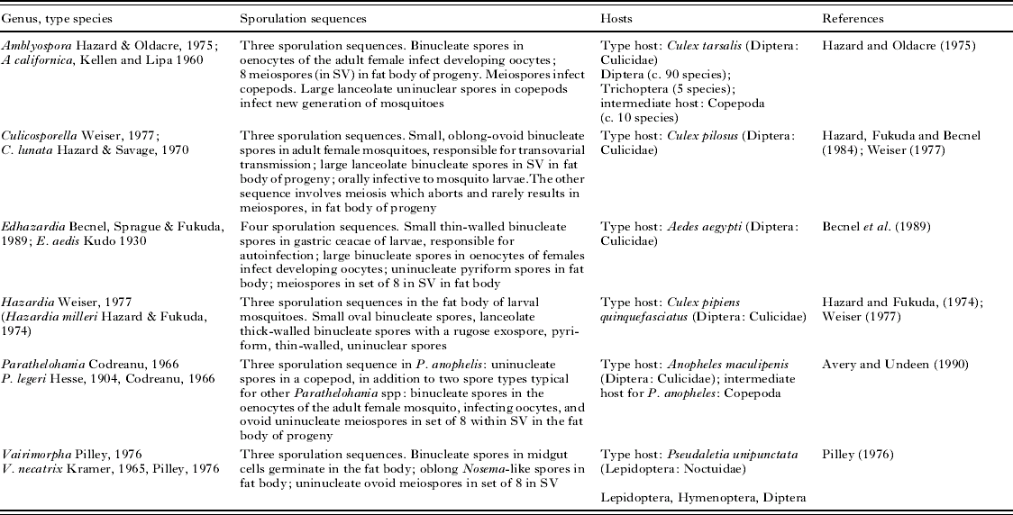
T. solenopsae and polysporous microsporidia Table 4
All other species currently assigned to the genus Thelohania are monomorphic (Hazard and Oldacre, Reference Hazard and Oldacre1975; Larsson, Reference Larsson1999; Lom et al. Reference Lom, Nilsen and Dykova2001). Disporous sporogony is somewhat common among microsporidia, for example, the production of 2 types of spores is characteristic of the genera Nosema (Maddox et al. Reference Maddox, Baker, Jeffords, Kuras, Linde, Solter, McManus, Vavra and Vossbrinck1999), Vairimorpha (Pilley, Reference Pilley1976), Burenella (Jouvenaz et al. Reference Jouvenaz, Lofgren and Allen1981), and Parathelohania (except P. anophelis, which produces uninucleate spores in copepods) (Avery and Undeen, Reference Avery and Undeen1990). Microsporidia producing 3 types of spores are not uncommon (Becnel and Andreadis, Reference Becnel, Andreadis, Wittner and Weiss1999). In Vairimorpha spp. (Pilley, Reference Pilley1976), in addition to diplokaryotic and monokaryotic spores produced in fat body, a third type of internally infective spore developing in the midgut has been recorded (Vavra et al. Reference Vavra, Hylis, Vossbrinck, Pilarska, Linde, Weiser, McManus, Hoch and Solter2006). P. anophelis and species of the genus Amblyospora have a third sporulation sequence in a copepod intermediate host; Culicosporella and Hazardia spp. have 3 sporulation sequences developing in one dipteran host; and as many as 4 types of sporulation have been described in Edhazardia spp. The pattern of sporulation modes of E. aedis (development in 1 host; thin-walled binucleate spores in larvae; large binucleate spores infecting oocytes; uninucleate pyriform spores for infection of fat body; and meiospores) is amazingly similar to T. solenopsae. This similarity favours the hypothesis that the pre-adaptive potential for multivariant sporulation programmes is a fundamental feature of Microsporidia, based on pecularities of their nuclear organization and nuclear cycle (Flegel and Pasharawipas, Reference Flegel and Pasharawipas1995; Vossbrinck and Debrunner-Vossbrinck, Reference Vossbrinck and Debrunner-Vossbrinck2005).
Polysporous genera of Microsporidia

T. solenopsae and microsporidia from hymenopterans
So far only 3 confirmed species of microsporidia have been recorded from ants: Burenella dimorpha (Jouvenaz and Hazard, Reference Jouvenaz and Hazard1978) from the tropical fire ant (Solenopsis geminatea), and Vairimorpha invictae (Jouvenaz and Ellis, Reference Jouvenaz and Ellis1986) and T. solenopsae (Knell et al. Reference Knell, Allen and Hazard1977) from the red imported fire ant (Solenopsis invicta). Until evidence is found to differentiate Vairimorpha sp. and Thelohania sp. (Moser et al. Reference Moser, Becnel, Maruniak and Patterson1998) from V. invictae and T. solenopsae (Knell et al. Reference Knell, Allen and Hazard1977; Jouvenaz and Ellis, Reference Jouvenaz and Ellis1986), respectively, we consider the former two species, also described from the red imported fire ants, as geographical isolates of the latter two due to structural similarity (Moser et al. Reference Moser, Becnel, Maruniak and Patterson1998). For example, a high rate of SSUrDNA sequence identity between T. solenopsae and Thelohania sp. strongly indicates that they probably belong to a single species; an SSUrDNA sequence of the type isolate of V. invictae is not available.
Host-parasite relationships in the B. dimorpha – S. geminata and T. solenopsae – S. invicta systems are very different. The dimorphous microsporidium B. dimorpha parasitizes the larval stage and causes pathological manifestations only in non-melanized pupae. Infection is almost inevitably fatal and has never been recorded in adults. Data suggest peroral transmissian via digestion of infective spores by larvae 4 as the main route (Jouvenaz and Hazard, Reference Jouvenaz and Hazard1978). In the B. dimorpha – S. richteri system, the host population ‘cleans’ itself through mortality of the infected pupae, so the infection does not significantly influence the reproduction capacity of the infected host. On the other hand, propagation and transmission of the parasites are supported by cannibalism of pupae and contamination of food fed to larvae 4, with invasive spores circulating throughout the nest. Sequence information on B. dimorpha is urgently needed, because the monotypic genus Burenella is the type genus for the family Burenellidae, the legitimacy of which is currently in question (Vavra et al. Reference Vavra, Hylis, Vossbrinck, Pilarska, Linde, Weiser, McManus, Hoch and Solter2006).
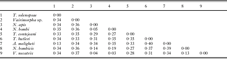
On the contrary, host-parasite relationships of T. solenopsae and V. invictae with their host S. invicta have much in common. In both species, diplokaryotic free spores and octospores developing inside sporophorous vesicles are produced in imagoes; both species cause hypertrophy of infected adipocytes, which become ‘cysts’ containing mature spores; sporogony is delayed until the late larval and pupal stages; and only free spores are produced in the pre-imaginal phase. Judging from different spore morphology and low sequence similarity (Table 5) (Moser et al. Reference Moser, Becnel, Maruniak and Patterson1998), T. solenopsae and V. invictae can only be distantly related. Thus, such striking similarity in manifestations of the infection in two parasites with similar types of development and stratagies of parasitism can be attributed to convergent evolution against similar defensive mechanisms of the host. The SSUrDNA gene of Vairimorpha sp. from fire ants has little similarity with V. necatrix (Table 5), and it is very probable that the former species will be assigned eventually to another, likely new, genus. Interestingly, Vairimorpha sp. from S. invicta forms a dichotomy with the Edhazardia-Amblyospora branch in the SSU-rDNA sequence-based phylogram.
Pairwise distances among 9 species of microsporida (from Phylip Distance Matrix)

With regard to other hymenopterans, microsporidia have been recorded from parasitoid wasps of the family Braconidae, bees and bumble bees of the family Apidae (Bombus spp., Apis melifera, and A. ceranae), a communal bee Andrena scoticae (Andrenidae), and from the wasp Vespula germanica (Vespidae). Braconids become infected with microsporidia via their hosts, lepidopteran or dipteran larvae, and are parasitized by the same species as the host, such as Vairimorpha sp. (Hoch et al. Reference Hoch, Schopf and Maddox2000), Nosema pyrausta (Andreadis, Reference Andreadis1980), and Tubulinosema kingi (Franzen et al. Reference Franzen, Futerman, Schroeder, Salzberger and Kraaijeveld2006a). Antonospora scoticae has been recorded from a solitary wild bee, Andrena scoticae (Fries et al. Reference Fries, Paxton, Tengo, Slemenda, da Silva and Pieniazek1999), and is closely related to the species of the genus Paranosema (Sokolova et al. Reference Sokolova, Dolgikh, Morzhina, Nassonova, Issi, Terry, Ironside, Smith and Vossbrinck2003). Other microsporidian parasites – N. apis (Fries, Reference Fries1993), N. bombi (McIvor and Malone, Reference McIvor and Malone1995; Fries et al. Reference Fries, de Ruijter, Paxton, da Silva, Slemenda and Pieniazek2001), and N. ceranae (Higes et al. Reference Higes, Martin and Meana2006, Reference Higes, Garcia-Palencia, Martin-Hernandez and Meana2007) – cluster within the Vairimorpha-Nosema group of species, which are primarily parasites of lepidopterans. The latter 3 are monomorphic species producing diplokaryotic spores of the Nosema-type. High virulence, parasitizion of epithelial cells of imagoes, simplified life-cycle, and solely peroral transmission make their strategy of parasitism completely different from that of T. solenopsae. ‘Nosema vespula’ (the tentative name of a yet-undescribed microsporidium) was initially isolated from a hymenopteran, V. germanica. All further studies of this microsporidium were performed on an isolate propagated in Helicoverpa armigera (Rice, Reference Rice2001).
Thus, according to SSUrDNA sequence analyses, comparison of life-cycles, types of host-parasite relationships, and morphology, T. solenopsae is not related to any of the described microsporidia parasitizing hymenopterans. T. solenopsae has evolved unique adaptations to its host, S. invicta, a highly successful species characterized by complicated social organization and complex reproductive biology (Tschinkel, Reference Tschinkel1998).
Does T. solenopsae belong to the family Tubulinosematidae? In all phylogenies, including that in Fig. 13, T. solenopsae falls into 1 clade with Tubulinosema spp. and Anncaliia (syn. Brachiola) spp., which are characterized by small tubuli on the parasite's surface at some life-cycle stages (Franzen et al. Reference Franzen, Fischer, Schroeder, Scholmerich and Schneuwly2005, Reference Franzen, Nassonova, Scholmerich and Issi2006b). The occurrence of such tubules may indicate a type of host-parasite interaction peculiar to this group of species. Could the flattened, elongated vesicles forming a multilayer sheath around certain stages of the T. solenopsae megaspore sequence be homologous to the tubules of tubulonosematids? Interestingly, some stages of Tubulinosema ratisbonensis (Fig. 20 in Franzen et al. Reference Franzen, Fischer, Schroeder, Scholmerich and Schneuwly2005) and T. kingi (Fig. 3d in Franzen, Reference Franzen, Nassonova, Scholmerich and Issi2006b) are also surrounded by similar flattened membrane contours. In our opinion, the question about placing T. solenopsae into the family Tubulinoematidae is premature, because the current state of knowledge on Microsporidia, especially regarding the biochemistry of host-parasite interactions and their influence on morphology, does not provide reliable criteria (characters) that would allow creating taxa higher than the generic level. Most current families and classes are polyphyletic, require revison, and can be considered only as tentative.
Taxonomic summary
Kneallhazian. g.
Heterosporous microsporidia, primarily parasites of terrestrial insects, with up to 4 sporulation sequences, at least 1 of which develops in the pre-imaginal stage of the host. Octospores, if produced, are enclosed in a sporophorous vesicle and develop in mesodermal tissues of imagoes as a result of octosporoblastic sporogony via formation and subsequent division of sporogonial plasmodia. We anticipate that the number of developmental sequences and their morphology might differ in new potential members of this genus, depending on the host life-cycle. We include in the diagnosis of the new genus Kneallhazia the SSU rDNA sequence of Thelohania solenopsae (Acc. no. AF134205) as a reference sequence. Because the pairwise distance of T. solenopsae and the most closely related microspridium, Anncaliia meligheti, was calculated as 0·13 in the Phylip distance matrix, we suggest that dissimilarity in the SSU rDNA sequence of novel species should not exceed 13% to be assigned to the genus Kneallhazia. This generic name is proposed in remembrance of Drs J. D Knell, G. E. Allen and E. I. Hazard, who discovered and described this microsporidium. The type species is K. solenopsae (by monotypy).
Kneallhazia solenopsae(Knell, Allen and Hazard, 1977)
Synonymy. Thelohania solenopsae Knell, Allen and Hazard, 1977 (Knell et al. Reference Knell, Allen and Hazard1977).
Characters. Those of the genus. Heterosporous microsporidia, a parasite of fire ants Solenopsis invicta, with 4 sporulation sequences. The first sequence results in production of Type 1 diplokaryotic internally infective spores in larvae 4 and pupae. These spores are oval, thin-walled, with prominent posterior vacuoles and short isofilar polar filaments with 3–4 coils (Shapiro et al. Reference Shapiro, Becnel, Oi and Williams2003). They measure 4·5±0·10×2·3±0·05 μm when fresh (Oi et al. Reference Oi, Becnel and Williams2001) and 3·9±0·33×2·0±0·24 μm when methanol-fixed (Sokolova et al. 2004 b). The second sequence occurs in fat body of imagoes of all castes and consists of diplokaryotic stages developing in direct contact with adipocyte cytoplasm and resulting in production of Type 2 diplokaryotic spores via disporoblastic sporogony. These spores readily discharge polar filaments on slides and serve for autoinvasion of imagoes. Type 2 diplokaryotic spores measure 4·93±0·58×1·85±0·16 μm when fresh (Knell et al. Reference Knell, Allen and Hazard1977), and 4·6±0·29×2·3±0·20 μm when fixed (Sokolova et al. 2004 b). The isofilar polar filament is arranged in 1 row and forms 11–13 coils. In ultrathin sections spore envelopes measure 0·13±0·045 μm thick with smooth 0·04±0·014 μm thick exospores (Sokolova et al. 2004). The third (‘megaspore’) and fourth (‘octospore’) sequences develop concurrently in imagoes of all castes and include karyogamy of nuclei of the diplokaryotic meront and subsequent meiosis. The megaspore is produced in ovaries of females and is specialized for transovarial transmission; this spore type can be formed in moderate numbers in abdominal muscles and fat body in imagoes of all castes and in the last larval and pupal stages as well. Oval megaspores with 2 nuclei measure 7·3±0·17×3·7±0·13 μm when fresh and 6·2±0·41×3·6±0·29 μm when methanol fixed. The polar filament is slightly anisofilar; it is arranged in 2–3 rows and 18–23 coils. Megaspore envelopes are 0·4±0·10 μm wide with thick exospores 0·14±0·053 μm wide (Sokolova et al. 2004 b). An ‘octospore sequence’ produces pyriform uninucleate spores within slightly elongated sporophorous vesicles (ca. 9·0 μm in the larger diameter) in all castes of imagoes, predominantly in workers and alates, but never in brood. Octospores measure 3·32±0·48×1·95±0·20 μm when fresh (Knell et al. Reference Knell, Allen and Hazard1977) and 3·1±0·38×2·1±0·23 μm, when methanol-fixed (Sokolova et al. 2004 b). Ultrastructurally, octospores are characterized by an isofilar polar filament arranged in 1 row of 9–12 coils; spore envelope 0·15±0·062 μm thick, consisting of a wide endospore and thin (0·04±0·011 μm) undulating exospore; and the elongated polar sac often embracing the anterior part of the polaroplast (Sokolova et al. 2004 b). Sporophorous vesicles contained two types of secretion: electron-dense tubules deriving from the envelope of the sporogonial plasmodium, and granulated ‘labyrinth-like’ secretion; both types of secretion disappeared from the central part of the SV after sporoblasts and spores formed.
Pathology. There is no sign of external pathology. K. solenopsae causes false hypertrophy of the infected adipocyte; dissection of abdomens of the infected insects reveals 1–20 ‘cysts’, 70–260 μm in diameter, filled with octospores.
Type host. Solenopsis invicta Buren
Type locality. Cuabá, Mato Grosso, Brazil (Knell et al. Reference Knell, Allen and Hazard1977).
Type specimens. Holotype slides have been sent to the United States National Museum by the authors of the species (Knell et al. Reference Knell, Allen and Hazard1977).
We are indebted to Arthur Richter for his skillful assistance in the field and laboratory experiments. We are grateful to Irma Issi for fruitful discussions and critically proof reading the first version of the manuscript, to Maynard Milks for advice on molecular studies, to Olga Borkhsenius for her assistance in electron microscopy, and to two anonymous reviewers for greatly improving the text. We acknowledge the use of the Sokolovsky Microscopy Center, Department of Biological Sciences, and Laboratory of Electron microscopy of the Department of Comparative Biomedical Sciences, School of Veterinary Medicine, Louisiana State University, Baton Rouge. This research was supported by the Louisiana Fire Ant Research and Management Legislative Grant. The paper was approved for publication by the Director of Louisiana Agricultural Experimental Station.