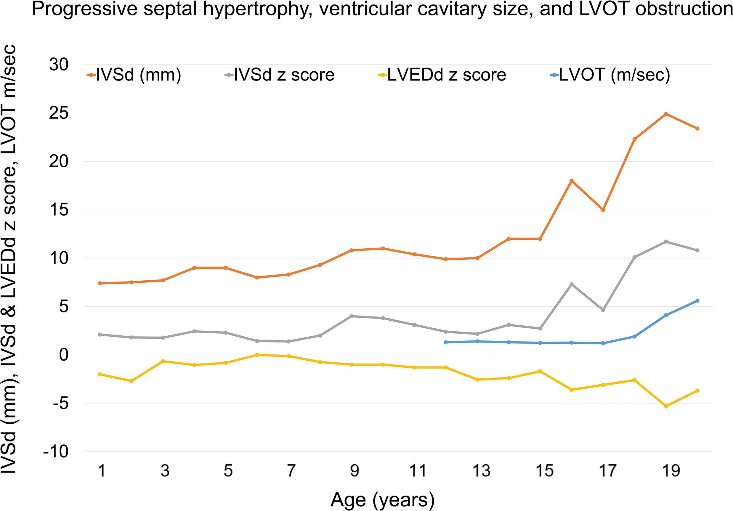
A male infant was found to carry a pathogenic MYBPC3 gene mutation following cascade genetic screening. He started to show mild septal hypertrophy at age 9 years old, which became progressive following his 15th birthday (Figure 1).
Development of progressive septal hypertrophy and symptomatic LVOT obstruction in a child with MYBPC3 from cascade screening. Serial transthoracic echocardiographic measurements across childhood and adolescence demonstrating progressive interventricular septal hypertrophy with a concomitant decline in left ventricular end-diastolic dimension (LVEDd Z score). LVOT peak velocity (m/s) increases in late adolescence, consistent with development of dynamic LVOT obstruction. IVSd = interventricular septal thickness in diastole; LVEDd = left ventricular end-diastolic dimension; LVOT = left ventricular outflow tract; VS = ventricular septum. Boston Z scores were utilised.

Figure 1. Long description
A line graph illustrates the progressive septal hypertrophy, ventricular cavitary size, and left ventricular outflow tract obstruction in a male infant with a pathogenic MYBPC3 gene mutation. The x-axis represents age in years, ranging from 1 to 19 years. The y-axis represents the interventricular septal thickness in diastole (IVSd) in millimeters, IVSd and left ventricular end-diastolic dimension (LVEDd) z scores, and left ventricular outflow tract (LVOT) peak velocity in meters per second. The graph includes four data lines: IVSd in millimeters (orange line), IVSd z score (gray line), LVEDd z score (yellow line), and LVOT in meters per second (blue line). The IVSd line shows a progressive increase in septal hypertrophy, particularly after age 9. The IVSd z score line also shows an upward trend, indicating progressive hypertrophy. The LVEDd z score line remains relatively stable with slight fluctuations. The LVOT line shows an increase in peak velocity during late adolescence, consistent with the development of dynamic LVOT obstruction. All values are approximated.
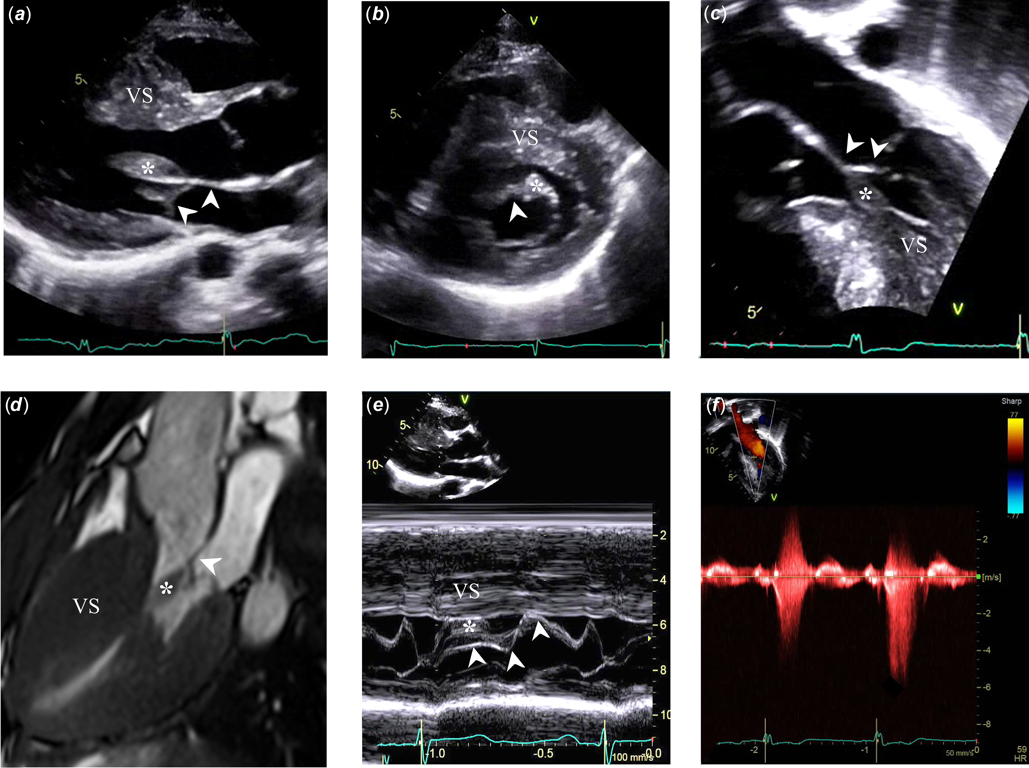
Two-dimensional transthoracic echocardiography revealed an anomalous papillary muscle inserting directly into the anterior mitral leaflet, without apparent chordal attachment to the leaflet tip, consistent with a type I variant (Figure 2 a – d ). Reference Lentz Carvalho, Schaff and Morris1 M-mode assessment demonstrated systolic anterior motion of the abnormal papillary muscle (Figure 2 f ), together with severe septal hypertrophy, causing outflow tract obstruction. Over the following two years, obstruction of the outflow tract progressed to a peak systolic velocity of 5.6 m/s (Figure 2 f ).
Multimodality imaging demonstrating mechanism of obstructive hypertrophic cardiomyopathy phenotype. ( a – c ) Transthoracic echocardiography in multiple views shows marked ventricular septal (VS) hypertrophy and abnormal insertion of a hypertrophied papillary muscle (*) into the anterior mitral valve leaflet (arrowheads) contributing to LVOT narrowing. ( d ) Cardiac MRI long-axis cine view confirms severe septal hypertrophy (VS) with LVOT narrowing, attributed to by abnormal papillary muscle insertion (*) into the anterior mitral valve leaflet (arrowhead). ( e ) M-mode echocardiography across the LVOT demonstrates systolic anterior motion of the hypertrophied papillary muscle (*), together with septal hypertrophy (VS), causing LVOT obstruction. The arrowheads depicted the motion of the anterior mitral valve leaflet. ( f ) Continuous-wave Doppler interrogation across the LVOT demonstrates a late-peaking systolic “dagger-shaped” jet, consistent with dynamic LVOT obstruction. LVOT, left ventricular outflow tract; VS, ventricular septum.

Figure 2. Long description
The image displays multiple transthoracic echocardiography views highlighting marked ventricular septal hypertrophy. The abnormal insertion of a hypertrophied papillary muscle into the anterior mitral valve leaflet is visible, contributing to left ventricular outflow tract narrowing. The cardiac MRI long-axis cine view confirms severe septal hypertrophy and LVOT narrowing due to the abnormal papillary muscle insertion. M-mode echocardiography across the LVOT shows systolic anterior motion of the hypertrophied papillary muscle and septal hypertrophy causing LVOT obstruction. Continuous-wave Doppler interrogation across the LVOT demonstrates a late-peaking systolic dagger-shaped jet, indicating dynamic LVOT obstruction.
During this period, the patient developed exertional dyspnoea with limitation of daily activities, corresponding to NYHA class II–III symptoms, and exercise stress testing demonstrated a blunted blood pressure response. Reference Maron, Olivotto and Betocchi2 Given progressive symptoms and haemodynamic significance of the obstruction, the patient underwent surgical septal myectomy with targeted thinning and partial resection of the anomalous papillary muscle. Recognition of this variant is critical, as failure to address anomalous papillary muscle insertion during myectomy may result in persistent obstruction, whereas targeted resection or repositioning of the papillary muscle can optimise surgical outcomes. Reference Ommen, Ho and Asif3,Reference Nagueh, Phelan and Abraham4 Postoperative echocardiography demonstrated a marked reduction in the outflow tract gradient and resolution of papillary muscle motion into the outflow tract, with improvement in functional status.
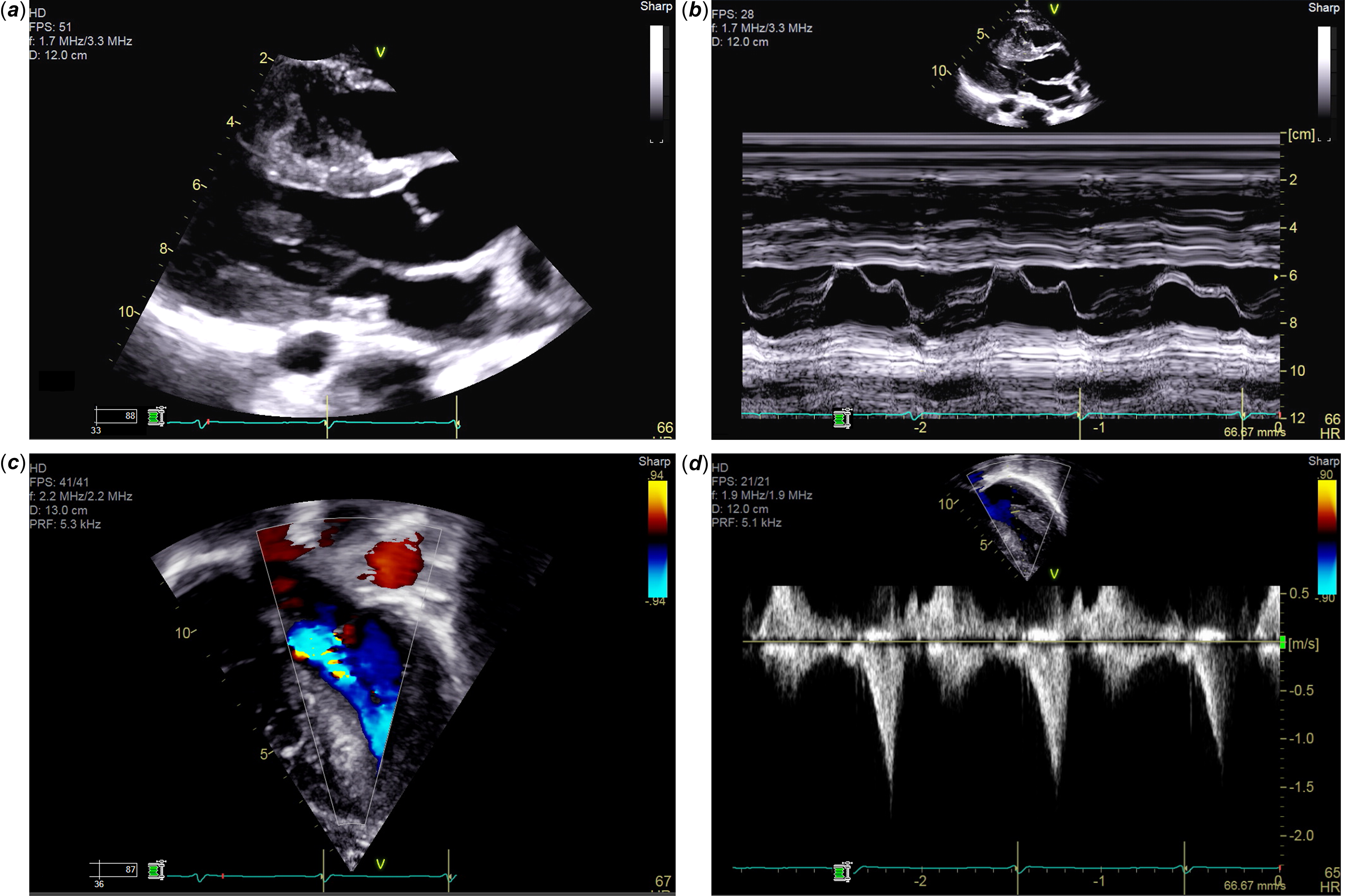
This case illustrates the variable age-dependent expression of hypertrophic cardiomyopathy in individuals with MYBPC3 mutations and highlights the importance of careful assessment of the mitral valve apparatus in obstructive disease. Reference Lentz Carvalho, Schaff and Morris1,Reference Ommen, Ho and Asif3 Anomalous papillary muscles are reported in approximately 10–15% of patients with obstructive hypertrophic cardiomyopathy, with type I and type II variants more commonly associated with outflow tract obstruction due to direct insertion into the body of the anterior leaflet. This anatomy represents a distinct systolic anterior motion mechanism of outflow tract obstruction, which was demonstrated by two-dimensional and M-mode imaging, differing from the more typical leaflet motion. Reference Nagueh, Phelan and Abraham4 Comprehensive preoperative identification of subvalvular abnormalities on transthoracic echocardiography or cardiac MRI has important surgical implications, as targeted resection at the time of septal myectomy is recommended to ensure adequate relief of obstruction. Reference Lentz Carvalho, Schaff and Morris1 The hypertrophied papillary muscle was selectively thinned at the time of septal myectomy to relieve left ventricular outflow tract obstruction. The postoperative echocardiogram showed no evidence of systolic anterior motion, no significant mitral valve regurgitation, and no significant left ventricular outflow tract obstruction (Figure 3).
Postoperative transthoracic echocardiogram following septal myectomy and papillary muscle thinning. Parasternal long-axis imaging demonstrates relief of left ventricular outflow tract narrowing ( a ) with no systolic anterior motion of the mitral valve ( b ). Colour Doppler demonstrates no significant mitral regurgitation ( c ). Continuous-wave Doppler across the left ventricular outflow tract shows a dynamic spectral Doppler profile without significant residual obstruction, with peak velocity 1.7 m/s ( d ).

Figure 3. Long description
The image consists of four panels labeled (a), (b), (c), and (d), each showing different aspects of a postoperative transthoracic echocardiogram. Panel (a) displays a parasternal long-axis view demonstrating relief of left ventricular outflow tract narrowing. Panel (b) shows no systolic anterior motion of the mitral valve. Panel (c) uses color Doppler to demonstrate no significant mitral regurgitation. Panel (d) features continuous-wave Doppler across the left ventricular outflow tract, showing a dynamic spectral Doppler profile without significant residual obstruction, with a peak velocity of 1.7 meters per second.
Supplementary material
The supplementary material for this article can be found at https://doi.org/10.1017/S1047951126113626.
Financial support
This research received no specific grant from any funding agency, commercial, or not-for-profit sectors.
Competing interests
None.
Ethical standard
Informed consent was obtained from the patient and family for publication of this case and accompanying images. The study was conducted in accordance with institutional and international ethical standards.