INTRODUCTION
The tunicate Ascidiella aspersa (O.F. Müller, 1776) is the host for a number of organisms (Svavarsson, Reference Svavarsson1990; Svavarsson et al., Reference Svavarsson, Svane and Helgason1993) including the mytilid Modiolarca subpicta (Cantraine, Reference Cantraine1835). Since 1850–1900, M. subpicta has received little attention although Roberts & Breen (Reference Roberts and Breen1985) undertook a modest study of its anatomy (as M. tumida) and Bodger & Allen (Reference Bodger and Allen2008) made a more comprehensive examination of its reproductive cycle (also as M. tumida) in North Sea waters off the north-eastern coast of England and illustrated (Bodger & Allen, Reference Bodger and Allen2008; figure 2) how it ‘recessed’ into the tunic, or test, of its ascidian host, in this case Ascidiella scabra (O.F. Müller, 1776).
Several mytilids are known to be gregarious and form dense beds on the seafloor and provide habitats for a diverse associated fauna (Ong Che & Morton, Reference Ong Che and Morton1992; Service & Magorrian, Reference Service and Magorrian1997; Seed et al., Reference Seed, Richardson, Smith, Harper, Taylor and Crame2000; Van Dover, Reference Van Dover2002; Tendal & Dinesen, Reference Tendal and Dinesen2005). It is, however, unusual for a mytilid to have adopted an epizoic lifestyle and this is known for only a few mussel species, all belonging to Modiolarca. These include Modiolarca subpicta in the north-eastern Atlantic boreal, Lusitanian and Mediterranean waters, M. lateralis (Say, 1922) in the western Atlantic and M. lebourne (White, Reference White1949) in the Indian Ocean.
The shell morphology and anatomy of Modiolarca subpicta have been described and illustrated by Jeffreys (Reference Jeffreys1863) and List (Reference List1902), and its ontogeny by Lovén (Reference Lovén1848). Apart from those studies and a few others that mention the relationship between M. subpicta and various ascidians, all other papers are exclusively taxonomic and some of these will be identified herein as we: (i) attempt to stabilize the complex nomenclature of this small north-eastern Atlantic bivalve. Other aims of this study were to: (ii) examine in more detail the relationship and degree of intimacy exhibited between Ascidiella aspersa and M. subpicta, particularly with regard to their life histories; (iii) understand the functional morphology of M. subpicta in relation to other mytilids; and (iv) attempt to reveal how its unusual (for a mussel) lifestyle has evolved.
MATERIALS AND METHODS
Living tunicates, including Ascidiella aspersa, were collected from Ålbæk Bay, western Kattegat, Denmark, in late August 2009. The ascidians were sorted into three size-categories and preserved in 80% ethanol. In the laboratory, all individuals with a tunic height of ≥5 mm were identified to species and their contracted heights from the basal attachment to the inhalant siphon measured to the nearest 0.5 mm using Vernier calipers.
The external surfaces of the tunics of each ascidian were inspected for Modiolarca subpicta individuals under a dissecting microscope. Each tunic was then cut open along the basal and ventral surfaces to the inhalant siphon and loosened for inspection of its inner face. The latter was particularly helpful in detecting M. subpicta individuals within the tunic in larger ascidians as the host's outer papillation and lightly-adhering detritus, red filamentous algae, foraminiferans, bryozoans, barnacles, sponges and other bivalves obscured their presence externally. For each host, the location of each M. subpicta individual was mapped on master diagrams (see Figure 7). In addition, the shell lengths, heights and widths of each individual were measured under the dissecting microscope to the nearest 0.1 mm (or 0.05 or 0.03 mm, depending on magnification). Developmental stage and sex were also determined.
Some individuals were kept alive within 5 ml glass tubes held in 500 ml dishes of seawater in a refrigerator kept at 10°C. The seawater was changed every four days and the animals examined for aspects of their behaviour.
Two individuals were fixed in 5% neutral formalin, decalcified, sectioned transversely at 4 µm and stained in Ehrlich's haematoxylin and eosin.
RESULTS
ORIGINAL DESCRIPTION
Modiola subpictus in Cantraine F.J. (1835) Diagnoses ou description de quelque espèce nouvelle de mollusques. Bulletin de l'Académie Royale des Sciences et Belles-lettres de Bruxelles 2, 380–411.
SYNONYMY
Mytilus marmoratus Forbes, Reference Forbes1838non Schroeter, 1803.
Modiola tumida Hanley, Reference Hanley1843.
Modiolaria poliana Philippi, 1844.
Modiolaria europaea Weinkauff, 1868 ex. d'Orbigny ms.
Mytilus discors sensu auct. non Linnaeus, 1767.
Modiola discrepans Lamarck, 1819 non Montagu, 1803.
TYPE LOCALITY
Found in the Gulf of Venice.
REMARKS
This ascidian-associated mussel species has been described independently by several authors as seen from the synonymy above. The description by Cantraine predates that of Forbes and other authors, however, and the species taxon subpicta has been acknowledged recently as being valid by other authors, for example, Poppe & Goto (Reference Poppe and Goto1993) and Rodríguez & Sánchez (Reference Rodríguez and Sánchez1997).
Adding to the confusion regarding its specific affinity, the species has been assigned to several genera, that is, initially, Mytilus Linnaeus, 1758, Modiolus (Lamarck, 1799) (Leach, Reference Leach1815; Forbes, Reference Forbes1838; Hanley, Reference Hanley1843) and Crenella T. Brown, 1827 (Forbes & Hanley, Reference Forbes and Hanley1853). Most often, however, it has been ascribed to Musculus Röding, 1798 (p. 156, type: Mytilus discors Linnaeus, 1767, Iceland, subsequent designation by Iredale, Reference Iredale1915) (e.g. Thiele, Reference Thiele1934; Tebble, Reference Tebble1966; Poppe & Goto, Reference Poppe and Goto1993) and its junior synonym, Modiolaria Beck ms. in Roberts, 1838 (e.g. Lovén, Reference Lovén1846, Reference Lovén1848; Jeffreys, Reference Jeffreys1863; List, Reference List1902; Dodge, Reference Dodge1952). Modiolaria has been used as the generic name for Linnaeus's Mytilus discors, as it was questioned whether Musculus Röding, 1798 was valid and, further, was not occupied by Musculus Martyn, 1787 (Dodge, Reference Dodge1952).
Several authors have considered the taxon subpicta (and its junior taxon tumida) to be a species of Modiolarca Gray, Reference Gray and Dieffenback1843 (p. 259, type: Mytilus impactus Hermann, 1782, New Zealand) (e.g. Roberts & Breen, Reference Roberts and Breen1985; Bodger & Allen, Reference Bodger and Allen2008; Jensen & Knudsen, Reference Jensen and Knudsen1995; Rodríguez & Sánchez, Reference Rodríguez and Sánchez1997). Some authors have recognized Modiolarca as a valid genus (e.g. Vokes, Reference Vokes1980; Jensen & Knudsen, Reference Jensen and Knudsen1995), while others (e.g. Højsæter, Reference Højsæter1986; Poppe & Goto, Reference Poppe and Goto1993; Hansson, Reference Hansson1998) have continued to consider it to be a subgenus of Musculus.
Some authors have questioned whether Modiolarca is a valid taxon or, rather, a mis-spelling of Modiolaria and, thus, a junior synonym of Musculus (Iredale, Reference Iredale1914; Thiele, Reference Thiele1934; Soot-Ryen, Reference Soot-Ryen1955, Reference Soot-Ryen and Moore1969). This confusion may have come about when Gray (Reference Gray1840, p. 151) assigned Modiolarca, but without a species designation (nomen nudum), to the Crenellidae and did so again subsequently (Gray, Reference Gray1842, p. 92), but this time noting in the text under Crenellidae (p. 82): ‘The Crenellae are sub-orbicular, and the Modiolariae ovate elongated shells’. In Gray (Reference Gray and Dieffenback1843), the genus Modiolarca was based on Mytilus impactus and a number of other species. As pointed out by Iredale (Reference Iredale1914), however, four years later, Gray (Reference Gray1847) erected Lanistina for M. impactus.
Soot-Ryen (Reference Soot-Ryen1955) separated four species from Musculus, that is, M. marmoratus (Forbes, Reference Forbes1838), M. impactus, M. cumingianus (Reeve, 1857) and M. lebourae—the latter newly described and designated to Musculus by White (Reference White1949)—and placed them in Lanistina Gray, Reference Gray1847. While shell shape and sculpture of the four species were similar to that of Musculus discors and their close allies, the four differ anatomically in being more inflated and having elongated posterior retractor muscles attached along the dorsal margin of the shell with a single branch above the posterior adductor muscle.
Fleming (Reference Fleming1959) discovered that Lanistes Swainson, 1840 (Humphrey, MS) (preoccupied by de Montefort, 1810) and its new name Lanistina Gray, Reference Gray1847 was based on Musculus discors (Linnaeus, 1767), and were thus junior synonyms of Musculus Röding, 1798 and, instead, erected Ryenella as the genus for the four species. Soot-Ryen (Reference Soot-Ryen1963, Reference Soot-Ryen and Moore1969) considered Ryenella to be a subgenus of Musculus.
Herein, we consider Modiolarca Gray, Reference Gray and Dieffenback1843 to be a valid genus with the junior synonyms of Lanistina Soot-Ryen, Reference Soot-Ryen1955 (non Gray, Reference Gray1847) and Ryenella Fleming, Reference Fleming1959. Based on shell morphology and anatomy (to be described herein) we also consider this genus to belong to the Musculinae.
Thiele (Reference Thiele1934) included two distinct subgenera in Musculus Röding s.l., that is, Musculus s.s. and Gregariella Monterosato, 1884. Thiele further divided the latter into two sections, that is Gregariella s.s. and Botulina Dall, 1889 (with its junior synonym, Trichomusculus Iredale, 1924). Soot-Ryen (Reference Soot-Ryen1955) suggested that Botulina Dall and Trichomusculus Iredale should be considered junior synonyms of Gregariella. As a consequence, some species have been referred to both of the latter two genera. For example, the epibyssate Trichomusculus semigranata from the Açores (Morton, Reference Morton1995) has been subsequently referred to as Gregariella semigranata (Frias Martins et al., Reference de Frias Martins, Borges, Ávila, Costa, Madeira and Morton2009). However, Soot-Ryen (Reference Soot-Ryen and Moore1969) recognized Trichomusculus as a valid genus distinct from both Musculus and Gregariella. Until further revisions are made, we adopt the view of Soot-Ryen (Reference Soot-Ryen and Moore1969).
DISTRIBUTION AND HABITATS
Ascidiella aspersa
Ascidiella aspersa is a solitary, pear-shaped tunicate up to 12 cm long that sometimes forms un-fused colonies. The species occurs from the shallow subtidal (–5 m) to a depth of 90 m on soft muds but is more common attached by its base and left side to hard substrata, such as rocks and harbour piers, docks and other man-made structures. Ascidiella aspersa is particularly common in low energy habitats, such as estuaries, lochs and semi-enclosed embayments, including harbours, and can tolerate salinities of between 18 and 40‰.
Ascidiella aspersa is naturally distributed in the waters of the western Baltic Sea, Kattegat, Skagerrak, the North Sea, southern and western Norway, the Irish Sea, English Channel, the Mediterranean and the coast of north-west Africa (Hayward & Ryland, Reference Hayward and Ryland1995). It has, however, also been introduced into the waters of the east coast of North America, India, New Zealand and southern Australia, including Tasmania (Kott, Reference Kott1985). Introductions were probably via fouled ships' hulls, ballast water discharges and along with other species destined for mariculture (Carlton, Reference Carlton2003). Ascidiella aspersa is common in the Kattegat, where it lives attached to firm substrata, including large pebbles and boulders, vertical rock faces, gas seep pockmarks (Dando et al., Reference Dando, O'Hara, Schuster, Taylor, Clayton, Baylis and Laier1994), the ‘bubbling reefs’ of Jensen et al. (Reference Jensen, Aagard, Burke, Dando, Jørgensen, Kuijpers, Laier, O'Hara and Schmalljohan1992), and nestling within the holdfasts of laminarian kelps (McKay & Smith, Reference McKay and Smith1979).
Ascidiella aspersa has a thick, transparent, cartilaginous and nodulose tunic that covers the body of the adult organism. The rough, greyish-black to brown to yellow surface of the tunic is papillate and has a terminal, 6–8 lobed, brachial siphon and a 6 lobed atrial siphon located ~two-thirds up the length of the body from its base. Both siphons are retractile. Ascidiella aspersa is a protandric hermaphrodite. Gametes are released in spring and the larvae go through a brief free-swimming stage and then quickly metamorphose into a settled juvenile (Niermann-Kerkenberg & Hofmann, Reference Niermann-Kerkenberg and Hofmann2006). Ascidiella aspersa has a lifespan of 18 months, lasting from mid-summer to winter of the following year. The testes begin to mature at ~30 mm, the ovaries later. At ~40 mm, both sexes are mature. A single spawning occurs usually one year after larval settlement (Millar, Reference Millar1952). Larval settlement and metamorphosis occur in early- to mid-summer and take approximately 24 hours at 20°C. Ascidian biology, including that of Ascidiella aspersa has been reviewed by Millar (Reference Millar1971).
Modiolarca subpicta
Ascidiella aspersa is the host for a number of organisms (Svavarsson, Reference Svavarsson1990; Svavarsson et al., Reference Svavarsson, Svane and Helgason1993), including Modiolarca subpicta (Roberts & Breen, Reference Roberts and Breen1985; Bodger & Allen, Reference Bodger and Allen2008), which has an eastern Atlantic boreal (except Iceland), Lusitanian and Mediterranean distribution. Modiolarca subpicta was reported by Tebble (Reference Tebble1966), as Musculus marmoratus) to occur from northern Norway and the Baltic, south to Spain and Portugal and the Mediterranean (Poppe & Goto, Reference Poppe and Goto1993). It also occurs along the Atlantic coast of Morocco, south to Guinea and Angola.
Jensen & Knudsen (Reference Jensen and Knudsen1995) have recorded Modiolarca subpicta and its junior synonym Musculus marmoratus from the northern and middle part of the Øresund but not the Kattegat (on the Swedish west coast) to Bergen (Norwegian Sea, also on the Norwegian west coast). Hansson (Reference Hansson1998) reported the species to occur in the North Sea, the Kattegat (Bohuslän, Sweden) and the Baltic Sea, but it was not known from east of Bornholm. Collin (Reference Collin1880) first reported it (as Modiolaria marmorata) in Danish waters, from near Hellebæk, Zealand (in the Øresund). Subsequently, Collin (Reference Collin1884) reported the species as being new to the Limfjord (northern Jutland), which it was suggested to have invaded from the Kattegat in the east, and where the species was living in the ascidian Dendrodoa grossularia (van Beneden, 1846), but with never more than one mussel individual in each host. Jensen & Spärck (Reference Jensen and Spärck1934) reported it to occur also in the western Baltic as far south as to Kiel, and as being common in Danish fjords and bays, particularly where calmer waters prevail, and here it may live either non-symbiotically attached to kelps and shells, or symbiotically inhabiting the tunic of ascidians, such as Dendrodoa grossularia, Corella parallelogramma (O.F. Müller, 1776) and Ascidiella aspersa, in the latter often in groups of 5–6 mussel individuals.
Modiolarca subpicta is usually collected from the lower shore down into the subtidal and attaches to the under-surfaces of stones, shells and other seabed objects including kelp holdfasts and the tests of various ascidians. It has been recorded from the Canary Islands by Rodríguez & Sánchez (Reference Rodríguez and Sánchez1997) and from where these authors report it as occurring in the tests of large tunicates, particularly Ascidia mentula O.F. Müller, 1776, Ciona intestinalis (Linnaeus, 1758) and Ascidiella aspersa and the epidermis of the holothurian Holothuria tubulosa (Gmelin, 1778).
In Northumberland waters (north-east coast of England), Bodger & Allen (Reference Bodger and Allen2008) recorded Modiolarca subpicta (as M. tumida) as occurring most frequently in Ascidia scabra and Polycarpa pomaria (Savigny, 1817), but also, exceptionally, in Dendrodoa grossularia, Corella parallelogramma and in colonies of Ascidia conchilega (O.F. Müller, 1776). Roberts & Breen (Reference Roberts and Breen1985) add Styela plicata (Lesueur, 1823), Microcosmus sulcatus (Coquebert, 1797), Ascidia sordida (O.F. Müller, 1776) and Phallusia mamillata (Cuvier, 1815) to this list. Modiolarca subpicta is thus known to occur in the tests of twelve species of ascidians and one holothurian, although it is also a free living, albeit cryptic, little mussel recorded just as widely from non-living substrata in the shallow sublittoral.
THE ASCIDIELLA–MODIOLARCA RELATIONSHIP
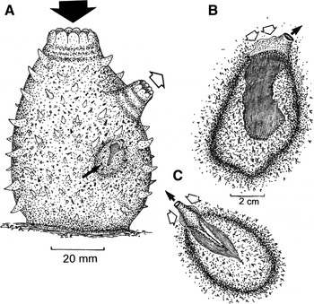
Figure 1A is an illustration of Ascidiella aspersa in life position with inhalant and exhalant currents indicated by closed and open arrows, respectively, and with a single individual of Modiolarca subpicta buried within its test (small arrow). More detailed illustrations of M. subpicta individuals buried within the test of A. aspersa (Figure 1B & C) show that the bivalve lives on its back and its inhalant and exhalant currents are represented by open and closed arrows, respectively.
(A) An illustration of Ascidiella aspersa in life position with inhalant and exhalant currents indicated by closed and open arrows, respectively, and with a single individual of Modiolarca subpicta buried within its test (small arrow); (B & C) individuals of M. subpicta buried in life positions within the test of A. aspersa and showing the bivalves' inhalant (open arrows) and exhalant (closed arrow) currents.

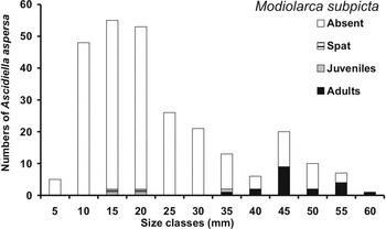
A total of 265 Ascidiella aspersa individuals with tunic heights ≥5 mm were examined. Of these, 24 were inhabited by either one or more individuals of Modiolarca subpicta. Figure 2 shows the size–frequency distribution of the collected A. aspersa individuals divided into 5 mm size-classes. Most individuals (with preserved, contracted tunics) ranged in height between 10 and 20 mm and most had tests that were either unoccupied or occupied by the spat and juveniles of M. subpicta individuals. Adults of M. subpicta were obtained only from A. aspersa tunics ranging in height from 35 to 60 mm. It thus appears that newly-settled individuals of A. aspersa attract newly-settled M. subpicta and that the pair grow and mature together, both possibly, therefore, completing their life cycles within the life span of ~18 months attributed to the host by Millar (Reference Millar1952; see above).
The size–frequency distribution of Ascidiella aspersa individuals divided into 5 mm size (shell length) categories and either unoccupied or occupied by Modiolarca subpicta individuals nestling in the tunics and divided into post-metamorphic age-categories of spat, juveniles and adults.

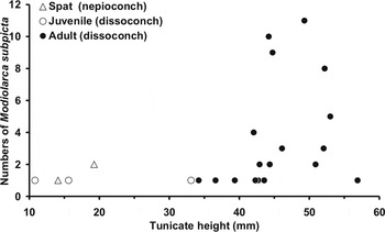
Figure 3 shows the numbers of post-metamorphic stages (spat, juveniles and adults) of M. subpicta individuals inhabiting the tunics of A. aspersa individuals of different sizes. Modiolarca subpicta spat with a shell length of <1 mm were found in A. aspersa individuals between 10 and 20 mm in height. Larger juveniles of M. subpicta with a shell length of 0.7–2.0 mm were obtained from ascidians with tunic heights of 14–33 mm. Adult M. subpicta individuals with shell lengths >3 mm were collected from A. aspersa individuals with tunic heights of 34–57 mm. The highest number of M. subpicta occurred within the tests of A. aspersa individuals of 40–55 mm height.
The number of post-metamorphic stages (spat, juveniles and adults) of Modiolarca subpicta individuals nestling externally in the tunics of Ascidiella aspersa individuals of different sizes.

The majority of Ascidiella aspersa individuals with a tunic height <35 mm did not host Modiolarca subpicta individuals and, typically, only one mussel inhabited each ascidian. A total of 73 M. subpicta individuals were retrieved from 24 A. aspersa individuals (Figure 3). Three spat and one juvenile were found alone, while two of the juveniles shared their host. Of the adults, seven individuals were found alone, while the other 60 were found in groups of 2–11 individuals occupying a single host.
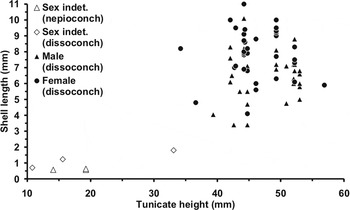
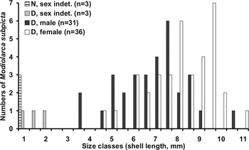
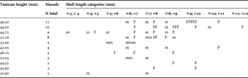
The largest M. subpicta individuals with shell lengths >7 mm, comprising both males and females, occurred in medium-sized ascidians with tunic heights of 40–53 mm (Figure 4). A clear tendency was seen for males and females of M. subpicta to live separately in smaller groups of between 2 and 5 individuals (Figure 5). In one case (A. aspersa No. 12 with a tunic height of 42 mm), three males of M. subpicta were situated together near the exhalant siphonal area of A. aspersa whereas one female individual was situated on the opposite side in the middle. All larger groups of between 8 and 11 M. subpicta individuals comprised both males and females, with the largest individual in each of the groups being female (Table 1; Figure 5).
A plot of Ascidiella aspersa tunic height (mm) and the shell lengths and sexes of the nepioconchs, dissoconchs and identifiable male and female individuals of Modiolarca subpicta occurring with them.

A histogram showing the distribution of Modiolarca subpicta individuals divided into shell length-categories in individuals of Ascidiella aspersa. The groups are also separated into sexually immature nepioconch (N) and dissoconch (D) individuals and sexually mature males and females.

The shell length-categories and sexes of Modiolarca subpicta living in groups of two or more individuals in 12 individuals of Ascidiella aspersa.

N, number; m, male; F, female.
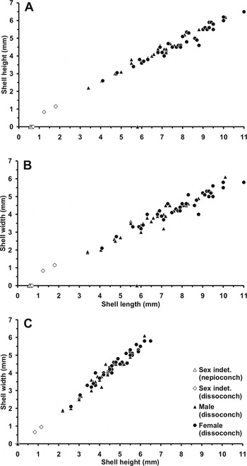
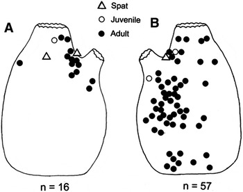
Shell allometry of Modiolarca subpicta showed linear relationships between shell height and length (Figure 6A), width and length (Figure 6B), and width and height (Figure 6C). No sexual dimorphism was detected. Adult Modiolarca subpicta (•) were mostly distributed in the left side of the host tunic (Figure 7B, N = 55), rather than the left (Figure 7A, N = 13). This may be because, as noted above, A. aspersa typically attaches to the substratum by its base and left side. The three spat (Δ) and the three juveniles (O) occupied positions around the exhalant siphons of A. aspersa.
Plots of the shell dimensions of Modiolarca subpicta in terms of (A) shell height versus shell length; (B) shell width versus shell length (C) shell width versus shell height. The plots also identify nepioconchs and dissoconchs (where sex was indeterminable) and male and female adults.

(A & B) Master outlines of left and right lateral views of Ascidiella aspersa showing the positions of: (i) spat (Δ); (ii) juvenile (O); and (iii) adult (•) individuals of Modiolarca subpicta.

FUNCTIONAL MORPHOLOGY
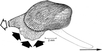
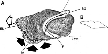
The living animal: a living individual of M. subpicta is illustrated in Figure 8 from the right side with the inhalant aperture and exhalant siphon and their ciliary currents represented by closed and open arrows, respectively. Also shown is the long foot that is used for locomotion in the direction arrowed by its extension and then contraction. Once a site for habitation is selected, however, the animal typically attempts to turn onto its back. In this action, once again, the foot is of importance and Figure 9A shows a living individual of M. subpicta, again shown from the right side, with siphons and the long, mobile, foot extended over the shell for the placement and attachment of byssal threads. In the figure, the inhalant aperture (IA) and exhalant siphon (ES) and their respective currents are represented by closed and open arrows but also shown are the currents emanating from the infra-branchial chamber and that take rejected material from the mantle cavity and which is expelled from the vicinity of the mussel with the assistance of the exhalant stream from the supra-branchial chamber. The foot (F) is shown in transverse section in Figure 9B, in outline only, but in Figure 9A it (F) is shown extended over the shell and attempting to plant from its inflated end a byssal thread arising from the byssal groove (BG). Once attached, in this case to the internal surface of a narrow (5 ml) glass tube, the shell can be turned over.
Modiolarca subpicta. A living individual shown from the right side with siphons and the long, mobile, foot extended for locomotion. Inhalant and exhalant currents are represented by closed and open arrows, respectively.

(A) Modiolarca subpicta. A living individual shown from the right side with siphons and the long, mobile, foot extended over the shell for the placement and attachment of byssal threads. Inhalant and exhalant currents are represented by closed and open arrows, respectively. Also shown as small arrows are the rejectory currents arising from the infra-branchial chamber; (B) the foot as shown in transverse section (outline only). For abbreviations see Appendix.

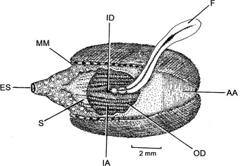
Such a living individual of M. subpicta is illustrated in Figure 10. The individual is shown from the ventral aspect with the exhalant siphon (ES) extended, the inhalant aperture (IA) wide open thereby exposing the extensive siphonal septum (SE) posteriorly and the anterior adductor muscle (AA) beneath the anteriorly fused mantle margins. Because the valves gape so widely, the ventral margins of the inner (ID) and outer (OD) demibranchs of the ctenidia are also clearly visible within the infra-branchial chamber. The long, mobile, foot (F) is extended over the shell for the placement and attachment of byssal threads.
Modiolarca subpicta. A living individual shown from the ventral aspect with siphons and the long, mobile, foot extended over the shell for the placement and attachment of byssal threads. For abbreviations see Appendix.

The shell: the diameter of the eggs of M. subpicta were ~60 µm. This corresponds well with the measurement by Thorson (Reference Thorson1946) of 56 µm; he further described the larval shell of M. subpicta (as Modiolaria marmorata) as being strongly orange with distinctly purple-reddish umbones and hinge. Ockelmann (Reference Ockelmann, Cox and Peake1965) reported the diameter of the yolk mass of ripe eggs to be ~60 µm in diameter, and the initial larval shell, the prodissoconch I, to be ~95 µm in diameter. The latter is highly correlated with egg size. The dimensions of the nepioconch and dissoconch shell stages of the M. subpicta individuals (Figure 6) showed that after metamorphosis of the triangular prodissoconch II (larval shell stage), the shell becomes more modioline. The nepioconch (early juvenile shell stage) is transparently iridescent, with fine concentric growth lines and a shell length of ~0.7 mm. It lacks the coloration and radial ribs of the final shell stage, the dissoconch (the adult shell stage). The largest M. subpicta individual retrieved was 11 mm in shell length. There appear to be three age-classes represented in the sample of M. subpicta, that is: (i) newly settled 1–2 mm juveniles; (ii) individuals with a median shell length of ~7–8 mm; and (iii) those with a median shell length of ~9–10 mm (Figure 5).
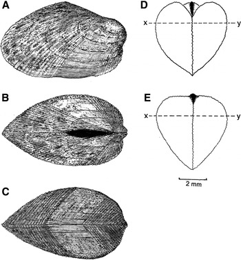
The adult shell of Modiolarca subpicta is equivalve, reaches a maximum length of ~20 mm and is delicately fragile. In external lateral appearance (Figure 11A), the shell is inequilateral, weakly heteromyarian and covered in a shiny, often iridescent, yellow-green-turquoise periostracum with a darker, loosely radial, external patterning of brown to red-brown flecks, blotches and chevrons. The shell is rhomboidal in overall appearance with a steeply down-curved postero-dorsal margin that tapers to a rounded point postero-ventrally. The umbones are prominently swollen and slightly overhang the anterior margin, which is inwardly curved towards the posterior. The shell has fine concentric commarginal growth lines and is characterized by 15–18 fine radial ribs anteriorly and 20–35 posteriorly according to size (and age). The crenulate anterior and posterior shell margins make the left and right valves interlock weakly in these positions. In life, each individual is attached by delicate byssal threads within its chosen host and the shell is mid-ventrally smooth and weakly concave.
Modiolarca subpicta. The shell characteristics. The shell seen in (A) right lateral view; (B) dorsal view; (C) ventral view; (D) anterior view; (E) posterior view. x–y represents the greatest shell width.

When seen from the dorsal aspect (Figure 11B), the shell is inflated anteriorly and roundly pointed posteriorly. The external, opisthodetic, ligament is deeply inset and extends posteriorly from the umbones for approximately half the length of the shell. Ventrally (Figure 11C), the valve margins are straight, that is, the shell is equivalve, and they interlock anteriorly and posteriorly. From the anterior aspect (Figure 11D), the shell is roundly inflated dorsally, giving a swollen outline left and right, roundly tapering to a point mid ventrally. The shell is heart-shaped when viewed from the anterior and posterior (Figure 11E) aspects, the inflated antero-dorsal area of the shell being lower than the more elevated postero-dorsal. The shell diameter is greatest between x and y, towards the upper one-fifth of the dorso-ventral height of the shell.
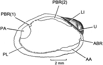
Internally, the shell of Modiolarca subpicta (Figure 12) is thin and transparent. The inner nacreous layer is bluish-turquoise-purple but lighter antero-ventrally and darker elsewhere. The umbones (U) lie posterior to the anterior shell margin and the opisthodetic ligament (LI) is external but deeply inset on a weak hinge plate. The muscle scars of M. subpicta (Figure 12) are difficult to see but comprise a large posterior adductor muscle (PA) scar located posteriorly and adjacent to which is a small scar of the posterior component of the posterior byssal retractor muscle (PBR(1)). Anterior to this is a gap, which (as will be seen) is where the heart is located, and anterior to this again is the anterior component of the byssal retractor muscle scar (PBR(2)) that extends along much of the length of the dorsal region of the shell. The pallial line (PL) is thin and situated postero-ventrally. The anterior adductor muscle (AA) is relatively large and located antero-ventrally. The anterior byssal retractor muscle scar (ABR) is located just above the end of the adductor muscle (AA) and, thus, just beneath the shell umbo.
Modiolarca subpicta. Internal shell characteristics. For abbreviations see Appendix.

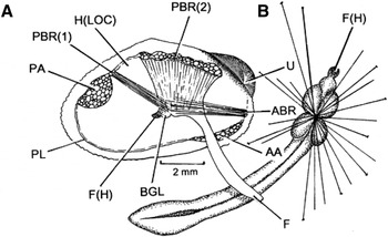
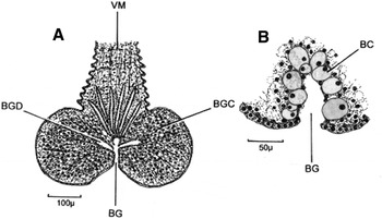
Internal anatomy: as described above, the muscle scars on the internal surface of the shell of Modiolarca subpicta are accurately indicative of the musculature as illustrated in Figure 13A from the right side. This figure also identifies the tiny ‘heel’ (F(H)) of the foot and anterior to this the four-lobed byssal gland (BGL). Figure 13B also illustrates the foot in ventral view showing an array of byssal threads attached by the plantar tip of the foot and the swollen four lobes of the byssal gland posterior to which is the tiny, violet-brown, heel of the foot (F(H)). The structure of the byssal gland is illustrated in transverse section in Figure 14A. The four swellings comprise a large number of basophilic gland cells (BGC), approximately 30 µm in diameter, that presumably secrete the byssal threads because they discharge into the byssal groove (BG) via byssal gland ducts (BGD).
Modiolarca subpicta. (A) The musculature as seen from the right side; (B) the foot as seen in ventral view showing an array of placed and attached byssal threads. For abbreviations see Appendix.

Modiolarca subpicta. (A) A tansverse ection through the foot showing the swollen byssal glands; (B) a more detailed transverse section through the byssal groove. For abbreviations see Appendix.

Figure 14B is a more detailed illustration through the foot of M. subpicta showing the byssal groove. The byssal groove is lined by eosinophilic mucous cells that are up to 40 µm in diameter and presumably lubricate the byssal threads as they are hardening and prior to their attachment by the, swollen, plantar tip of the foot.
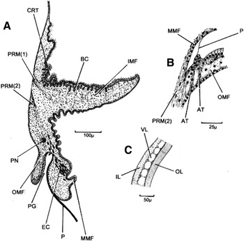
When expanded, the mantle margin of Modiolarca subpicta extends beyond the edge of the shell and is patterned with alternating small patches of light brown and cream marks. Figure 15A is an illustration of a transverse section through the right mantle lobe. Each lobe comprises the usual three folds—a large inner (IMF), a swollen middle (MMF) and a small outer one (OMF). Arising from the pallial line on the shell, the pallial retractor muscle is divided into two components: the innermost (PRM(1)) sending muscle fibres into the inner mantle fold, the outermost (PRM(2)) delivering muscle fibres into the middle and outer folds. Between the mantle under the shell and the inner mantle fold, there is a ciliated rejection tract (CRT), responsible for clearing the mantle cavity of pseudofaeces. In the inner mantle fold are sub-epithelial basophilic cells (BC) ~15 µm long and which possibly secrete mucus to bind up pseudofaecal particles into strings. In the middle mantle fold are sub-epithelial eosinophilic cells (EC) ~ 20 µm long with a granular cytoplasm. The outer mantle fold is but ~125 µm long and the inner surface of which is lined with tall (20 µm) columnar cell. This surface is in close apposition to the outer surface of the middle mantle, which has a very low (a few μm) epithelium. This creates the periostracal groove (PG) that contains basal origin of the periostracum (P).
Modiolarca subpicta. (A) A transverse section through the right mantle lobe; (B) a more detailed section through the base of the periostracal groove; (C) a section through the periostracum. For abbreviations see Appendix.

The periostracal groove is shown in more detail in Figure 15B. The periostracum (P) is seen to arise from between the base of the middle mantle fold (MMF) and a small ridge of the outer mantle fold deep in the recess of the periostracal groove. Tiny fibrils (AT) attach this structure to the inner mantle surface and the lower branch of the pallial retractor muscle (PRM(2)) secures the whole structure to the shell.
Figure 15C is a yet more detailed section through the periostracum. It is of the basic mytilid plan (Beedham, Reference Beedham1958). The periostracum comprises an amorphous inner layer (IL) that is basophilic and a maximum of ~30 µm wide. There is also a non-staining outer layer (OL) that is also a maximum of ~30 µm wide and the two are separated by a vacuolated layer ~40 µm wide, which may or may not have once contained calcareous spicules prior to decalcification.
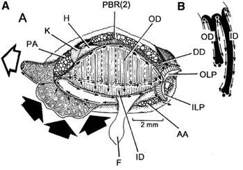
The organs of the mantle cavity of Modiolarca subpicta are shown in Figure 16A as seen from the right side after removal of the right shell valve and mantle. Also shown are the exhalant siphon and the expanded flap like inhalant aperture. The exhalant siphon comprises a low cone with a large internal siphonal valve, as seen in ventral view in Figure 10. The mantle margins are brown with cream blotches and the mantle edges are similarly cream. Water enters the mantle cavity via the long inhalant aperture and exits via the small exhalant aperture. Between the two, lie the large, dorso-ventrally long, ctenidia, each comprising longer inner (ID) and shorter outer demibranchs (OD). These lead to short labial palps (ILP, OLP) with the mouth (not illustrated) lying between the left and right pairs of these. The digestive diverticulae (DD) lie under the umbones and ligament in the usual position but the heart (H) is located, unusually, between the posterior adductor muscle (PA) and the posterior byssal retractor muscles. Posterior to the heart are the paired kidneys.
Modiolarca subpicta. (A), The organs of the mantle cavity as seen from the right side after removal of the right shell valve and mantle lobe; (B) the left ctenidium as seen in diagrammatic transverse section showing the surface ciliary currents. For abbreviations see Appendix.

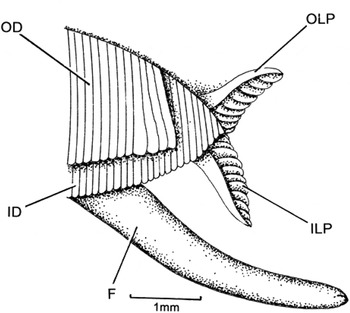
The left ctenidium is as seen in the diagrammatic transverse section in Figure 16B and shows the ciliary currents of the surfaces of the inner (ID) and outer (OD) demibranchs. They are of the usual mytilid plan. The anatomy of the ctenidial–labial palp junction of Modiolarca subpicta is illustrated in Figure 17. They are of the typical mytilid arrangement, that is, the outer demibranch (OD) is anteriorly foreshortened and its anterior-most filaments somewhat larger than the others (Morton & Dinesen, Reference Morton and Dinesen2010). Hence, only the anterior end of the longer inner demibranch (ID) passes into the food grooves between the inner (ILP) and outer (OLP) labial palps.
Modiolarca subpicta. The anterior end of the right ctenidium and right pair of labial palps. For abbreviations see Appendix.

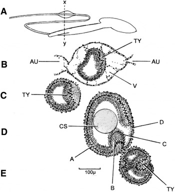
As noted above, the heart in Modiolarca subpicta is located between the posterior adductor muscle (PA) and its adjacent posterior component of the posterior byssal retractor muscle (PBR(1)) and the anterior component of the posterior byssal retractor muscle (PBR(2)). This has important effects on the arrangement of the gut and the course of the intestine. That is, since the stomach is located under the ligament, the course of the gut is entirely between the left and right elements of the anterior component of the posterior byssal retractor muscle. The basic arrangement of the intestine M. subpicta is illustrated in Figure 18A. This shows that the conjoined style sac and mid-gut separate posteriorly, the former terminating, the latter continuing as the mid-gut that eventually loops back on itself as the hind gut which then loops again posteriorly to penetrate the ventricle of the heart, pass between the left and right elements of the posterior component of the posterior byssal retractor muscle and over the top of the posterior adductor muscle to terminate on the posterior side of this as an anal papilla.
Modiolarca subpicta. (A) A simplified outline representation of the gut as seen from the right side (x–y = the sections below though); (B) the rectum inside the ventricle of the heart; (C) the hind gut; (D) the separated style sac; (E) the separated mid-gut. For abbreviations see Appendix.

The structure of the various components of the intestine, as seen from the right side and through a vertical section of the organs (x–y) are illustrated in Figure 18B, C, D & E. The rectum (Figure 18B) inside the ventricle (V), with auricles (AU) to left and right, of the heart is ~200 µm in diameter and possesses a bi-lobed typhlosole composed of ciliated cells some 20 µm, the others, also ciliated being 10 µm tall. The hind gut (Figure 18C), is ~200 µm in diameter and has a uni-lobed typhlosole. This comprises ciliated cells 50 µm tall the others being some 10 µm tall. The crystalline style sac (Figure 18D) has an overall diameter of ~300 µm containing a style some 200 µm in diameter. The majority of the style sac lining comprises cells of Type A that are 15 µm tall with cilia 15 µm long. The Type B cells that project into the lumen of the style sac are 40 µm tall and similarly possess cilia 15 µm long. Type C cells in the ‘gutter’ of the style sac are no more than a few microns tall as are the opposite D type cells and both possess a weak fringe of shorter 10 µm cilia. The separated mid-gut (Figure 18E) comprises a tube ~150 µm in diameter with an un-lobed typhlosole comprising cells some 50 µm tall with cilia 15 µm long. The other cells are some 10 µm tall.
Reproduction: the gonads (G) of Modiolarca subpicta are located in the mantle dorsally and extend laterally and ventrally within the left and right mantle lobes occupying the upper 2/3 and anterior 4/5 of the latter. Unusually, the gonads do not invade the visceral mass. Sex could not be determined in individuals with a shell length of <2 mm. The smallest males and females had shell lengths of 3.4 mm and 4.1 mm, respectively (Figure 5). Males thus appear to mature at a slightly smaller size than females and, further, females appear to attain a larger size than males. Spawning is free, and often initiated by male individuals (G.D., personal observation), and development is by a planktotrophic larva that lives for 4–6 weeks before settlement and metamorphosis (Lovén, Reference Lovén1848; Thorson, Reference Thorson1946; Ockelmann, Reference Ockelmann, Cox and Peake1965).
DISCUSSION
Modiolarca subpicta individuals living within their ascidian host are camouflaged by the external papillation of the tunic but also by the lightly adhering detritus, red filamentous algae, bryozoans and sponges, which obscure their presence externally. It seems clear that the epizoic (ectosymbiotic) lifestyle of M. subpicta with ascidians, notably Ascidiella aspersa (Roberts & Breen, Reference Roberts and Breen1985), is for protection and can thus be defined as aegism (Morton, Reference Morton1988). Host specificity is loose, however, and M. subpicta is known to occur in the tests of twelve species of ascidians and one holothurian throughout its wide range. Bodger & Allen (Reference Bodger and Allen2008, p. 97) suggest that free-living individuals of M. subpicta are ‘likely…in the process of transferring to another host’. The results of this study, however, suggest that colonization of a chosen host individual occurs at an early stage in the life histories of both species and that the two remain together throughout their equally short lifespans of perhaps 18–24 months (Millar, Reference Millar1952; Bodger & Allen, Reference Bodger and Allen2008). Larsen et al. (Reference Larsen, Frischer, Ockelmann, Rasmussen and Hansen2007) have reported, for example, that in the Danish Isefjord, the temporal occurrence of bivalve larvae show two patterns. Larvae from species known to exhibit a protracted annual spawning period, such as Mytilus edulis (Linnaeus, 1758), were present in the water column throughout the year whereas others appeared at clearly defined periods including M. subpicta (as Musculus marmoratus) in the summer months, as does A. aspersa (Millar, Reference Millar1952). The life histories of the two species are, therefore, closely intertwined and it seems more likely that free-living individuals of M. subpicta and those living with unusual hosts such as the holothurian Holothuria tubulosa (Rodríguez & Sánchez, Reference Rodríguez and Sánchez1997) have been forced to settle in the absence of their real ascidian host(s). Forbes & Hanley (Reference Forbes and Hanley1853) cited Clark (unpublished notes from 1835 and 1852) as suggesting that M. subpicta is able to change location/position only when settled on stones, but not when enveloped within an ascidian tunic. Adult individuals have, however, been observed to vacate their host after a few hours in vitro (G.D., personal observation) (Roberts & Breen, Reference Roberts and Breen1985). Our findings of the largest M. subpicta individuals occupying ascidians of medium sizes (Figure 4) suggest that some individuals do outlive their initially occupied ascidian, and are able to relocate themselves to a new host, perhaps belonging to a subsequent generation. The findings that all smaller groups of between 2 and 5 M. subpicta individuals (except for one group) comprised either males or females, suggest that relocation may be advantageous, perhaps at maturity, to enhance spawning success. Modiolarca subpicta has been described as being dioecious (Jeffreys, Reference Jeffreys1863). The fact, however, that the majority of individuals in the >7.5 mm shell length-size category were females suggests the intriguing possibility that either (i) the species is a protandric consecutive hermaphrodite or (ii) that females grow faster or (iii) that females live longer. This apparent size dimorphism between the two sexes is highlighted in Table 1 and we suggest that further studies are needed to clarify which of the three options identified above best explains it, or, indeed, a combination of them.
Roberts & Breen (Reference Roberts and Breen1985, p. 441) reported that the smallest individuals of Modiolarca subpicta were to be found in the region of the oral (inhalant) aperture of the Ascidiella mentula specimens studied by them. This is in contrast to our findings of spat and juveniles occurring around the exhalant siphon of A. aspersa (Figure 7). If attraction to an ascidian were by the detection of either a current flow or the discharge of ‘tunicin’ as suggested by Bourdillon (Reference Bourdillon1950), the exhalant stream would intuitively seem to be the obvious source of such an attractant, either physical or chemical, or both. It is possible, that the competent larvae of M. subpicta react to cues from a potential host rather than from conspecifics, at least initially. This is in contrast to the settlement behaviour of another gregarious mussel, Modiolus modiolus (Linnaeus, 1758), that also lives subtidally in boreal waters and which settles in response to pheromone cues emanating from conspecific adults (Dinesen & Ockelmann, in preparation).
Meissner (Reference Meissner1893) suggested that Modiolarca subpicta ‘bores’ into its host ascidian test but, as described by Roberts & Breen (Reference Roberts and Breen1985, p. 438) and better illustrated by Bodger & Allen (Reference Bodger and Allen2008, figure 2), it actually pulls itself, using byssal threads, into ever-increasing intimacy with the host epidermis resulting in the latter forming a pocket into which the bivalve nestles, dorsal side down.
Merrill & Turner (Reference Merrill and Turner1963, p. 56) describe how epifaunal Musculus discors (Linnaeus, 1767) builds a byssal nest among algae that resembles ‘a hollow ball of twine’. The infaunal, colonial, Musculista senhausia (Benson, 1842) does the same thing but, in this case, upon and within the mud surface (Morton, Reference Morton1973, figure 1). Another mytilid, Arcuatula elegans (Gray, 1828), does the same thing but in subtidal muds (Morton, Reference Morton and Morton1980, figure 1) but perhaps the closest similarity with M. subpicta is provided by Gregariella coralliophaga (Gmelin, 1791) which nestles in vacated Lithophaga bore holes in corals and weaves a matted, byssal cocoon, particularly posteriorly, to protect itself from predators (Morton, Reference Morton, Morton and Tseng1982, figure 1).
Yonge (Reference Yonge and Bayne1976) thought that the primitive mytiloidean was probably similar to something like the modern-day species of Musculus. That is, an approximately equivalve, equilateral, globose, thin-shelled, solitary, nestling animal. Morton (Reference Morton, Morton and Tseng1982) shared this view and imagined that the shell shape of the ancestors of this seemingly uninspiring and unpretentious group of widely distributed bivalves radiated into the diverse variety of byssally-attached mussels that are so familiar to us today. Much of the success of the mytiloidean body form was related to the neotenous retention of the byssus into adult life (Yonge & Campbell, Reference Yonge and Campbell1968; Yonge, Reference Yonge and Bayne1976) and, subsequently, the evolution of the heteromyarian form (Morton, Reference Morton and Gosling1992). It was thought that early modiolines ‘emerged’ (in evolutionary terms) from within sediments and that the adoption of a progressively more ventrally flattened, dorsally peaked form allowed for the adoption of a shell that, notably, allowed colonization of the rocky intertidal and, in Asia, flowing freshwaters (Morton & Dinesen, Reference Morton and Dinesen2010). The taxonomic affinities of the Mytiloidea have never been resolved satisfactorily and the evolutionary concepts created by Yonge (Reference Yonge and Bayne1976) and Morton (Reference Morton and Gosling1992) are not meant to represent phylogenetic lineages but, rather, ideas as to how the highly successful heteromyarian form might have evolved.
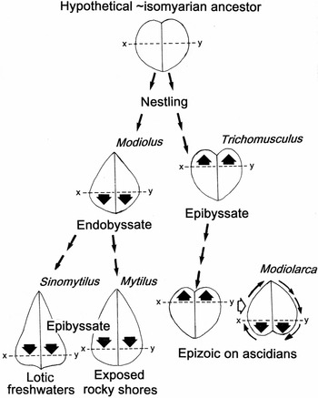
Figure 19 attempts to provide more information with regard to the evolution of the heteromyarian form within the Mytilidae. From a hypothetical, rotund, equivalve and isomyarian, musculine-like ancestor, the left lineage of Figure 19 examines transverse sections through the shells of the endobyssate mytilids Modiolus, Sinomytilus and Mytilus to suggest how their forms have evolved by the greatest width of the shell (x–y) having moved progressively ventrally to create a more stable form for life in flowing freshwaters and on exposed rocky shores. Conversely, the right lineage of Figure 19 suggests that from the same hypothetical ancestor, Modiolarca, epizoic in the tests of ascidians, could have evolved from an epibyssate, more heteromyarian, Trichomusculus-like (Morton Reference Morton1995, figure 4) mussel with the x–y axis moving dorsally instead of ventrally. But, in Modiolarca, the ventral stability characteristic of the modiolid–mytilid lineage has been achieved through living dorsal side down.
Left lineage: transverse sections through the shells of the endobyssate mytilids Modiolus, Sinomytilus and Mytilus have evolved from a hypothetical, approximately isomyarian and globose ancestor, showing how the greatest width of the shell (x–y) has moved progressively ventrally to create a more stable form for life in flowing freshwaters and on exposed rocky shores. Right lineage: conversely, in Modiolarca, symbiotically epizoic within the tests of ascidians, which is also thought to have evolved from a hypothetical, approximately isomyarian, globose, ancestor via the intermediary of an epibyssate, nestling, Trichomusculus-like animal (with the x–y axis moving dorsally instead of ventrally), ventral stability has been achieved through living dorsal side down.

Another mytilid associated with ascidians occurs on the west coast of the Atlantic Ocean and is identified by Andrews (Reference Andrews1981) as Musculus (Ryenella) lateralis (Say, 1822). Regardless of the authenticity of the generic and subgeneric names assigned to this species, it too occurs within the tests of ascidians and has a distributional range from North Carolina, Florida, the Gulf of Mexico, and the Caribbean, south to Brazil. Several mytilids have evolved symbiotic relationships with other organisms, such as species of Gregariella, Botula, Lithophaga and Leiosolenus boring into corals (Morton, Reference Morton and Morton1990). In general terms, a small size is prerequisite for such relationships and it seems possible that these coral-associated species and M. lateralis and, now, Modiolarca subpicta, have achieved this via the nestling and byssal camouflage route and, in the case of the latter species, a more intimate relationship with ascidians.
ACKNOWLEDGEMENTS
We thank Professor R.S.S. Wu, City University of Hong Kong, Hong Kong Special Administrative Region, China, for organizing the sectioning of specimens for light microscopy, and Phil Dr R. Dinesen and P. Dinesen, Denmark, for assistance with translations of French and Latin species descriptions.
Appendix. Abbreviations used in the figures
- A
A cell epithelium of style sac
- AA
Anterior adductor muscle (or scar)
- ABR
Anterior byssal retractor muscle (or scar)
- AT
Attachment tissues of periostracal groove to mantle
- AU
Auricle of heart
- B
B cell epithelium of style sac
- BC
Basophilic cells
- BG
Byssal groove
- BGC
Byssal gland cells
- BGD
Byssal gland duct
- BGL
Byssal gland
- C
C cell epithelium of style sac
- CRT
Ciliary rejection tract
- CS
Crystalline style
- D
D cell epithelium of style sac
- DD
Digestive diverticula
- EC
Eosinophilic cells
- ES
Exhalant siphon
- F
Foot
- F(H)
‘Heel’ of the foot
- H
Heart
- H(LOC)
Location of the heart
- IA
Inhalant aperture
- ID
Inner demibranch
- IL
Inner layer of periostracum
- ILP
Inner labial palp
- IMF
Inner mantle fold
- K
Kidney
- LI
Ligament
- MM
Mantle margin
- MMF
Middle mantle fold
- OD
Outer demibranch
- OL
Outer layer of periostracum
- OLP
Outer labial palp
- OMF
Outer mantle fold
- P
Periostracum
- PA
Posterior adductor muscle (or scar)
- PBR(1)
Posterior component of the posterior byssal retractor muscle (or scar)
- PBR(2)
Anterior component of the posterior byssal retractor muscle (or scar)
- PG
Periostracal groove
- PL
Pallial line
- PN
Pallial nerve
- PRM(1) & PRM(2)
Components of the pallial retractor muscle
- SE
Septum
- TY
Typhlosole
- U
Umbo
- V
Ventricle of heart
- VL
Vacuolated layer of periostracum
- VM
Visceral mass