

Introduction
White adipose tissue of adult cattle was recently shown to contain cells expressing genes characteristic for brown and beige adipocytes. The physiological role of these cells remains unclear. As brown adipocytes are able to dissipate energy, their occurrence may exert negative effects on feeding efficiency in cattle (Asano et al., Reference Asano, Yamada, Hashimoto, Umemoto, Sato, Ohwatari, Kanamori, Terachi, Funaba and Matsui2013). Initial investigations targeted effects of feeding interventions on the expression of genes characteristic for brown and beige fat. Activity of uncoupling protein 1 (UCP1) as marker for brown adipocytes increased in subcutaneous fat (SCF) of cattle fed a concentrate-based diet, whereas vitamin A deficiency did not affect its expression (Asano et al., Reference Asano, Yamada, Hashimoto, Umemoto, Sato, Ohwatari, Kanamori, Terachi, Funaba and Matsui2013; Kanamori et al., Reference Kanamori, Yamada, Asano, Kida, Qiao, Abd Eldaim, Tomonaga, Matsui and Funaba2014). It is unknown, however, whether the abundance of marker genes for beige and brown adipocytes varies between breeds or adipose tissue depots.
We analysed at messenger RNA (mRNA) and protein level markers for white (leptin (LEP)), beige and brown adipocytes (UCP1, transmembrane protein 26 (TMEM26) and Zic family member 1 (ZIC1)) in five different adipose tissue depots of cattle highly divergent in fat deposition.
Material and methods
Animals
Japanese Black (n=6), Holstein (n=5) and Charolais (n=6) steers were fattened on a high-energy diet and were slaughtered at 26 months of age. Details are described by Albrecht et al. (Reference Albrecht, Gotoh, Ebara, Xu, Viergutz, Nürnberg, Maak and Wegner2011). Briefly, intramuscular fat (IMF) content was determined by Soxhlet extraction method using petroleum ether as the solvent. Area of the M. longissimus dorsi (MLD) was measured with a planimeter at the 12th rib. A slice of the MLD (2 to 3-cm thick) was taken from the same position and fixed in 10% neutral buffered formalin for at least 3 weeks. Slices of 2-mm thickness from the fixed samples were stained with Oil-Red-O (Chroma, Münster, Germany) and semi-automatically analysed for marbling parameters (e.g. average marbling fleck size, marbling fleck number) with Cell^D image analysis software. Samples of SCF, IMF, intermuscular, perirenal fat (PF) and visceral fat were taken at slaughter and used for mRNA and protein isolation, and immuno-histochemical analyses. Perirenal brown adipose tissue (BAT) of a new-born Holstein calf and mouse BAT were used as controls.
Analyses
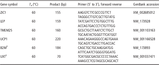
Abundance of mRNA was measured by quantitative reverse transcription PCR (RT-qPCR) and normalized to expression of UXT and B2M with the REST 2009 software (v. 2.0.13, Pfaffl et al., Reference Pfaffl, Horgan and Dempfle2001). This software takes into account the efficiency of reference and target genes. Efficiency is calculated from a dilution series for each gene. Primers for all genes are listed in Table 1. The procedures for RNA isolation, complementary DNA synthesis, and qPCR were essentially the same as described by Komolka et al. (Reference Komolka, Albrecht, Schering, Brenmoehl, Hoeflich and Maak2014). Proteins were detected by Western blot and localized in cryo-sections of tissues with commercially available antibodies (UCP1: LS-C146466-50, TMEM26: ASB-ARP71769_P050; Biozol, Eching, Germany; ZIC1: ab72694; Abcam, Cambridge, UK). The procedures for protein isolation, Western blotting and immuno-histochemistry were identical to those described by Albrecht et al. (Reference Albrecht, Gotoh, Ebara, Xu, Viergutz, Nürnberg, Maak and Wegner2011).
Primers for quantitative reverse transcription PCR

LEP=leptin; TMEM26=transmembrane protein 26; UCP1=uncoupling protein 1; ZIC1=Zic family member 1.
1 Reference genes.
Statistical analysis
Data were analysed with ANOVA using the GLM procedure of SAS (v.9.2) with breed as fixed factor. Values obtained for every adipose tissue were considered as repeated measurements and animal was added as random effect. REST 2009 (v. 2.0.13, Pfaffl et al., Reference Pfaffl, Horgan and Dempfle2001) was used for group-wise comparison of normalized, efficiency corrected RT-qPCR data.
Results and discussion
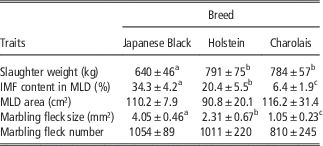
The three breeds differed significantly in slaughter weight and IMF content but not in muscle area (Table 2). The large IMF differences derived from larger marbling flecks in the order Japanese Black > Holstein > Charolais but not from different numbers of marbling flecks (Table 2).
Phenotypic traits of steers (mean±SD)

IMF=intramuscular fat; MLD=M. longissimus dorsi.
a,b,cValues within a line with different superscripts differ significantly at P<0.05.
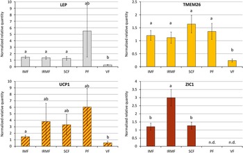
Markers for white (LEP) and beige adipocytes (TMEM26, UCP1) were expressed in all investigated fat depots with high individual variability for LEP in PF and UCP1 in most depots. In contrast, ZIC1 mRNA was detected only in muscle associated fat and in SCF (Figure 1). The expression of the investigated genes did not differ significantly between breeds within adipose depots (data not shown). Although, all animals received a concentrate-based diet, there was no general up-regulation of UCP1 mRNA in SCF as described by Asano et al. (Reference Asano, Yamada, Hashimoto, Umemoto, Sato, Ohwatari, Kanamori, Terachi, Funaba and Matsui2013). Instead, UCP1 varied strongly between animals regardless of breed. This may be caused by the fact that beige and brown adipocytes are not evenly distributed in the depots and were arbitrarily included or excluded by sampling from longissimus muscle. UCP1 is now considered as marker for brown as well as for beige adipocytes, whereas ZIC1 is more specific for brown adipocytes (Walden et al., Reference Walden, Hansen, Timmons, Cannon and Nedergaard2012). We could demonstrate that ZIC1 is expressed in some depots providing additional support for the existence of brown adipocytes in adult cattle.
Expression of genes specific for white, beige and brown fat cells in adipose depots of steers at an age of 26 months. IMF=intramuscular fat; IRMF=intermuscular fat; SCF=subcutaneous fat; PF=perirenal fat; VF=visceral fat; n.d.=not detectable. a,bDifferent superscripts indicate significant differences between depots, P⩽0.05.

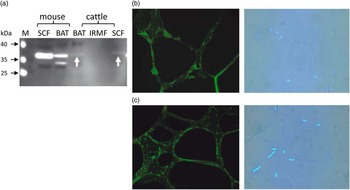
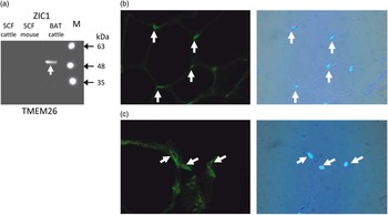
Protein expression could be shown for UCP1 and ZIC1 in BAT of a new-born calf by Western blot (Figures 2a and 3a), whereas TMEM26 was not detectable. Moreover, UCP1 was demonstrated in SCF of adult cattle. These results are partly attributable to the relatively low sensitivity of Western blots. However, the antibodies for UCP1, ZIC1 and TMEM26 were sufficiently sensitive to localize the respective proteins in muscle-related adipose depots (Figures 2b and c and 3b and c), even though no multivacuolar adipocytes were present.
Detection of uncoupling protein 1 (UCP1) in murine and bovine adipose tissues by Western blot (a), immune-localization of UCP1 in intermuscular fat (IRMF) (b, left) and intramuscular fat (c, left), and respective overlays of bright field and nuclear staining with Hoechst 33258 (b, c, right). Scale bar is 50 µm. SCF=subcutaneous fat; BAT=perirenal brown adipose tissue.

Detection of Zic family member 1 (ZIC1) in murine and bovine adipose tissues by Western blot (a), immune-localization of ZIC1 in bovine intramuscular fat (b, left) and transmembrane protein 26 (TMEM26) in bovine intermuscular fat (c, left), and respective overlays of bright field and nuclear staining with Hoechst 33258 (b, c, right). Scale bar is 50 µm. SCF=subcutaneous fat; BAT=perirenal brown adipose tissue.

We found specific staining for UCP1 protein in the cytoplasm of univacuolar cells at mitochondria in IMRF (Figure 2b), IMF (Figure 2c) and in SCF. A weak ZIC1 signal was detected in nuclei of IMF adipocytes (Figure 3b), whereas TMEM26 signals were found in the cytoplasm of cells without vacuoles in IMRF (Figure 3c).
Collectively, these results confirm the existence of beige and brown adipocytes in muscle-related fat depots and in SCF of cattle at 26 months of age. The occurrence of brown or beige adipocytes in peripheral rather than in internal adipose depots is in accordance with results in mice (Fisher et al., Reference Fisher, Kleiner, Douris, Fox, Mepani, Verdeguer, Wu, Kharitonenkov, Flier, Maratos-Flier and Spiegelman2012) and may indicate a common feature in mammals (Asano et al., Reference Asano, Yamada, Hashimoto, Umemoto, Sato, Ohwatari, Kanamori, Terachi, Funaba and Matsui2013). This is supported by the fact that we did not find differences in the abundance of brown/beige markers at mRNA and protein level in cattle with different IMF content. Consequently, a relationship between the abundance of brown or beige adipocytes and the degree of fat deposition in different cattle breeds could not be established. It remains subject to further investigations whether brown and beige adipocytes are only involved in the dissipation of excess energy from feeding or whether they exert more extensive influence on metabolic processes.
Acknowledgements
The authors are grateful to E Schwitulla and S Foß for expert technical assistance