Implications
As a traditional Chinese food, superior flavor in Peking duck is of vital importance. Force-feeding of these ducks can improve growth performance and lead to a high proportion of abdominal fat making their meat more delicious. However, force-feeding can destroy the integrity of intestinal epithelial cells. Moreover, force-feeding has been shown to induce severe intestinal inflammation and oxidative stress injury. In summary, these findings suggest it is advisable to limit the practice of force-feeding due to its adverse effects on the duck welfare.
Introduction
Pekin duck is a famous, popular and traditional food in China. It is famous for meat production and is the unique raw material for roast duck (Xu et al., Reference Xing, Jiang, Wang, Lei, Zhi, Wu, Zhu, Huang, Xia and Chen2015). High-proportioning of abdominal fat in Pekin ducks can make the ducks become more flavorful during the process of roasting (Xu et al., Reference Xu, Gu, Huang, Xia, Zhang, Zhang, Rong, Schachtschneider and Hou2017). Growers have thus tried many different methods to increase the abdominal fat of these ducks, using strategies such as feed additives and force-feeding.
Numerous studies have demonstrated that force-feeding for a short time was the effective way to rapidly increase body weight and abdominal fat deposition in Pekin ducks (Chartrin et al., Reference Chartrin, Bernadet, Guy, Mourot, Hocquette, Rideau, Duclos and Baeza2006). In foreign countries, particularly in Hungary, the main object of force-feeding is geese. Force-feeding during goose breeding facilitates the deposition of fat to the liver to produce ‘foie-gras’. Whole goose ‘foie-gras’ is a highly valued foodstuff, even broken goose ‘foie-gras’ can be used to produce foie gras sauce, the most famous food in France (Rodríguez et al., Reference Rodríguez, García, González, Asensio, Mayoral, Lópezcalleja, Hernández and Martín2014; Murphy, Reference Murphy2018). In China, force-feeding is mainly used for breeding ducks because force-feeding promotes the rapid increase of lipid deposition in the skin and abdomen of Pekin ducks, making them the best source for roast duck dishes (Salichon et al., Reference Salichon, Guy, Rousselot and Blum1994).
Force-feeding has been demonstrated to significantly increase the body weight, weight gain capacity and feed conversion ratio of ducks (Guy, Reference Guy2004). Other beneficial effects to force-fed ducks are evident through their increased respiration rate and body temperature compared with the controls. Force-feeding leads to a rapid accumulation of lipids in the skin and abdomen (1.5- to 1.7-fold) which is mainly induced by triglyceride deposition (Chartrin et al., Reference Chartrin, Bernadet, Guy, Mourot, Hocquette, Rideau, Duclos and Baeza2006; Guémené et al., Reference Guémené, Guy, Noirault, Destombes and Faure2006). However, recently a considerable amount of scientific research has revealed that force-feeding does not meet the animal welfare standard morally (Guémené et al., Reference Guémené, Guy, Noirault, Destombes and Faure2006). Performance records, as well as behavioral, physiological and clinical parameters are considered to be good indicators for evaluating animal welfare (Lundmark et al., Reference Lundmark, Berg and Röcklinsberg2018). Force-feeding is a mandatory operation that can induce some adverse effects for the welfare of the ducks; in support of this hypothesis, it was demonstrated that problems such as stress injury resulting during driving and fetching, damage caused by insertion of the tube into the esophagus, abnormal gait and liver dysfunction (Mohammed et al., Reference Mohammed, Abdelrahman and Darwish2014). Moreover, various painful injuries to the esophagus, including hemorrhagic inflammation and perforations of the esophagus, which can be compounded by subsequent growth of opportunistic germs and fungal growth have been induced by force-feeding (Han et al., Reference Han, Wang, Xu, Li, Ye, Jiang and Zhuo2008). Force-feeding could affect lipid deposition in the liver to form fatty liver in Pekin ducks and also could improve meat quality, change blood composition, disorder the hormone metabolism, induce an inflammatory response and increase serum corticosterone (CORT) at the same time (Guemene et al., Reference Guémené, Guy, Noirault, Garreau-Mills, Gouraud and Faure2001). The intestinal tract, especially the intestinal villus, is not only the main digestive organ that absorbs nutrients in an organism, but also an important barrier between the internal organs and foreign substances (usually entering from the mouth). Force-feeding has been shown to affect the digestive function of the gut and disrupt gastrointestinal homeostasis (Hu et al., Reference Hu, Jia and Edelblum2018). However, scarcity of studies is not sufficient to justify the research because little literature has focused on the effect on digestive function and mucosa immune function in the duodenum and jejunum for force-fed Pekin ducks. The aim of this study was to determine the effects of force-feeding on duodenal and jejunal villus conditions, mucosa inflammatory cytokines, digestive enzyme activities and the oxidative stress status of Pekin ducks by comparing force-feeding and ad libitum feeding. This study provides a theoretical basis for the banishment of force-feeding in the Pekin duck industry.
Material and methods
Animals and experimental design
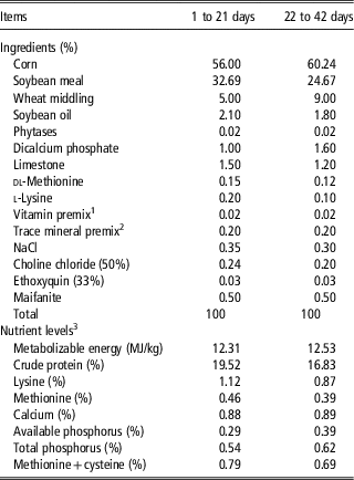
All experimental procedures were approved by the China Agricultural University Animal Care and Use Committee (Beijing, China, permit no. CAU20180329-2). All animal treatment procedures during the experiment followed Experimental Animal Care and Use Guidelines of China. A total of 500 female Pekin ducks (37 days-old) were purchased from a local commercial hatchery (Beijing, China) and reared on the net at the Experimental Center of China Agricultural University. Ducks were raised in environmentally controlled housing while being exposed to 24 h of constant light before and during the experiment. The ducks were randomized into two groups, each made up of five replicates with 50 ducks. One group served as the control, receiving no additional treatment, and the other was the force-feeding group. Diet nutrient levels were formulated to meet or exceed the standards set by the National Research Council (1994) (Table 1). In the control group, ducks were allowed free access to feed and water. In the force-fed group, each duck was grabbed by the neck, and had a plastic feeding tube 8 to 10 inches long inserted down its esophagus and force-fed three times a day (every 8 h) from 37 to 42 days (in total 6 days). These ducks were forced to ingest a greater amount of food (0.30 kg/bird per time) than what they would normally eat voluntarily but were allowed free access to water. Feed intake was recorded by each replicate from 37 to 42 days of age. Ducks were weighed after feed deprivation for 6 h to calculate average body weight at 37 day (ABW37), average body weight at 42 day (ABW42), average daily gain (ADG) and average daily feed intake (ADFI) at 42 day of age.
Composition and nutrient levels of the basal diets (air-dry basis) for Pekin ducks

1 The vitamin premix provided the following per kg of diet: vitamin A, 12 500 IU; vitamin D3, 3500 IU; vitamin E, 20 IU; vitamin K3, 2.65 mg; thiamin, 2.00 mg; riboflavin, 6 mg; pyridoxin, 3 mg; vitamin B12, 0.025 mg; biotin, 0.0325 mg; folic acid, 12 mg; pantothenic acid, 50 mg; nicotinic acid, 50 mg.
2 The mineral premix provided the following per kg of diet: Cu, 6 mg; Fe, 80 mg; Zn, 40 mg; Mn, 100 mg; Se, 0.15 mg; I, 0.35 mg.
3 Calculated values.
Sample collection and preparation
Blood samples were taken aseptically from the wing vein of 10 randomly selected ducks (two ducks per replicate) from each group at 42 days of age after 6 h fasting into commercial blood collection tubes containing no additive and then centrifuged at 3500×g for 10 min to obtain supernatant serum. The serum was stored in 0.5-ml Eppendorf tubes at −80°C for further study. Subsequently, all ducks were sacrificed using sodium pentobarbital solution (30 mg/kg BW) anesthesia. Immediately after bleeding, the abdominal cavity was dissected along the midline of the abdomen, and 10 cm segments of duodenum (2-cm away from the gastro-duodenal junction) and 10 cm segments of the jejunum (middle of the small intestine) were cut transversally and washed with a 0.9% NaCl solution and quickly frozen in liquid nitrogen and stored at −80°C for further analysis. In all, 8 cm subsegments of duodenum and jejunum were then cut longitudinally, flushed the chyme with a sterile 0.9% NaCl solution and scraped the mucosa layer (do not scrape the muscular layer) using a sterile glass slide. Subsequently, the mucosa was quickly moved into 5-ml Eppendorf tubes and stored at −20°C for further study. The intestine mucosa (200 mg) of duodenum and jejunum, respectively, was homogenized with 1800 ml ice-cold isotonic physiological saline (0.9%, v/w, NaCl) using a Handheld Tissue Homogenizer (Polytron PT1200, Kinematica, Luzern, Switzerland). The homogenates were centrifuged at 3500 to 4000×g for 10 min at 4°C. The supernatants were carefully collected from the tube and used directly for digestive enzyme activities, inflammatory cytokines parameters and oxidative stress parameters assays. The last 2 cm duodenum and jejunum tissues were then cut and fixed with 10% ineutral-buffered formalin for histological measurements.
Measurement of experimental parameters
Serum diamine oxidase (DAO), D-lactic acid (d-Lac), endotoxin and CORT of 10 ducks from each group were tested using respective ELISA kits (catalog number: A088-1, H263, L00350 and H205, respectively; Nanjing Jiancheng Bioengineering Institute, Nanjing, China) for each parameter measured (R&D Systems, Minneapolis, MN, USA) following the manufacturers’ instructions. Levels of the inflammatory cytokines indexes, such as interleukin (IL)-2, IL-6, IL-10, IL-17, tumor necrosis factor (TNF)-α and interferon (IFN)-γ were determined using commercial duck ELISA kits (catalog number: H003, H007, H009, H014, H025 and H052, respectively; Nanjing Jiancheng Bioengineering Institute) according to the manufacturers’ instructions. The activity of the digestive enzymes (sucrose, maltase, chymotrypsin, amylase and lipase) was determined using commercial kits (catalog number: A099-2, A082-3, A080-3, C016-1 and A054-2, respectively; Nanjing Jiancheng Bioengineering Institute) according to the manufacturers’ instructions. To test the oxidative stress status of ducks, total antioxidant capacity (T-AOC), superoxide dismutase (SOD), glutathione peroxidase (GSH-PX), catalase (CAT) and malondialdehyde (MDA) were determined using commercial kits (catalog number: A015-2, A001-3, A005, A007-1 and A003-1, respectively; Nanjing Jiancheng Bioengineering Institute) according to the manufacturers’ instructions. The content of total duodenal and jejunal proteins were determined by the Coomassie brilliant blue method by the commercial kit A045-2 (Nanjing Jiancheng Bioengineering Institute) according to the manufacturers’ instructions. All measurements of the samples were independently performed in triplicates.
Histological examination of intestinal segments and villus conditions
The last 2 cm duodenum and jejunum were sectioned for evaluation of the epithelium lesions. Each piece was fixed in 10% ineutral-buffered formalin at room temperature for 24 h and embedded in paraffin. Sections of 5-μm thickness were then stained with hematoxylin and eosin. Intestinal tissue slices were observed under a conventional light microscope (Olympus BX41; Olympus Corporation, Tokyo, Japan). A total of 10 straight and complete villi and crypts were selected from the mucosal site of each sample to measure the villus length and crypt depth using Image-ProPlus 6.0 (Media Cybernetics, Silver Spring, MD, USA), and the villus length and crypt depth (V/C) ratios were calculated. The number of goblet cell was also calculated in villous epithelial cells of the duodenum and jejunum.
Statistical analysis
Experimental data was analyzed using SPSS for Windows (version 19.0; SPSS Inc., Chicago, IL, USA). The figures were processed using Adobe Photoshop CS6 Software (Adobe Systems Incorporated, San Jose, CA, USA). The results were analyzed using one-way ANOVA with post hoc Bonferroni’s test. Data are showed as means±SEM. Statistical differences were considered at P<0.05.
Results
Growth performance and serum parameters
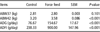
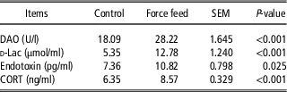
At the age of 37 day, the ABW37 of the ducks was nearly the same (P=0.101) between the two groups. However, at the age of 42 day, in comparison with the control group, increased ABW42, ADG and feed conversion ratio were observed significantly (P<0.001) in the force-feeding group (Table 2). The levels of DAO, d-Lac, endotoxin and CORT in the serum of force-fed Pekin ducks are shown in Table 3. Compared with the control group, there was a significant (P<0.001) increase in serum DAO, d-Lac and CORT levels of ducks in the force-feeding group. The endotoxin level also increased significantly (P=0.025) in the force-feeding group in comparison to the control group.
Effects of force-feeding on growth performance in Pekin ducks

ABW37=average body weight at 37 day; ABW42=average body weight at 42 day; ADG=average daily gain; ADFI=average daily feed intake.
Values are mean of 10 independent determinations.
The levels of diamine oxidase (DAO), d-lactic acid (d-Lac), endotoxin and corticosterone (CORT) in serum of Pekin ducks by force-feeding

Values are mean of 10 independent determinations.
Intestinal structure injury
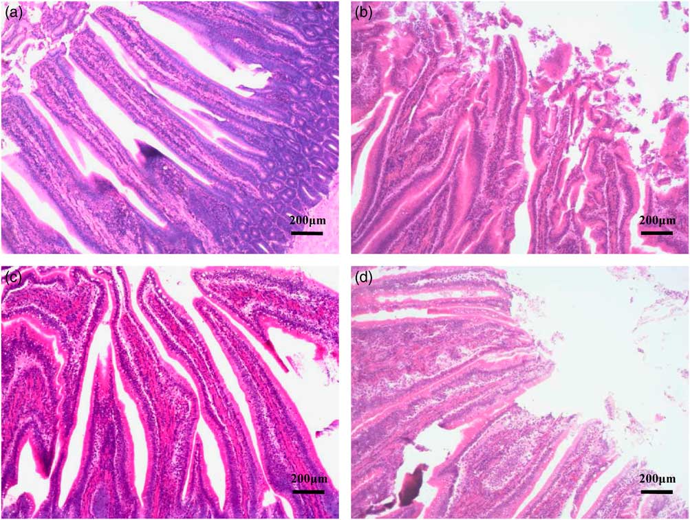
Intestinal histopathology analysis revealed that degeneration and necrosis of intestinal villus epithelium cells were found in the force-feeding group (Figure 1b and d) but not the control group (Figure 1a and c). Intestinal villus epithelium cells had been severely removed in force-feeding group. Compared with the control group, the villus length in duodenum and jejunum was not significantly different (P=0.401, Table 4), but the crypt depth was significantly decreased (P<0.001, Table 4). In addition, the mucosal regular V/C ratio was significantly higher (P<0.001) in the jejunum while there was no significant difference in the duodenum.
Histological changes of duodenum and jejunum villus (magnification 100×) in Pekin ducks. (a) Duodenum of control group, (b) duodenum of force-feeding group, (c) jejunum of control group and (d) jejunum of force-feeding group. Degeneration and necrosis of crypt cells, lymphocytic infiltration, and degeneration of the mucosal epithelial cells and pyknosis are shown in (b) and (d). Measurements of villus length and crypt depth are presented in Table 4.

The levels of villus length and crypt depth in duodenum and jejunum of Pekin ducks by force-feeding

V/C=villus length/crypt depth.
Values are mean of 10 independent determinations.
1Goblet cell is the number per 100 intestinal villus.
Digestive enzyme activities
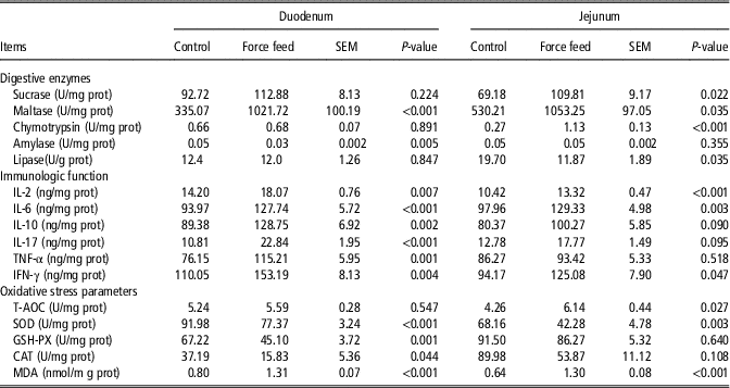
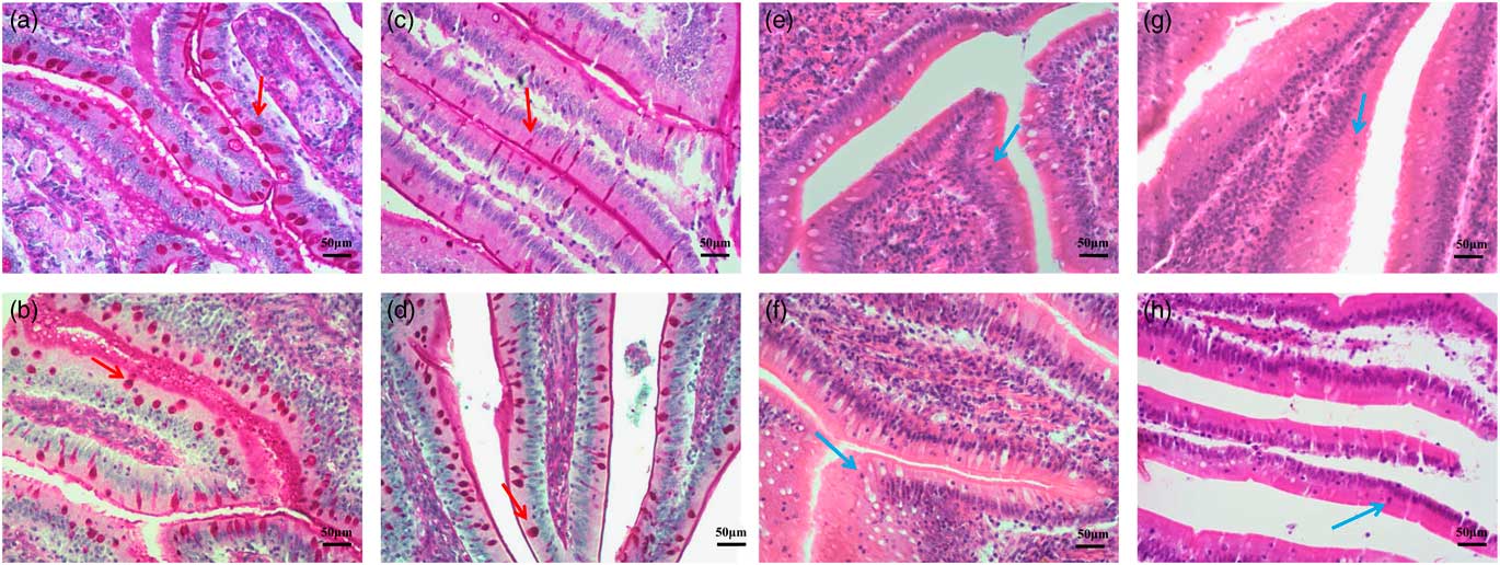
The activities of digestive enzymes in the duodenal and jejunal mucosa of force-fed Pekin ducks are shown in Table 5. In the duodenum, the activity of maltase increased significantly (P<0.001) in the force-feeding group compared with the control group while the activities of amylase decreased significantly (P=0.005). In the jejunum, the activities of sucrose (P=0.022), maltase (P=0.035) and chymotrypsin (P<0.001) increased significantly in the force-feeding group compared with the control group while the activities of lipase decreased significantly (P=0.035). Intestinal histopathology analysis showed that goblet cell number was only reduced significantly (P=0.019) in the duodenum in the force-feeding group (Figure 2c, Table 3) compared with the control group (Figure 2a).
The levels of digestive enzymes, immunologic function and oxidative stress parameters in duodenal and jejunal mucosa of Pekin ducks by force-feeding

IL=interleukin; TNF=tumor necrosis factor; IFN=interferon; T-AOC=total antioxidant capacity; SOD=superoxide dismutase; GSH-PX=glutathione peroxidase; CAT=catalase; MDA=malondialdehyde.
Values are mean of 10 independent determinations.
The distribution of goblet cells and lymphocytes in villus epithelial cells of the duodenum and jejunum (magnification 400×) in Pekin ducks. Goblet cells are indicated by the red arrows and lymphocytes are indicated by the blue arrows. (a) Goblet cells in duodenum epithelial cell of control group, (b) goblet cells in duodenum epithelial cell of force-feeding group, (c) goblet cells in jejunum epithelial cell of control group, (d) goblet cells in jejunum epithelial cell of force-feeding group, (e) lymphocytes in duodenum epithelial cell of control group, (f) lymphocytes in duodenum epithelial cell of force-feeding group, (g) lymphocytes in jejunum epithelial cell of control group and (h) lymphocytes in jejunum epithelial cell of force-feeding group.

Inflammatory cytokines parameters
The levels of immunologic function in the duodenal and jejunal mucosa of force-fed Pekin ducks are shown in Table 5. In the duodenum, the levels of IL-2 (P=0.007), IL-6 (P<0.001), IL-10 (P=0.002), IL-17(P<0.001), TNF-α (P=0.001) and IFN-γ (P=0.004) increased significantly in the force-feeding group compared with the control group. However, in the jejunum, only the levels of IL-2 (P<0.001) and IL-6 (P=0.003) increased significantly and the level of IFN-γ increased significantly (P=0.047) in the force-feeding group compared with the control group. Finally, partial lymphocytic infiltration was observed in mucosal epithelial cells in the force-feeding group (Figure 2g and h) compared with the control group (Figure 2e and f).
Oxidative stress parameters
Levels of T-AOC, SOD, GSH-PX, CAT and MDA in the duodenal and jejunal mucosa of force-fed Pekin ducks are shown Table 5. In the duodenum, the activities of SOD (P<0.001), GSH-PX (P=0.001) and CAT (P=0.044) decreased significantly in the force-feeding group compared with the control group. However, in the jejunum, only the activities of SOD decreased significantly (P=0.003) in the force-feeding group. Similarly, in the force-fed ducks, MDA content increased significantly (P<0.001) in both the duodenal and jejunal mucosa in the force-feeding group compared with the control group.
Discussion
In the current study, ducks were force-fed 0.90 kg food every day from 37 to 42 day, leading to a significantly increased ABW42, ADG and ADFI compared to the control group, which indicated that force-feeding could indeed improve the growth performance of the ducks in a short time. However, force-feeding was shown to destroy the normal intestinal morphology, decrease villus height and lower crypt depth of duodenum and jejunum in Pekin ducks. The small intestine is the main bodily location for absorption and transport of nutrients; absorption is the main function of villus that make up the walls of the small intestine (Li et al., Reference Li, Lu, Hu, Sun and Yao2002). An increase in the height of intestinal villus can increase the area of the small intestine to absorb nutrients, so the length of intestinal villus directly affects the growth and development of animals (Wilson et al., Reference Wilson, Cummings, Barbosa, Williams, Gerard and Peebles2018). The height of the villus is significantly correlated with the number of cells, and only mature villus can absorb nutrients (Umemura et al., Reference Umemura, Komura and Hoshino2018). When the gut is attacked or stressed, villi are damaged and shortened. Therefore, the short villus of force-fed Pekin ducks showed low nutrient absorption capacity. Furthermore, crypt of the intestine is a tubular gland formed when the epithelium of the villus root reaches the proper layer. Crypt depth reflects the production rate of epithelial cells, if this process is slowed down, the production rate of basal cells is decreased, making the crypt shallower (Xing et al., Reference Xing, Jiang, Wang, Lei, Zhi, Wu, Zhu, Huang, Xia and Chen2015). The depth of the crypt indicates the maturation rate of intestinal epithelial cells. The ratio of villus height to crypt depth comprehensively reflected the functional state of the intestine (He et al., Reference He, Lu, Ma, Zhang, Li, Jiang, Zhou and Gao2018). A decrease of such a ratio indicated a decrease in digestion and absorption function. In this study, the villus height and crypt depth values in the force-feeding group were lower than in the control group, indicating that force-feeding damaged the normal villus structure of the duodenum and jejunum of Pekin ducks and affected their intestinal absorption of nutrients.
Diamine oxidase is a highly activate enzyme localized to the intestinal mucosa epithelial cells (Biegański and Ulatowska, Reference Biegański and Ulatowska1983). Normally, DAO cannot enter into the blood stream, therefore the content of DAO in the blood is very low, but once the integrity of intestinal mucosa is destroyed, a large number of DAO can be released into the bloodstream increasing the level of DAO in the blood (Xin-Rong et al., Reference Xin-Rong, Rao and Liu2018). As such, DAO is a reliable indicator for the integrity of intestinal mucosal epithelial cells. In addition, once intestinal permeability is increased or its integrity destroyed, d-Lac and endotoxin can also enter into the blood through the damaged mucosa (Zhang et al., Reference Zhang, Zhao and Lian2018). We found the contents of DAO, d-Lac and endotoxin in Pekin duck serum significantly increased in the force-feeding group rather than the control group. This suggests that force-feeding severely damaged the integrity of the intestinal epithelial cells and changed the osmotic pressure of the gut. Consistent with our results, researchers have also found higher d-Lac and endotoxin contents in serum in the intestinal mucosa lesions caused by sepsis or excessive feeding (Gao et al., Reference Gao, Jiang, Xiao, Peng, Xiao and Yang2015).
The activities of maltase, sucrase and chymotrypsin were significantly increased in the duodenum and jejunum mucosa of force-fed ducks. Force-feeding damaged the integrity of the duodenum and jejunum villus epithelial cells and the intestinal barrier function; thus cells produced more maltase and sucrase to digest more food. Significantly increased activity of chymotrypsin in the duodenum and jejunum after force-feeding has been shown to result from forced feeding of large amounts of food in a short period of time (Nakano et al., Reference Nakano, Inoue, Alpers, Akiba, Katayama, Shinozaki, Kaunitz, Ohshima, Akita and Takahashi2009). For this reason, we speculated that when large amounts of food are force-fed to Pekin ducks, their normal digestion function is disturbed to control their metabolism. Nitsan et al. (Reference Nitsan, Nir, Dror and Bruckental1973) also found that the activities of the digestive enzymes such as trypsin and chymotrypsin increased with the increasing forced feeding of food, consistent with our results. This study also reported that the content of amylase decreased in the small intestine of geese after force-feeding, which was consistent with the results of the present experiment.
Lymphocytes, a type of white blood cell, participate in the body’s immune regulation (Hu et al., Reference Hu, Jia and Edelblum2018). The intestinal tract was the first line of contact with food and pathogenic microorganisms. Intestinal epithelial lymphocytes (IELs) play an important role in maintaining the intestinal mucosa integrity (Montalbanarques et al., Reference Montalbanarques, Chaparro, Gisbert and Bernardo2018). Siegmund et al. (Reference Siegmund, Lehr and Fantuzzi2002) found that the number of intestinal lymphocytes was reduced when the symptoms were alleviated in mice with colitis, thus the quantity of IELs can somewhat reflect the integrity of the small intestine. An increased number of lymphocytes may serve as an indicator for the diagnosis of inflammatory and autoimmune diseases (Wu and Hamilton, Reference Wu and Hamilton1999). The increased number of lymphocytes in force-fed Pekin ducks showed inflammation of intestinal mucosa. Interleukins play an important role in fighting against virus, anti-proliferation and immune regulation as well as inflammatory reaction (Kanuri et al., Reference Kanuri, Spruss, Wagnerberger, Bischoff and Bergheim2011). In our study, IL-2 and IFN-γ content in the duodenum and jejunum mucosa were significantly higher in the force-feeding group compared with the control group; IL-10, IL-17 and TNF-α were found to be significantly increased only in the duodenum; a result that may be associated with intestinal inflammatory disease. Force-feeding for 6 days might lead to the inflammatory response in the duodenum and jejunum mucosa of Pekin ducks.
After force-feeding for 6 days (from 37 to 42 day), the serum CORT content increased significantly of the ducks indicated the oxidative injury in the body (Kv et al., Reference Kv, Madhana, Js, Lahkar, Sinha and Naidu2018). The activities of SOD, CAT and GSH-PX in the duodenum and jejunum mucosa decreased whereas the MDA content increased after force-feeding are related to the body’s antioxidant levels. Cells can produce reactive oxygen species (ROS) in their metabolic processes; these ROS can destroy the cell membrane, lipid protein and DNA damage (Urso and Clarkson, Reference Urso and Clarkson2003). There is also a molecular antioxidant system designed to fight ROS damage in the body. This system can be divided into two parts: the enzymatic and non-enzymatic systems. Enzymatic antioxidants mainly include SOD, GSH-PX and CAT (Liu et al., Reference Liu, Liu, Cheng, Fan, Zhao and Liu2015). Under normal conditions, oxidant–antioxidant concentration in the body is in equilibrium, but stress can stimulate an increased free radical release (Cheng et al., Reference Cheng, Fan, Zhao, Liu, Cheng, Liu and Liu2016). Antioxidant activity was significantly decreased in the duodenum and jejunum mucosa of force-fed Pekin ducks, suggesting that force-feeding induced severe oxidative stress in the gut of Pekin ducks. In the present study, the MDA content of the duodenal and jejunal mucosa in Pekin duck was increased as well as T-AOC which might be attributable to the increasing ability of the intestine to resist lipid peroxidation. The increased CORT meant the ducks were in oxidative stress status; MDA is usually used as a marker to detect the degree of lipid peroxidation (Wu et al., Reference Wu, Cui, Peng, Fang, Zuo, Deng and Huang2014). Another indicator is T-AOC, which indicates the overall ability of tissues or blood to resist oxidative stress (Liu et al., Reference Liu, Zhang, Guan, Zhang and Xu2013). Michiels et al. (Reference Michiels, Tagliabue, Akbarian, Ovyn and De Smet2014) found that the content of MDA in the proximal jejunum mucosa and plasma decreased in chickens after feeding restriction, which are consistent with our findings. Our experiment showed that force-feeding for 6 days could induce the oxidative stress of Pekin ducks and abnormally change the antioxidant mechanism in the duodenum and jejunum mucosa of Pekin ducks; hence, the activities of antioxidant enzymes decreased, whereas that of lipid peroxide increased.
In conclusion, force-feeding at the end of the fattening period for 6 days has a substantial effect on digestive function in the duodenal and jejunal mucosa of Pekin ducks by improving the activities of maltase and chymotrypsin while reducing the amylase and lipase activities. However, force-feeding also destroys the integrity of intestinal epithelial cells. Moreover, force-feeding can induce severe intestinal inflammation and oxidative stress injury in affected ducks. It is therefore advisable to limit force-feeding of Pekin ducks during raising due to considerable adverse effects on the duck welfare.
Acknowledgments
This research was supported by the System for Poultry Production Technology, Beijing Innovation Research Team of Modern Agriculture (BAIC04-2019).
Declaration of interest
The authors declare that they have no competing interests.
Ethics statement
All experimental procedures were approved by the China Agricultural University Animal Care and Use Committee (Beijing, China, permit no. CAU20180329-2). All animal treatment procedures during the experiment followed Experimental Animal Care and Use Guidelines of China.
Software and data repository resources
All data are available upon request.