Introduction
Vestibular neuritis is a common cause of peripheral vestibular vertigo; in one study, it accounted for 7 per cent of presentations to out-patient clinics specialising in the treatment of dizziness.Reference Brandt1
While the exact aetiology is unknown, it is well accepted that vestibular neuritis is caused by viral infection.
The symptoms and signs of vestibular neuritis usually include sudden onset of prolonged vertigo, spontaneous nystagmus, absence of other auditory and neurological findings, and reduced or absent caloric test responses. In routine clinical practice, the diagnosis of vestibular neuritis mainly depends on the medical history and, particularly, on caloric test results, which can offer a reliable assessment of vestibular function.Reference Goebel2 However, caloric testing is only able to evaluate the horizontal semicircular canals, reflecting the function of the superior vestibular nerve. Therefore, patients who present with similar symptoms but with normal caloric test results pose a diagnostic challenge.
In recent years, vestibular evoked myogenic potential testing, which assesses the saccule and the inferior vestibular nerve, has been used to assist the diagnosis of such conditions.Reference Lesmas Navarro, Pérez Garrigues, Morera Pérez and Piqueras3 In cases of vestibular neuritis, it is generally believed that the superior vestibular nerve is involved but the inferior nerve is usually preserved, due to anatomical differences.Reference Gianoli, Goebel, Mowry and Poomipannit4, Reference Goebel, O'Mara and Gianoli5 To date, there have been only two reports of vestibular neuritis involving only the inferior vestibular nerve, including a total of five patients worldwide.Reference Halmagyi, Aw, Karlberg, Curthoys and Todd6, Reference Monstad, Økstad and Mygland7 These patients had symptoms of traditional vestibular neuritis but presented with normal caloric test results and abnormal vestibular evoked myogenic potential test results, indicating involvement of the inferior vestibular nerve.
In this study, we report eight patients with inferior vestibular neuritis, out of a series of 216 patients with vestibular neuritis, directing special attention to the clinical features of this novel subtype of vestibular neuritis.
Materials and methods
Patients
A total of 216 patients (104 males, 112 females; age range 10–64 years; mean age 38.4 years) with full clinical documentation who had attended our hospital's vertigo clinic between May 2007 and December 2008 were enrolled in the study. All patients underwent radiological investigation and hearing, caloric and vestibular evoked myogenic potential testing within 15 days of onset of symptoms.
The study was approved by the ethics committee of Shandong University, and written, informed consent was obtained from all patients.
Caloric testing
The bithermal caloric test was performed according to the method of Fitzgerald and Hallpike. Each ear was aerated with a constant flow of air alternating between 24 and 49°C, for 40 seconds. The patient's response was recorded over 3 minutes, and a 7-minute interval was allowed between each stimulus to avoid cumulative effects. A video-based system was used (Ulmer VNG version 1.4; Synapsys, Marseille, France) to record and analyse the eye response. The head position was checked before each aeration to maintain the horizontal semicircular canal close to ‘earth-vertical’, and the patient's alertness was maintained by having them perform mental arithmetic throughout the recording. Aerations were delivered randomly in terms of both temperature and side, and no fixation was allowed while recording. The maximum slow-phase velocity of nystagmus after each aeration was calculated, and unilateral weakness was determined according to Jongkee's formula. In our laboratory, a unilateral weakness value of less than 20 per cent was considered normal.
Vestibular evoked myogenic potential testing
Vestibular evoked myogenic potentials were recorded using a Smart EP device (Intelligent Hearing Systems, Miami, FL, USA). Patients lay supine on a bed, and the electromyographic activity of the sternocleidomastoid muscle was recorded while they were asked to raise their head off the bed (in order to activate their neck flexors bilaterally and saccular receptors were acoustically stimulated). The recording electrode was placed at the middle third of the muscle ipsilateral to the stimulated ear, the reference electrode was placed on the upper edge of the sternum, and the ground electrode was placed on the sternocleidomastoid muscle contralateral to the stimulated side. Care was taken to place bilateral electrodes symmetrically. The amplifier gain was set to 100 000, and signals and bandpass were filtered from 10 to 3000 Hz. Short tone bursts (100 dBnHL, 500 Hz, with 1 millisecond raise-fall time and 5 millisecond plateau time) were delivered monaurally via TDH 49P earphones (Telephonics Corp., Huntington, NY). The stimulation rate was 5 Hz and the analysis time 60 milliseconds. A total of 128 responses to stimuli were averaged, and measurements were repeated twice to check test wave reproducibility. We analysed the amplitudes of the first positive-negative peak, p13-n23 and the peak latencies of p13 and n23. The mean of two runs was used to calculate amplitudes and latencies. An amplitude ratio between the two ears of over 1.61 was considered abnormal. A p13 latency of over 17.3 milliseconds and an n23 latency of over 24.6 milliseconds was considered abnormal.
Results
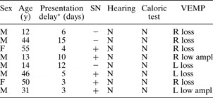
This study included 216 patients with vestibular neuritis, who all presented with sudden onset of severe, prolonged vertigo despite normal hearing. Central lesions were excluded by careful neuro-otological examination and cerebral computed tomography or magnetic resonance imaging (MRI) on admission. Of these 216 patients, eight had a normal caloric test result but an abnormal vestibular evoked myogenic potential test result (unilateral loss was found in six cases and low amplitude in two cases) (Table I). Based on comprehensive analysis of all test data, a diagnosis of inferior vestibular neuritis was clearly established in these eight cases.
Clinical data for 8 patients with inferior vestibular neuritis

*Time from onset to presentation. Y = years; SN = spontaneous nystagmus; VEMP = vestibular evoked myogenic potential testing; M = male; F = female; −= absent; += present; N = normal; R = right; L = left; ampl = amplitude
All patients were given anti-viral and steroid medication and vestibular rehabilitation therapy, and their symptoms disappeared within six months. Follow-up times ranged from six months to two years. During the follow-up period, two of the six patients with no initial vestibular evoked myogenic potential responses showed recovery of these responses. One of these two patients showed recovery of responses to within the normal range in four months, while the other still showed a low amplitude response after one year. Of the two patients who initially showed a low amplitude vestibular evoked myogenic potential response, one showed recovery of response to within the normal range in six months, whereas the other showed no recovery.
Typical case
A 50-year-old woman presented with a three-day history of sudden onset, intense vertigo with nausea and vomiting. In the clinic, she was nauseated and unable to walk.
Standard clinical examination showed no limb ataxia or other obvious pathological findings. Spontaneous nystagmus with a fast phase beat to the left was found. This nystagmus was completely suppressed by fixation.
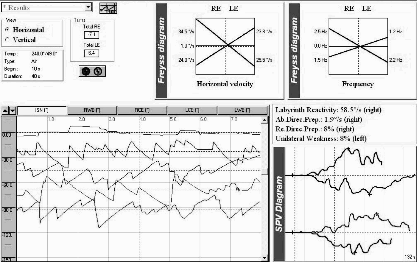
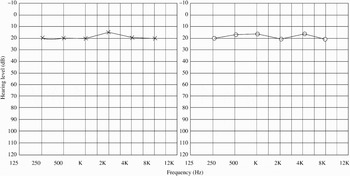
Caloric testing demonstrated no significant asymmetry (Figure 1). Otoscopy and audiometry were normal (Figure 2). Vestibular evoked myogenic potentials were normal on the left side but absent on the right (Figure 3). An MRI scan of the brain was normal.
Screengrab showing normal caloric test results.

Audiogram showing normal pure tone hearing test results.

Vestibular evoked myogenic potential test results, indicating normal responses on the left but absent responses on the right.

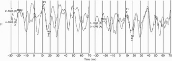
The patient recovered completely in four months, with disappearance of all symptoms. Notably, vestibular evoked myogenic potentials recovered to within the normal range (Figure 4).
Vestibular evoked myogenic potential test results recorded four months after onset of symptoms, indicating a return to normal.

Discussion
It had previously been believed that vestibular neuritis mainly affects the superior vestibular nerve.Reference Fetter and Dichgans8 Most patients with vestibular neuritis lose function in the superior portion of the vestibular nerve, due to the longer, narrower osseous canal of the superior nerve portion, compared with the inferior branch canal.Reference Goebel, O'Mara and Gianoli5 This anatomical difference makes the superior vestibular nerve more susceptible to entrapment during inflammatory swelling. However, vestibular evoked myogenic potential testing has revealed that vestibular neuritis can involve not only the superior vestibular nerve but also the inferior nerve. Halmagyi et al. were the first to propose the concept of inferior vestibular neuritis, and presented two patients with probable inferior vestibular neuritis.Reference Halmagyi, Aw, Karlberg, Curthoys and Todd6 Later, Monstad et al. reported three patients with clinical features of acute vestibular neuritis, normal caloric test results and loss of vestibular evoked myogenic potentials.Reference Monstad, Økstad and Mygland7 In the current study, we identified eight patients, out of 216 with vestibular neuritis, who presented with sudden onset of severe, prolonged vertigo and symptoms similar to routine vestibular neuritis, along with normal hearing. The unique features of these eight patients were normal caloric test results but abnormal vestibular evoked myogenic potential test results, in agreement with previous reports.Reference Halmagyi, Aw, Karlberg, Curthoys and Todd6, Reference Monstad, Økstad and Mygland7 This established a firm diagnosis of inferior vestibular neuritis in these eight patients. Our data, together with those of others, provide objective evidence that inferior vestibular neuritis does indeed exist, as a novel subtype of vestibular neuritis.
Interestingly, we encountered two cases with low amplitude vestibular evoked myogenic potentials. In the five cases of inferior vestibular neuritis discussed in two previous reports, there was complete loss of vestibular evoked myogenic potential responses on the side of the lesion.Reference Halmagyi, Aw, Karlberg, Curthoys and Todd6, Reference Monstad, Økstad and Mygland7 Our results indicated that patients with inferior vestibular neuritis might show either disappearance or reduced amplitude of vestibular evoked myogenic potential responses, just as patients with routine vestibular neuritis may show complete or partial unilateral loss of caloric response. In addition, there were no reports of benign positional paroxysmal vertigo in our eight inferior vestibular neuritis patients during follow up, perhaps due to normal function of the utriculus. This may be an important feature of inferior vestibular neuritis, which differentiates it from routine vestibular neuritis.
All our patients received anti-viral and steroid medication and vestibular rehabilitation therapy, and their symptoms disappeared over a six-month period. Follow-up time ranged from six months to two years. During the follow-up period, two patients whose vestibular evoked myogenic potential responses had been absent showed recovery of responses. One of these two patients showed recovery of responses to within the normal range in four months, while the other still showed low amplitude responses after one year. Of the two patients who initially showed low amplitude vestibular evoked myogenic potential responses, one patient's responses recovered to within the normal range in six months, while the other's responses failed to recover. Murofushi et al. reported 13 patients with vestibular neuritis, of whom five showed vestibular evoked myogenic potential recovery over a period of six months to two years.Reference Murofushi, Iwasaki and Ushio9 The time course of vestibular evoked myogenic potential recovery observed in our study was consistent with previous reports, suggesting that the clinical character and prognosis of inferior vestibular neuritis is similar to those of routine vestibular neuritis. This further confirms the theory that inferior vestibular neuritis is a new subtype of vestibular neuritis.
• Vestibular neuritis is a common cause of peripheral vestibular vertigo; in one study, it accounted for 7 per cent of patients presenting to an out-patient clinic specialising in the treatment of dizziness
• This study reports eight cases of possible inferior vestibular neuritis, evaluated by vestibular evoked myogenic potential testing
• Six cases had unilateral loss of vestibular evoked myogenic potential responses, whereas two had a unilateral reduced amplitude of responses
• Inferior vestibular neuritis is a novel subtype of vestibular neuritis, which involves the inferior vestibular nerve alone
Two of our patients presented to the clinic more than 10 days after the onset of vertigo. Therefore, we cannot deny the possibility that these patients' caloric tests were abnormal during their initial bout of vertigo but normalised due to central compensation. However, the majority of authors have reported that, in patients with vestibular neuritis, caloric responses recover slowly. Okinaka et al. studied 60 patients with vestibular neuritis, and found canal paresis in approximately 90 per cent of patients one month after onset, in 80 per cent after six months, and in 50 per cent after five to 10 years.Reference Okinaka, Sekitani, Okazaki, Miura and Tahara10 Kim et al. found that otolith-related test abnormalities improved more rapidly than canal-related test abnormalities after vestibular neuritis.Reference Kim, Hong, Lee, Yi, Lee and Lee11 In our two patients presenting more than 10 days (but less than 15 days) after onset of vertigo, caloric tests were normal while vestibular evoked myogenic potential responses were abnormal; thus, it would have been almost impossible for these patients' caloric tests to be abnormal during their initial bout of vertigo. Therefore, we believe that these two patients also had inferior vestibular neuritis.
In addition, different levels of vestibular compensation may explain the fact that five of our patients showed spontaneous nystagmus while three did not.
Conclusion
Inferior vestibular neuritis is a novel subtype of vestibular neuritis. The diagnostic criteria are as follows: (1) sudden, severe, prolonged vertigo; (2) absence of auditory and neurological symptoms or findings; (3) normal caloric responses; and (4) ipsilateral loss or low amplitude of vestibular evoked myogenic potential responses. Vestibular evoked myogenic potential testing should be routinely used in patients with vestibular neuritis.