Introduction
The family Epidermoptidae (Acariformes: Analgoidea) includes parasitic skin mites associated with birds. Some epidermoptid species of the subfamily Myialginae (Archemyialges Mironov, Bochkov, and Fain; Hemimyialges Mironov, Bochkov, and Fain; Myialges Trouessart), and Microlichus Trouessart and Neumann (Epidermoptinae), are unusual in that they have hyperparasitic relationships with parasitic chewing lice (Phthiraptera) and louse flies (Diptera: Hippoboscidae) on birds (Fain Reference Fain1965).
The taxonomy of the Epidermoptidae and the genus Myialges have varied over time. Fain (Reference Fain1965) divided epidermoptids into two subfamilies, Epidermoptinae and Dermationinae, and placed the genus Myialges in the former. In addition, Fain (Reference Fain1965) divided Myialges into three subgenera, Myialges, Promyialges Fain, and Metamicrolichus Fain. Gaud and Atyeo (Reference Gaud and Atyeo1996) elevated Promyialges to generic status, and placed Metamicrolichus as a subgenus of Microlichus; Metamicrolichus was later elevated to generic status by Mironov et al. (Reference Mironov, Bochkov and Fain2005). The subfamily Myialginae was resurrected by Mironov et al. (Reference Mironov, Bochkov and Fain2005), and they placed Myialges and two new described monotypic genera, Archemyialges and Hemimyialges, in the myialgine subfamily.
The life cycle of these hyperparasitic mites has been described for only a few species. Non-gravid females, males, and immatures are found in burrows in the skin of their hosts (Myialginae) or in feather bulbs (Microlichus) feeding on epidermal tissue and body fluids. Ovigerous females attach to the soft integument of a louse or hippoboscid fly using their ambulacra and chelicerae, or by a large anchor-like process on tarsus I (Myialginae), and feed on haemolymph (Evans et al. Reference Evans, Fain and Bafort1963; Fain Reference Fain1965). Eggs are laid in a cluster around the female mite and each egg is attached by a thin thread. Larvae eventually hatch and begin to feed on their avian host (Fain Reference Fain1965).
The host specificity of Myialginae species varies. Those hyperparasitic on hippoboscids tend to be generalists reported from several bird families, while species on lice show relatively more host specificity for their avian hosts and they are typically only associated with one genus of lice (Fain Reference Fain1965). However, avian host range of Myialginae species is likely determined by the level of host specificity of the hippoboscid or louse host species. Epidermoptid species can cause considerable response in tissues of their hosts. Some Microlichus species have been reported to cause severe lesions in feather bulbs (Fain Reference Fain1965). Myialgine species burrow into the superficial layers of the skin causing mange-like lesions and inflammation, which may lead to death of the host (Gilardi et al. Reference Gilardi, Gilardi, Frank, Goff and Boyce2001). The attachment of female Myialginae produces strongly sclerotised scars in the integument of their hippoboscid or louse hosts, and heavy infestation on the wings of hippoboscids may impede flight (Bequaert Reference Bequaert1953; Fain Reference Fain1965).
Myialgine specimens are typically collected as females from their insect hosts and are infrequently found on birds. As a result, the other life stages are not known for most species. There are 12 described species of Myialginae and the only species in which the male has been described is Hemimyialges macdonaldi (Evans, Fain, and Bafort). Radford (Reference Radford1949) purportedly described the male of Myialges trinotoni (Cooreman), but as Fain (Reference Fain1965) suspected Radford’s identification was incorrect. The male described by Radford (Reference Radford1949) was actually a female feather mite, Rectijanua Gaud (Rectijanuidae) species (identified by H.C. Proctor, University of Alberta, Edmonton, Alberta, Canada). Cooreman (Reference Cooreman1944) correctly described the female of Myialges trinotoni (as Myialgopsis trinotoni) from the louse Trinoton querquedulae (Linnaeus) (as Trinoton luridum Burmeister) on Mergus merganser Linnaeus (Aves: Anatidae) in Belgium. Herein, we describe the male of M. trinotoni, summarise avian host records and infestation parameters for this species on Trinoton querquedulae (Linnaeus), and sequence the barcode region of cytochrome oxidase subunit I (COI) from females of M. trinotoni.
Materials and methods
Sampling and identification
Most birds were casualties from the Wildlife Haven (Manitoba Wildlife Rehabilitation Organization, Ile des Chênes, Manitoba, Canada) and the Prairie Wildlife Rehabilitation Centre (Winnipeg, Manitoba, Canada); a small number of birds were obtained from hunters. Date of death and location were recorded for each bird. Birds were individually bagged and stored at −20 °C until processing in the laboratory at the University of Manitoba (Winnipeg, Manitoba, Canada). To collect lice and mites, birds were thawed and placed in a 4–40-L container, depending on the size of the bird, and submerged in warm water containing an appropriate amount of liquid dish detergent. Each bird was agitated vigorously for 3–10 minutes, depending on the size of the bird, then removed from the container and rinsed thoroughly. The washing solution was then passed through a 90-µm sieve. This process was repeated with warm, soapy water, and a third time with just warm water. The filtrate from all three washes was preserved in 70% or 95% ethanol, the latter when specimens were to be used for molecular analysis. All samples were examined for lice using a dissecting microscope; however, only one sample was examined specifically for the presence of male Myialges, that being from a common merganser (Mergus merganser) found in Winnipeg on 28 August 2014. The location of each mite found attached to a louse was noted, the number of mites and lice were counted, and any mites present were removed and retained. Imaging of lice was done using a Canon (Tokyo, Japan) 7D Mk II DSLR with a 65-mm MP-E lens, and imaging of mites was done using a Leica (Wetzlar, Germany) DM2500 compound microscope and Leica Application Suite v4.8. All mites and lice were collected and preserved in 70% or 95% ethanol for later identification and/or molecular analysis.
Mites were cleared in 85% lactic acid, mounted in polyvinyl alcohol medium (6371A, BioQuip Products, Rancho Dominguez, California, United States of America), and cured on a slide warmer at 40 °C for three to four days. Slide-mounted specimens were examined using a Leica DM2500 compound microscope with differential interference contrast illumination. Host lice were identified using characters suggested by Clay and Hopkins (Reference Clay and Hopkins1960). Species level identifications of mites were made using keys (Fain Reference Fain1965) and species descriptions from the primary literature. Voucher specimens (CNC564584 (female) and CNC564585 (male)) are deposited in the Canadian National Collection of Insects, Arachnids, and Nematodes (Agriculture and Agri-Food Canada, Ottawa, Ontario, Canada). Slide-mounted specimens of Epidermoptidae in the Canadian National Collection were also examined for Myialges. Scientific permits were issued to T.D.G. for migratory birds by officers of the Canadian Wildlife Service, Environment Canada.
Initial drawings of mites were made with pencil on paper using a camera lucida. These were later merged in Adobe (San Jose, California, United States of America) Photoshop CS3 and redrawn in Adobe Illustrator CS3 using an Intuos 3 Graphics Tablet from WACOM Company (Saitama, Japan). Chaetotaxy was based on Griffiths et al. (Reference Griffiths, Atyeo, Norton and Lynch1990). All measurements are in micrometres (µm) and for paired structures they are presented in the order of right, left.
Molecular techniques
Genomic DNA was extracted from whole specimens for 24 hours using a DNeasy Tissue kit (Qiagen, Santa Clara, California, United States of America). Following extraction, mites were removed from the extraction buffer, vouchers were slide mounted, and genomic DNA was purified following the DNeasy Tissue kit protocol. Polymerase chain reaction amplifications were performed in a total volume of 25 µL, with 14.7 µL ddH2O, 2.5 µL 10× ExTaq buffer, 0.65 µl 25 mM MgCl2, 1.0 µL of each 10 µM primer, 2.0 µL 10 mM dNTPs, 0.15 µL ExTaq DNA polymerase, and 3 µL genomic DNA template. Primer pairs LCO1490+HCO2198 (Folmer et al. Reference Folmer, Black, Hoeh, Lutz and Vrijenhoek1994) were used to amplify a 679 base-pair fragment of the 5'-end of COI. Polymerase chain reaction amplification was performed on an Eppendorf ep Gradient S Mastercycler (Eppendorf, Hamburg, Germany), using the following protocol: initial denaturation cycle at 94 °C for three minutes, followed by 45 cycles of 94 °C for 45 seconds, primer annealing at 45 °C for 45 seconds, 72 °C for one minute, and a final extension at 72 °C for five minutes. Amplified products and negative controls were visualised on 1% agarose electrophoresis gels, and purified using pre-cast E-Gel CloneWell 0.8% SYBR Safe agarose gels (Invitrogen, Carlsbad, California, United States of America). Sequencing reactions followed the protocol of Knee et al. (Reference Knee, Beaulieu, Skevington, Kelso and Forbes2012), and sequencing was performed at the Agriculture and Agri-Food Canada, Eastern Cereal and Oilseed Research Centre Core Sequencing Facility (Ottawa, Ontario, Canada). Sequence chromatograms were edited and contiguous sequences were assembled using Sequencher v5.4.6 (Gene Codes Corporation, Ann Arbor, Michigan, United States of America). Cytochrome oxidase subunit I sequences were aligned manually in Mesquite v3.10 (Maddison and Maddison Reference Maddison and Maddison2016) according to the translated amino acid sequence. Sequence for female Myialges trinotoni has been submitted to GenBank (KX060553).
Results
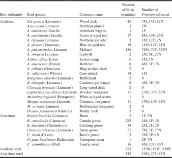
From 1993 to 2016, 27 species of waterfowl (Aves: Anatidae) have been examined for lice in the Galloway laboratory (post-downy stages only), including 20 species of ducks (n=323), five species of geese (n=328), and two species of swans (n=17) (Table 1). Six genera and at least 17 species of chewing lice were collected from these hosts (see in part, Galloway et al. Reference Galloway, Proctor and Mironov2014; Grossi et al. Reference Grossi, Sharanowski and Galloway2014). Of the 323 ducks examined, 104 (32% prevalence) were infested with at least one T. querquedulae. Of the 345 geese and swans examined, 15 (4%) were infested by at least one Trinoton anserinum (Fabricius). Myialges trinotoni infested only Trinoton querquedulae. Trinoton Nitzsch species were collected from all but six species of Anatidae (ring-necked duck, bufflehead, long-tailed duck, white-winged scoter, red-breasted merganser, and ruddy duck), all six of which were examined in small numbers. Trinoton anserinum were collected in relatively small numbers from trumpeter swans, and the five species of geese examined (Table 1). Trinoton anserinum were collected in large numbers on tundra swans (Table 1). Myialges specimens were not found on any of the Trinoton from geese.
Diversity and abundance of ducks (Aves: Anatidae: Anatinae) and geese/swans (Aves: Anatidae: Anserinae) (post-downy stages only) examined for lice (Trinoton querquedulae infesting ducks; Trinoton anserinum infesting geese/swans) in Manitoba from 1993 to 2016.

F, female; M, male; N, nymph.
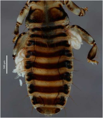
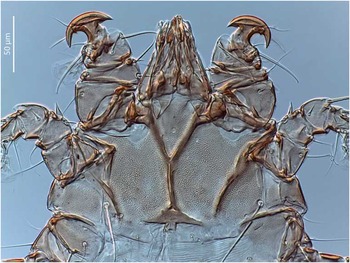
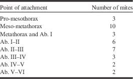
In total, 157 males, 191 females, and 539 nymphs of T. querquedulae were collected (Table 1). A total of 38 female M. trinotoni were collected, and these only infested adult T. querquedulae (15 adult lice with one mite each, seven with two each, and three lice with three mites each), and on only three birds: one mallard (Winnipeg, 18 November 2012; one of two female T. querquedulae infested with mites), one hooded merganser (Riverton, 13 October 2011; one female T. querquedulae infested), and one common merganser (Winnipeg, 28 August 2014; 10 of 10 female and 13 of 17 male T. querquedulae infested with mites). One M. trinotoni was attached to a teneral male T. querquedulae. One male mite of M. trinotoni was collected from the whole body washing of a common merganser that also had female mites, collected in Winnipeg, Manitoba. Female mites were attached to the intersegmental membranes along the lateral margins of their hosts (Fig. 1; Table 2), using anchor-like process on modified tarsus I (Fig. 2). There was no significant difference (bootstrap two-sample t-test, 2000 replicates, P=0.844) in mean intensity of infestation for males (mean intensity=1.54) versus female lice (mean intensity=1.60). Infestation parameters were calculated using Quantitative Parasitology 3.0 (Rózsa et al. Reference Rózsa, Reiczigel and Majoros2000).
Two female Myialges trinotoni with egg masses, attached to intersegmental membranes between abdominal segments I–II and III–IV of a female Trinoton querquedulae.

Female Myialges trinotoni dorsum showing modified tarsus I with anchor-like process.

Distribution of female Myialges trinotoni attached to intersegmental membranes along the lateral margins of the thorax or abdomen (Ab.) of adult Trinoton querquedulae (n=23) collected from one common merganser, one hooded merganser, and one mallard.

The barcode region of COI was successfully amplified from a female of M. trinotoni and deposited on GenBank (KX060553). Cytochrome oxidase subunit I has been amplified for only one other Myialges species, identified as M. caulotoon Speiser (GenBank numbers DQ503439–DQ503447) (Whiteman et al. Reference Whiteman, Sánchez, Merkel, Klompen and Parker2006), and these two species show considerable genetic divergence (25% uncorrected p-distance).
Myialges trinotoni (Cooreman, Reference Cooreman1944)
Material examined
Nine female mites (CNC564584) on Trinoton querquedulae from Mergus merganser, and one male mite (CNC564585) from M. merganser collected in Winnipeg, Manitoba, Canada; 28.viii.2014; T.D. Galloway, E.N. McNally, and W. Knee. One female mite on T. querquedulae from Anas platyrhynchos collected in Winnipeg, Manitoba, Canada; 18.xi.2012; T.D. Galloway and L.E. Peixoto. One female mite (CNC585101) on T. querquedulae from A. platyrhynchos collected 9 miles north of Juneau, Alaska, United States of America; 15.x.1949; R.B. Williams. One female mite on T. querquedulae from Lophodytes cucullatus collected in Riverton, Manitoba, Canada; 13.x.2011; T.D. Galloway and P. Snarr.
Diagnosis
Defining characters for female mites of this species are: propodosomal shield large; epimera I fused forming relatively long sternum divided posteriorly into two short branches; punctation absent between epimera I; epigynum thick and straight; legs I thick; T-shaped, anchor-like process on tarsus I bearing a tooth proximally (Fig. 2); tibia II retrorse process absent; ventral seta present on trochanter IV; bell-shaped pulvilli (Fain Reference Fain1965).
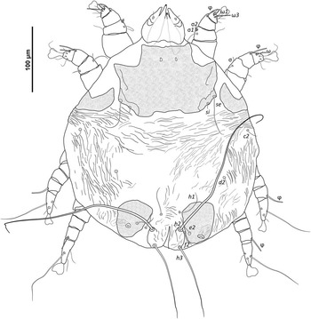
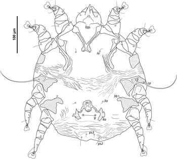
Defining characters for male mites of this species are: small, paired posterolateral hysterosomal shields present; genital setae g short and rounded; epimera I fused forming sternum divided posteriorly into two short branches; punctation absent between epimera I; extensive punctation on coxa III and IV; epimera IV poorly developed or absent; ventral seta present on trochanter IV; bilobate pulvilli. The following is the first description of the male of a Myialges species as defined by Mironov et al. (Reference Mironov, Bochkov and Fain2005).
Description
Male (Figs. 3, 4). Dorsum . idiosoma 313 µm long and 295 µm wide (level of cp seta), total length 368 µm. Cuticle striated, all dorsal seta simple. All dorsal shields finely punctate. Propodosomal shield 103 µm long at median and 156 µm wide (level of se seta). Setae se (right 54, left 65 µm long) and si (7, 6 µm) on lightly sclerotised, posterolateral margin of propodosomal shield. Paired lateral shields posterior of leg II wrapped around on dorsum and venter (57, 60 µm wide). Two small posterolateral hysterosomal shields present, right shield 62 µm long and 60 µm wide, left shield 59 µm long and 60 µm wide, seta e2 (30, 31 µm) on hysterosomal shields. Setae c2 (22, 21 µm), d2 (18, 19 µm), f2 (31, 30 µm), h1 (53, 54 µm), h2 (246, 261 µm), and h3 (113, 120 µm) in soft integument. Anus terminal.
Myialges trinotoni male dorsum.

Myialges trinotoni male venter.

Venter . Similar to known males of all other Myialginae species, male M. trinotoni lack adanal suckers and posterior lobes. Cuticle striate, all ventral setae simple. Epimera I fused forming sternum 22 µm long and 26 µm wide; sternum divided posteriorly forming two short branches. Epimera I and II with narrow punctate areas wider than those of female specimens. No punctation between epimera I. Extensive punctation on coxa III and IV. Epimera III narrow and divided (y shaped). Epimera IV poorly developed or absent. Two pairs of setae c3 (18, 22 µm) and cp (100, 95 µm) on coxa III, seta cp prominent and on lateral margin. Genital region with paired, lateral shields with fine punctation and seta 4a (7 µm) on the shield; seta g short (3, 2 µm), rounded and on small paired shieldlets. Setae la (17, 15 µm), 3a (9, 10 µm), 3b (18, 16 µm), ps2 (23, 26 µm), ps3 (13, 11 µm) in soft integument.
Gnathosoma . Gnathosoma 55 µm long and 67 µm wide. One pair of setae on subcapitulum and one pair of setae on dorsal palps. Chelicerae 69, 68 µm long. Palpal segmentation obscure.
Legs . Legs I and II similar to each other and without anchor-like tarsal process seen in female specimens, legs III and IV similar to each other and slimmer than legs I and II. Tibia II without a ventral retrorse process. Leg IV 116 µm long, trochanter IV 37 µm long. All leg setae simple, leg setation (solenidia) of coxa 0–0–2–0, trochanter 1–1–1–1, femur 1–1–0–0, genu 2(2)–2(1)–0–0, tibia 1(1)–1(1)–1(1)–1(1), tarsus 8(2)–8(1)–6–5. Tarsus I with solenidia ω1 (11, 9 µm) and ω3 (21, 18 µm), tarsus II with one solenidion (16, 14 µm). Tibia I and II with one long solenidion 22 µm (right) and 25 µm (left) in length. Tibia III with a long solenidion (24 µm), and tibia IV with a long solenidion (22, 21 µm). Genu I with a pair of solenidia in duplex, σ1 (5 µm) and σ2 (3 µm). Genu II with thick conical solenidion (7, 6 µm). Bilobate pulvilli.
Remarks . Of the few described males of Epidermoptidae, Myialges trinotoni males are most similar to Metamicrolichus nudus Fain (Epidermoptinae), described from Bombycilla garrulus (Linnaeus) (Aves: Bombycillidae) from Belgium. Both species have small posterolateral hysterosomal shields; however, the margins of the hysterosomal shields are more irregular in M. nudus. Setae si and se are on the propodosomal shield in M. trinotoni and off the shield in M. nudus. Coxa III and IV are almost entirely covered with punctures in M. trinotoni, while in M. nudus the coxa are partially covered with punctures. Epimera IV are poorly developed or absent in M. trinotoni, epimera IV are well developed in M. nudus.
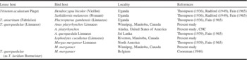
Radford (Reference Radford1949) described what he thought was the male of M. trinotoni, but this was actually a female feather mite, Rectijanua Gaud (Acariformes: Rectijanuidae) species (identified by H.C. Proctor). Considering that the male of M. trinotoni has been incorrectly described in the past, the question should be raised whether the male specimen described in this publication is the male of M. trinotoni. This specimen shares character states with the males of all other species of Myialginae and was identified as a Myialges species using keys in Fain (Reference Fain1965). This specimen was found in the material washed from a common merganser that had only M. trinotoni females parasitic on the associated host louse, T. querquedulae. Myialginae species hyperparasitic on lice show greater host specificity than those on hippoboscid flies (Fain Reference Fain1965); M. trinotoni has only been recorded from one genus of lice, Trinoton (Table 3). Taking this evidence into consideration, it is reasonable to assume this male specimen to be M. trinotoni.
Louse and bird host records for Myialges trinotoni (modified from Fain Reference Fain1965).

CNC, Canadian National Collection of Insects, Arachnids, and Nematodes.
Discussion
In this study, we report M. trinotoni from Canada (Manitoba) and United States of America (Alaska) for the first time. Although this mite was collected in North America before this study, specific localities were not provided (Thompson Reference Thompson1939; Fain Reference Fain1965). Myialges trinotoni has been collected from lice in five countries and on seven species of birds, two of which are novel host species reported in this study (Table 3). Comprehensive collections of Trinoton lice will likely reveal additional avian or louse host species records for this mite. Considering the broad geographic and avian host range of M. trinotoni, it is possible that future molecular investigations will reveal that this mite is actually a complex of cryptic species.
Myialges trinotoni appears to be quite rare in Manitoba. We found female mites attached to T. querquedulae on only three of 323 host ducks collected over a span of 23 years. The prevalence of M. trinotoni on lice from the three host ducks that had mites was high, 85% of adult T. querquedulae on the common merganser were infested, although only small numbers of adult Trinoton were present on the mallard (two lice) and the hooded merganser (one louse). Despite the high prevalence in the adult Trinoton on these three birds, the low overall prevalence on all T. querquedulae in this study invites speculation about the persistence of M. trinotoni in these populations. It is possible that dispersal on Trinoton is effective, and Myialges populations can maintain their survival. Each of the ovigerous Myialges may have many eggs suspended around them (Fig. 1), so total reproductive output may be sufficient to guarantee survival of a small population. On the other hand, these waterfowl species are all migratory, and it is possible the birds we found carrying Myialges were part of a more southern element of the host species, some part of North America where this mite is more prevalent. However, there are few records of M. trinotoni in North America (Thompson Reference Thompson1939; Fain Reference Fain1965), and given the large size of Trinoton, and the conspicuous nature of ovigerous Myialges, it is surprising it has not been reported more frequently if prevalence was higher elsewhere. Myialges trinotoni does infest other species of Trinoton (Table 3), though we did not find any T. anserinum from geese or swans infested in our study. This species of chewing louse was rarely collected, and given the low prevalence of Myialges associated with T. querquedulae on ducks, our sample sizes may have been too small to detect its occurrence.
The common merganser in our study was infested by 17 males, 10 females, and 23 nymphs of T. querquedulae, yet only adult lice (23 of 27) were infested with M. trinotoni. Female mites do not appear to differentiate between male and female lice, but we do not know whether mites are able to distinguish between adults and nymphs. If a female mite attached to a nymph, it could possibly be shed with the cuticle at the next moult. If this is the case, it is not surprising we found no infested nymphs, though it is still possible female mites are able to recognise adult lice and attach only to them. The factors that influence louse host selection by Myialges females are unknown, as are many ecological features in the life history of these fascinating mites.
Acknowledgements
The authors thank the hospital staff at the Wildlife Haven (Manitoba Wildlife Rehabilitation Organization) and the Prairie Wildlife Rehabilitation Centre for their care in receiving birds, and their careful triage and processing protocols to maintain specimens in the best condition possible for this study. Some of the mallards and Canada geese were examined by Alexandra Grossi for her M.Sc. thesis research. They also thank Dave Holder, Lisa Babey, and a long list of undergraduate summer research assistants for their help in washing birds; and the Department of Entomology and the Faculty of Agricultural and Food Sciences for their continued support. The authors thank Miles Zhang for imaging the louse infested with mites, and Pavel Klimov for his input on the illustrations. Funding was provided to T.D.G., in part, by a Discovery Grant from the Natural Science and Engineering Council of Canada.