Introduction
As part of ongoing studies on the world Agromyzidae, the author has been attempting to clarify taxonomic concepts and detail explicit synapomorphies at the genus-group level in order to construct more reliable and robust keys and diagnoses that are not as dependant on genitalic dissection or larval features, facilitating easier specimen identification. As a result of these studies, it has become apparent that existing generic concepts are partially inconsistent and overlapping, and as such, the definition and monophyly of many genera are in need of re-evaluation. In this paper, genus groups in the subfamily Agromyzinae are treated, more specifically, those in the Ophiomyia genus group (Dempewolf Reference Dempewolf2005), which is a single lineage in the subfamily encompassing all taxa except for Agromyza Fallén, 1810 and Japanagromyza Sasakawa, 1958. Cecidomyiaceltis Patton, 1897 is also a component of the basal Agromyzinae, but a synonymy of this genus is being prepared by M. vonTschirnhaus following the discovery of an adult male by R.J. Gagne of the Smithsonian (M. vonTschirnhaus, personal communication).
In the short term, this paper is necessary to establish more explicitely supported and easily diagnosed genus-level groups, which will facilitate the development of identification tools for publication in the upcoming Manual of Afrotropical Diptera. Genus keys will be provided in this book chapter and in an additional genus-level treatment of the family, which is presently in development. Over the long term, it is hoped that this introductory study will facilitate more explicitely quantitative phylogenetic investigations into the Agromyzinae by better elucidating synapomorphies and genus-level boundaries using the more complex, numerous, and historically reliable adult and male genitalic data sets. When a more robust phylogeny of this species-rich subfamily has finally been developed, the reconstruction of patterns in host choice and feeding habits should also prove to be rewarding.
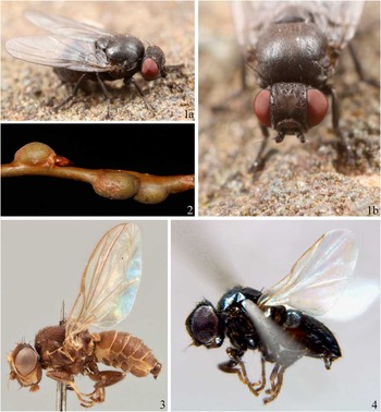
Hexomyza Enderlein, 1936, Ophiomyia Braschnikov, Reference Braschnikov1897, and Melanagromyza Hendel, Reference Hendel1920, which have been quite well studied in the north temperate regions, have been treated as a single lineage in many publications on the Agromyzinae. These genera consist of a number of mostly compact, dark species with a similar habitus (Figs. 1, 3–5); a large, bulging and well-sclerotised lunule with a medial groove (also seen in some Japanagromyza); an antenna inserted below the midpoint of the head; halters that are brown (white in the Afrotropical O. aberrans Spencer, Reference Spencer1959, white or partially white in some Melanagromyza, knob dorsally yellow in O. punctohalterata (Frost, 1936) and O. maculata Spencer, 1981, brown in a few Agromyza, and partially to entirely brown in many Japanagromyza); and no prescutellar acrostichal bristles (also absent in some Japanagromyza). Internally, the subepandrial sclerite is divided into two broad, bare, well-sclerotised plates that almost meet at a narrow point dorsomedially (Figs. 45, 47), the inner lobe of the hypandrium has partially separated into an arched, band-like sclerite (Fig. 12), and there is a small lobate projection basally on the stem of the ejaculatory apodeme (Fig. 15) (also found in Japanagromyza). Furthermore, the insertion point of the mesophallus is often shifted ventroapically on the distiphallus (compare subbasal insertion in Fig. 17 to ventromedial insertion in Fig. 49), although the ancestral state of a straight, linearly arranged mesophallus and distiphallus is seen in some species, including several Ophiomyia discussed below, likely representing a secondary recovery. Species, and sometimes genera, can often be difficult to differentiate, usually requiring the examination of genitalic and/or very subtle external characters that are often variable and/or morphometric in nature.
1a–b. Euhexomyza schineri (Giraud), Massachusetts, United States of America; 2. Twig galls on Poplar, Massachusetts, United States of America; 3. Ophiomyia aloes (Spencer), nontype male; 4. O. arabica (Deeming).

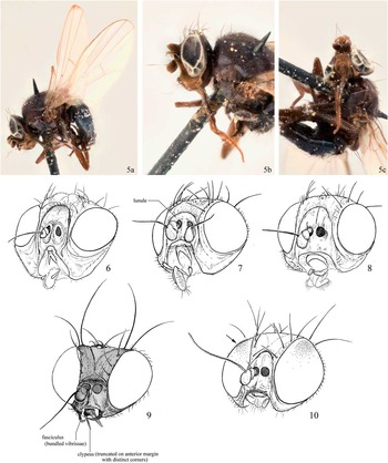
5a–c. Ophiomyia longilingua (Hendel), holotype; 6. Ophiomyia aloes (Spencer); 7. O. sarothamni (Hendel); 8. Euhexomyza simplicoides (Hendel); 9. Ophiomyia species [note gena produced anteriorly and facial carina with central bulb]; 10. Melanagromyza metallica (Thompson) [arrow pointing to pilosity on eye].

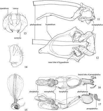
Ophiomyia phaseoli (Tyron) male genitalia; 11. hypandrium, epiphallus and phallapodeme, left lateral; 12. hypandrium, epiphallus and phallapodeme, ventral; 13. external genitalia, posteroventral; 14. external genitalia, left lateral; 15. ejaculatory apodeme [arrow indicating basal process on stem]; 16. phallus, ventral; 17. phallus, left lateral.

A number of more recently described genera from the Old World tropics are also attributable to this group, including Epidermomyia Ipe and Ipe, Reference Ipe and Ipe2004 (type species: Agromyza atomella Malloch, 1914), Kleinschmidtimyia Spencer, Reference Spencer1986 (type species: Melanagromyza pisi Kleinschmidt, 1961), Penetagromyza Spencer, Reference Spencer1959 (type species: P. aloes Spencer, Reference Spencer1959), and Tropicomyia Spencer, Reference Spencer1973 (type species: M. flacourthiae Séguy, 1951).
Two factors appear to be dominant in contributing to historical difficulties in the taxonomy of the Ophiomyia genus group, aside from a general paucity of useful morphological features and its pronounced species richness. The first is that definitions and higher relationships have rarely been discussed in a cladistic framework, which is very useful in locating clear, reliable synapomorphies useful for diagnosis. Contrary to this approach, authors have often used suites of homoplastic, nondiscreet, and ill-defined characters that must be observed in combination. These are often unfortunately subject to varied interpretation, providing a difficult gestalt for readers to use. The morphological analysis by Dempewolf (Reference Dempewolf2001), and molecular analyses such as those provided in Scheffer et al. (Reference Scheffer, Winkler and Wiegmann2007) and Winkler et al. (Reference Winkler, Scheffer and Mitter2009) are exceptions to the rule, and have done much to elucidate genus and species-level relationships. Many of these historical approaches can be understood, however, considering the difficulties associated with morphological studies in this group, and the author is still only able to provide a single character for the definition of Ophiomyia.
The second contributing factor is that plant host preference and feeding behaviour have been weighted disproportionately in establishing generic boundaries, with authors bending classification to reflect larval habits. The reliance on larval habits/hosts for use in classification over phylogenetic considerations is very evident in the placement of species in numerous agromyzine genera (as discussed below), as well as for assumptions of evolutionary “trends” in the family, which have led some authors to propose genus-level relationships not intuitive from morphology, affecting assumptions on homology and ancestral states. An insightful discussion on this latter topic is provided in Scheffer et al. (Reference Scheffer, Winkler and Wiegmann2007: 757). The added use of other more complex and often more informative data sets such as those of male genitalic morphology has already been discussed by others, including Griffiths (Reference Griffiths1974), who has greatly advanced our knowledge of phytomyzine systematics by incorporating these characters into classifications initially developed using immature features. As illustrated below, while broad patterns of feeding or larval behaviour are evident across genera, they are not necessarily predictive of evolutionary relationships and can be misleading when developing classification. Initial progress is now revealing that while clusters of species are indeed relatively loyal to a related group of host plants such as a genus or family, occassional shifts onto novel host groups followed by a period of subtle to pronounced adaptation and diversification appear to be expected, as illustrated in Winkler et al. (Reference Winkler, Scheffer and Mitter2009). There are also numerous difficulties in outlining discreet character states for feeding, mining, and life history strategies, as will be discussed in more detail below. Despite this, much groundwork has been made over the years, and minutiae associated with immature stages may eventually prove partially useful in redefining higher taxa in a phylogenetic context built on a framework also incorporating evidence derived from sources such as morphological character sets and molecular sequence data. These immature and life history characters, which have accumulated through decades of careful and laborious observation, are certainly more insightful towards terminal branches in the agromyzid phylogeny, and are key to understanding species-level dynamics. Towards this end, impressive contributions have been made by a number of Dipterists, including the prolific authors Griffiths, Hering, Hendel, Spencer, and deMeijere, to name only a few.
In an effort to clarify genus concepts in the Agromyzinae, evidence is provided below to more clearly delineate monophyletic entities in the Ophiomyia genus group. The placement of type species within these entities is then determined to provide appropriate genus names and junior synonyms. The most significant act made here is the redefinition of Ophiomyia, which has impacts on the status of Kleinschmidtimyia, Penetagromyza, and Hexomyza. Following the clarification of these genera, Melanagromyza can be more readily diagnosed, and one discreet lineage of gall-makers can be recognized as distinct and provided with a new genus name. Lastly, Tropicomyia and Epidermomyia are clarified because their boundaries are entwined, but full species-level resolution of Tropicomyia is outside the scope of this study and will necessitate additional examination, possibly with the description of one or more new genus groups.
Materials and methods
Genitalia were prepared by macerating the abdomen in hot potassium hydroxide (10% solution) for ~5–10 minutes, followed by washing in glacial acetic acid and demineralised water. Genitalia were stored in glycerin in microvials pinned with the specimen. Genitalic figures illustrate the internal components proportionally to each other, but the external components (epandrium, surstylus, subepandrial sclerite) are reduced. Terminology follows that outlined in Lonsdale (Reference Lonsdale2011), largely following the terminology of McAlpine (Reference McAlpine1981), except ori is an abbreviation for the inclinate anterior fronto-orbitals, and ors is an abbreviation for the reclinate posterior fronto-orbitals.
Material was examined from the following collections, or are collections in which relevant type material is deposited: The Natural History Museum, London, United Kingdom (BMNH); California Academy of Sciences, San Francisco, California, United States of America (CAS); The Canadian National Collection of Insects, Arachnids and Nematodes, Ottawa, Ontario, Canada (CNC); National Museum (Natural History), Prague, Czech Republic (NMPC); Naturhistorisches Museum Wien, Wien, Austria (NMW); National Museum of Wales, Cardiff, United Kingdom (NMWC); Landcare Research, New Zealand Arthropod Collection, Auckland, New Zealand (NZAC); Osaka Museum of Natural History, Osaka, Japan (OMNH); National Museum of Natural History, Washington, DC, United States of America (USNM); Museum für Naturkunde, Berlin, Germany (ZMHU). A full list of material examined is provided at the end of the manuscript.
Redefinition and diagnosis of Ophiomyia Braschnikov
Ophiomyia Braschnikov, Reference Braschnikov1897: 40 [as subgenus of Agromyza]. Type species: Agromyza maura Meigen, 1838 [misidentified as A. pulicaria Meigen, 1830 – see Hendel (Reference Hendel1931: 188) and clarification in Spencer and Martinez (Reference Spencer and Martinez1987: 256), who note that this has not been formally ratified by the Internation Commission on Zoological Nomenclature], by monotypy. Hendel Reference Hendel1920: 114 [status as genus], 1931: 180; Frick Reference Frick1952: 375; Spencer Reference Spencer1964: 775, 1966: 47, 1969: 81; Spencer and Steyskal Reference Spencer and Steyskal1986: 37; Černý Reference Černý1994: 455 [key to European species].
Tylomyza Hendel, Reference Hendel1931: 181 [as subgenus of Ophiomyia]. Type species: Madiza pinguis Fallén, 1820, by original designation. Enderlein Reference Enderlein1936a: 179 [as genus]. Synonymised in Spencer (Reference Spencer1964).
Stiropomyza Enderlein, Reference Enderlein1936a: 179. Type species: Phytomyza aeneonitens Strobl, 1893, by monotypy. Synonymised in Frick (Reference Frick1952).
Siphonomyza Enderlein, Reference Enderlein1936a: 179. Type species: Agromyza proboscidea Strobl, 1900 [now a junior synonym of A. curvipalpis Zetterstedt, 1848], by monotypy. Synonymised in Frick (Reference Frick1952).
Aulomyza Enderlein, Reference Enderlein1936a: 179. Type species: Melanagromyza longilingua Hendel, Reference Hendel1920, by monotypy. Frick Reference Frick1952: 375 [as synonym of Melanagromyza]. Synonymised in Spencer (Reference Spencer1966) [as synonym of Ophiomyia].
Siridomyza Enderlein, Reference Enderlein1936a: 179. Type species: Ophiomyia madizina Hendel, Reference Hendel1920 [now a junior synonym of A. nasuta Melander, 1913], by monotypy. Synonymised in Frick (Reference Frick1952) [as synonym of Tylomyza].
Solenomyza Enderlein, Reference Enderlein1936a: 179. Type species: Melanagromyza rostrata Hendel, Reference Hendel1920, by monotypy. Frick Reference Frick1952: 375 [as synonym of Melanagromyza]. Synonymised in Spencer (Reference Spencer1966) [as synonym of Ophiomyia].
Stirops Enderlein, Reference Enderlein1936a: 179 [nomen nudum – no type species designated].
Stirops Enderlein, Reference Enderlein1936b: 42. Type species: Ophiomyia submaura Hering, 1926, by original designation [Enderlein Reference Enderlein1936b: 42]. Synonymised in Frick (Reference Frick1952).
Triopisopa Enderlein, Reference Enderlein1936a: 179 [nomen nudum – no type species designated].
Triopisopa Enderlein, Reference Enderlein1936b: 42. Type species: Agromyza simplex Loew, 1869, by original designation. Frick Reference Frick1952: 376 [as synonym of Melanagromyza]. Synonymised in Spencer (Reference Spencer1966) [as synonym of Ophiomyia]. Synonymised in Tschirnhaus (Reference Tschirnhaus2000) [as synonym Hexomyza].
Carinagromyza Sasakawa, Reference Sasakawa1954: 23. Type species: Carinagromyza heringi Sasakawa, Reference Sasakawa1954 [now a synonym of Ophiomyia sasakawai Spencer and Martinez, Reference Spencer and Martinez1987], by original designation. Synonymised in Spencer (Reference Spencer1966).
Hexomyza Enderlein, Reference Enderlein1936a: 179 [nomen nudum – no type species designation].
Hexomyza Enderlein, Reference Enderlein1936b: 42. Type species: Melanagromyza sarothamni Hendel, 1923, by original designation. Hendel Reference Hendel1936: 570; Frick Reference Frick1952: 375 [as synonym of Melanagromyza]; Spencer Reference Spencer1966: 38, 1969: 79; Spencer and Steyskal Reference Spencer and Steyskal1986: 34; Tschirnhaus Reference Tschirnhaus2000: 137. New synonym.
Penetagromyza Spencer, Reference Spencer1959: 253. Type species: P. aloes Spencer, Reference Spencer1959, by original designation. Spencer Reference Spencer1990: 390, Spencer Reference Spencer1991: 57. New synonym.
Kleinschmidtimyia Spencer, Reference Spencer1986: 249. Type species: Melanagromyza pisi Kleinschmidt, 1961, by original designation. Spencer Reference Spencer1990: 391. New synonym.
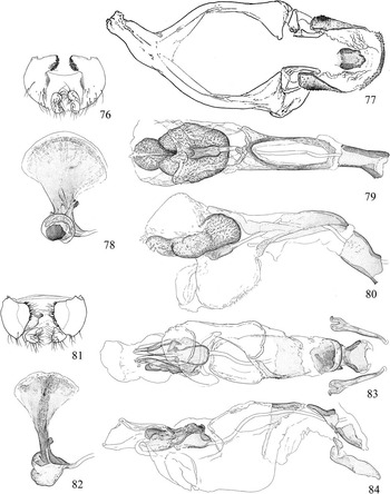
Ophiomyia can be a difficult genus to delineate. While many species can be readily placed following recognition of a produced male gena that often bears an apical “horn” of clustered vibrissae (also found in a reduced state in some females), sometimes called a “fasciculus” [Latin meaning bundle, sheaf] (Fig. 9); many more cannot be differentiated from similar genera externally aside from having a facial carina that sometimes has a bulging medial tubercle/knob. This carina is absent in a minority of species (Spencer Reference Spencer1964), however, and is sometimes seen as a faint or partial ridge in other genera. Complicating matters, some species have a faint metallic sheen, which is otherwise characteristic of many Melanagromyza. Internally, Ophiomyia is supposed to contrast Hexomyza and Melanagromyza in having an asymmetrical, sometimes rotated “distiphallus complex” and one pair of long basiphallus arms, but the former is also not always present, and the latter is not present in all species and sometimes found in other genera. The compartmentalised, bladder-like distiphallus seen in most Ophiomyia is also not cosmopolitan, with many unusual phallic forms included in the genus; this type of distiphallus often has the internal spinules reduced to rounded irregular mounds (Figs. 79–80), which are also found in some Melanagromyza (Figs. 49–50). Also present in many species is one pair of broad sclerotised lobes flanking the proepiphallus (also frequently seen elsewhere in the genus group) (Fig. 16), and a large, plate-like, subovate sclerite originating from the reduced metepiphallus (Figs. 30, 77), which in some Ophiomyia (Fig. 12) and other genera is developed as a box-like enclosure around the phallophorus and proepiphallus (Figs. 58–59), although a reduced metepiphallus and highly atrophied proepiphallus is seen in some species of Melanagromyza (Fig. 49).
As a result of this ambiguity in generic boundaries, the classification of Ophiomyia species has been subject to varied interpretation, beginning with the description of Tylomyza by Hendel (Reference Hendel1931) for two unusual species with proclinate orbital setulae and no upper orbital setae in males, and then an unfortunate proliferation of genus-level names by Enderlein (Reference Enderlein1936a, Reference Enderlein1936b) soon thereafter. Sasakawa’s (Reference Sasakawa1954) concept of the genus resulted in Carinagromyza, but this name, along with the others noted above, have since been synonymised by Frick (Reference Frick1952) and Spencer (Reference Spencer1964, Reference Spencer1966) to produce the single entity presently understood as Ophiomyia.
Following dissection and analysis of species from all biogeographic regions, the only character consistently found in species of Ophiomyia is an apically truncate clypeus, which is also often narrow and parallel-sided (Figs. 7, 9). This state is exaggerated in those species with a produced gena, where the clypeus has become extremely narrowed anteriorly, sometimes almost tapering to a point. In some species, the medial margin is also slightly concave to shallowly notched. This truncate clypeus contrasts with the rounded clypeus seen in the rest of the subfamily, which is often quite broadly arched and U-shaped (Figs. 8, 10). A few Melanagromyza have the anterior margin of the gena slightly to distinctly truncate, but these specimens are readily characterised – the anterior margin of the clypeus is usually without corners and the margins grade into a curve, and the entire body is distinctly metallic; furthermore, the eye is setose, often conspicuously so, being particularly dense dorsally in males (Fig. 10). In Ophiomyia, the eye is always either bare, or sparsely and inconspicuously setose. The recognition of this character is significant in that it allows for diagnosis of Ophiomyia without dissection, and has a number of important ramifications in the status of other agromyzine genera.
Exceptions to the rule. One important caveat to this new definition is that not all species of Ophiomyia have yet been examined. It is expected that exceptions will exist, and so far the females of O. orbiculata (Hendel, Reference Hendel1931) (host: several Fabaceae species) appear to have a clypeus that is slightly truncate to fully rounded. Furthermore, several taxa with elongate mouthparts, including the Australasian lineage containing O. centrosematis (Meijere, 1940) (external stem miner on some Fabaceae), O. mussauensis Spencer, Reference Spencer1966 and O. rotata Spencer, 1965, and the European lineage composed of O. longilingua (Hendel, Reference Hendel1920) and O. rostrata (Hendel, Reference Hendel1920), have a clearly rounded clypeus in both sexes with that of O. longilingua shallowly emargined anteromedially (almost heart-shaped – see Fig. 5c). While these species could be justifiably removed from Ophiomyia, leading to the resurrection of Aulomyza and/or Solenomyza, the current uncertaintly associated with their placement and their potential affinities to other more reliably placed Ophiomyia (discussed below) makes it more prudent to retian them in Ophiomyia until more extensive evidence can be gathered to support or counter this classification. As such, the rounded clypeus in this subset of taxa will be treated here as a secondarily recovered plesiomorphic state.
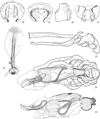
Of the Ophiomyia centrosematis lineage, males of O. rotata have been examined. The terminalia (Figs. 18–25) are revealing, suggesting a broader relationship with other Ophiomyia with a ventral connection between the arms of the basiphallus, a ventroapical plate-like extension of the basiphallus that encloses part of the mesophallus, a linear/subcylindrical mesophallus and distiphallus, and a distiphallus that is deeply to partially split apically, such as O. pisi, O. phaseoli (Tyron, 1895), and interestingly, O. orbiculata, which as mentioned above, includes some representatives with a truncated clypeus.
Ophiomyia rotata Spencer, male genitalia; 18. external components, posterior; 19. external components, anterior; 20. external components, left lateral; 21. external components, ventral; 22. ejaculatory apodeme; 23. hypandrium, phallapodeme, and epiphallus, left lateral; 24. phallus, hypandrium, and epiphallus, ventral; 25. phallus, left lateral.

Females of both species in the Ophiomyia longilingua lineage have been examined, plus the undissected male holotype of O. longilingua (Fig. 5a–c). Enderlein (Reference Enderlein1936a) designated O. longilingua (stem miner of Knautia (Dipsacaceae) with external pupation) as the type species for his genus Aulomyza based on the presence of elongate mouthparts, but these are commonly found elsewhere in Ophiomyia and the genus was not accepted by subsequent authors. Enderlein (Reference Enderlein1936a) also designated O. rostrata (host unknown) as the type species for Solenomyza based on the presence of three dorsocentrals, but this genus was also not accepted by previous authors, and examined specimens have revealed only two pairs of dorsocentrals. Both of these species were transferred to Melanagromyza by Frick (Reference Frick1952) and then to Ophiomyia by Spencer (Reference Spencer1966). Spencer (Reference Spencer1966) also supported a sister-group relationship between the two species, noting their similarly elongate mouthparts, anteriorly angled gena, and reduced chaetotaxy on the mid tibia (0–1 medial bristles). The phallus of O. rostrata was later described by Spencer (Reference Spencer1976) when he located a male, and the genitalia reinforce his proposed sister-group relationship: distiphallus dark, subcylindrical; mesophallus short, clear apically; hypophallus small, dark, distally directed; basiphallus short, broad. The linear orientation of the mesophallus and distiphallus may be phylogenetically significant, as this defining character of the Ophiomyia genus group is also absent in those species mentioned above in the O. centrosematis lineage and other putatively related Ophiomyia such as O. phaseoli.
This new genus concept of Ophiomyia unfortunately relies on a single character, albeit internally consistent, and so far, reliably predictive of generic standing excluding those few species mentioned above. It is hoped that additional characters can be found to test its use and the potential of parallel development or secondary loss, and should be accompanied by a phylogenetic analysis. One explaination for the presence of only a single defining character among an unusually diverse Ophiomyia can be found in the molecular results of Scheffer et al. (Reference Scheffer, Winkler and Wiegmann2007), which recovered two separate lineages of Ophiomyia. Hexomyza schineri (Giraud, Reference Giraud1861) and Tropicomyia theae (Cotes, 1896) were recovered with “Ophiomyia A” (which includes the type species and other “typical” Ophiomyia with a produced gena and usually no ventral connection of the basiphallus arms), and Melanagromyza was recovered with “Ophiomyia B”. “Ophiomyia B” includes O. phaseoli, which it may be noted, has only a very shallowly truncated clypeus, an “atypical” phallus including a symmetrical mesophallus/distiphallus and ventroapically united arms of the basiphallus (Figs. 16, 17), and was previously treated by several authors as Melanagromyza. In the event of a nonmonophyletic Ophiomyia, the synapomorphy of a truncate clypeus and facial carina would be added to the defining suite of characters here used for the genus group, and would be interpreted as secondarily lost in one or more genera.
Synonymy of Kleinschmidtimyia with Ophiomyia
One ramification of the redefinition of Ophiomyia is a change in the status of Kleinschmidtimyia. Because examined species (K. dianellae (Kleinschmidt, 1961), K. pisi, and K. wikstroemiae (Kleinschmidt, 1961)) have an anteriorly truncate clypeus, Kleinschmidtimyia new synonym is here determined to represent a component lineage of the larger Ophiomyia. Like most other Ophiomyia, examined specimens also have the supra-alar seta slightly larger than the intra post-alar (although both are relatively hair-like), the dorsum of the lunule is sharply bent inwards (as in Fig. 9), and in K. pisi the gena is slightly produced anteriorly (more so than some Agromyzinae such as Melanagromyza buccalis Spencer, Reference Spencer1969, but less than in those Ophiomyia with a produced gena). This lineage is morphologically atypical of most other representatives of the genus, and could be revealed as a basal offshoot of Ophiomyia when more is understood of the evolutionary history of the genus; this could result in subgeneric classification if the internal structure of Ophiomyia lends itself to subgeneric division. As a result of this synonymy, all eight species of Kleinschmidtimyia are here transferred to Ophiomyia (all have been classified as Ophiomyia at one point previously with the exception of the first taxon listed): O. arabica (Deeming, 2006) new combination; O. dianellae; O. gemina Spencer, Reference Spencer1977; O. indigoferae (Kleinschmidt, 1961); O. nuginiensis (Spencer, Reference Spencer1977); O. pisi; O. paramonovi (Spencer, 1963); and O. wikstroemiae.
The male terminalia of these species (Figs. 26–40) reveal most to represent a single derived lineage, with the exclusion of O. wikstroemiae. These species are almost entirely black with the calypter fringe and setae white, wing length is ~1.3–2.1 mm, most veins yellow to white behind the costa, there are three ors and one ori, the face is flat, the supra-alar seta and the intra post-alar are reduced (nearly absent in O. arabica), the tibiae have no medial bristles, and the larvae are miners in the upper leaf layer with pupation in the leaf. The male terminalia are characterised by a broad, heavily spinulose surstylus (Figs. 33–35), a distiphallus that emerges subapically from a narrow mesophallus (Figs. 38–39), a thin, nearly ring-like basiphallus, and a distiphallus with a bowl-like base, a small ventral plate (reduced in O. arabica), and one pair of thin apical tubules that usually fuse before the midpoint (at midpoint in O. paramonovi and near apex in O. nuginiensis; unified as a single tubule in O. arabica).
Ophiomyia arabica (Deeming), male genitalia; 26. external genitalia, anterior; 27. external genitalia, left lateral; 28. external genitalia, ventral; 29. hypandrium and epiphallus, left lateral; 30. hypandrium and epiphallus, ventral; 31. phallus, ventral; 32. phallus, left lateral; 33–40. O. pisi (Kleinschmidt), male genitalia; 33. external genitalia, anterior; 34. external genitalia, left lateral; 35. external genitalia, ventral; 36. hypandrium, epiphallus, and phallapodeme, left lateral; 37. hypandrium and epiphallus, ventral; 38. phallus, ventral; 39. phallus, left lateral; 40. ejaculatory apodeme; 41–43. O. aloes (Spencer), male genitalia; 41. external genitalia, ventral; 42. phallus, ventral; 43. ejaculatory apodeme.

The combination of an entirely white calypter and three ors+one ori is very uncommon, making this lineage relatively easy to diagnose. A white calypter is also seen in the European Ophiomyia aeneonitens (Strobl, 1983) (fronto-orbital number unknown), which has a similar phallus and is likely related. Nomenclaturally this is significant in that O. aeneonitens is the type species of the genus Stiropomyza (based on an absence of vein r-m and a costa that only extends to R4+5 (Enderlein Reference Enderlein1936a)); if a phylogenetic reconstruction of Ophiomyia finds Kleinschmidtimyia to be separate but inclusive of this species, the name Stiropomyza will have priority.
Despite these (putatively) apomorphic characters, erection of the genus appears to have been based primarily on the leaf mining habits of the species, which originally removed them to Tropicomyia (Spencer, Reference Spencer1973). When their clearly discordant morphology eventually forced Spencer (Reference Spencer1977) to remove them from that genus, they were accommodated in Ophiomyia because their leaf mining behaviour excluded the possibility of a return to Melanagromyza (the original combination of most included species), which by the time of Spencer (Reference Spencer1986) explicitely excluded all leaf-mining species. As unique as the species of Kleinschmidtimyia are, there is presently no evidence to support the lineage as anything other than an apomorphic (possibly even basal branch) of Ophiomyia, and it cannot be maintained as a separate entity while there is evidence to place it within the existing genus.
Other unrelated Ophiomyia species already accepted as leaf miners (mostly on dicots) are relatively numerous, suggesting that this life history trait is widespread, and that recurrent development of a leaf-mining taxon from a stem-mining ancestor (or vice versa) may be relatively unremarkable over small stretches of evolutionary space. New World taxa include O. camarae Spencer, 1963, which forms mines in Lantana Linnaeus (Verbenaceae) along the midrib with additional channels along the lateral veins; with the puparium in the mine. South American species of the O. pfaffiae group (see Spencer (Reference Spencer1990)) also pupate in the leaf, and include O. cabanae Valladares on Croton Linnaeus (Euphorbiaceae) and O. pfaffiae (Spencer, 1963) on Pfaffia Martius (Amaranthaceae). An unusual species of uncertain affinity from Guadeloupe is O. phaseoloides Martinez, 1992 (Spencer et al. Reference Spencer, Martinez and Etienne1992), which mines in Phaseolus Linnaeus (Fabaceae) and pupates on the lower surface of the leaf. The Japanese O. kwansonis Sasakawa, now invasive in the United States of America on Hemerocallis Linnaeus (Xanthorrhoeaceae) (Williams and Steck Reference Williams and Steck2011), forms irregular linear mines that sometimes cross the midrib; pupation is in the mine. The Old World O. phaseoli attacks numerous Fabaceae, and may represent a taxon inhabiting the transitional state between mining types, being found in both the stem and leaf; pupation is in the leaf near the base of the stem (Spencer Reference Spencer1973).
An unusual representative of the former Kleinschmidtimyia is Ophiomyia arabica (Figs. 4, 26–32), which despite its unusual appearance, appears to belong to the main lineage of species discussed above. This species has milky-white wings with vein dm-cu missing, the costa only extends to R4+5, the radial veins are inserted closer to the base of the costa, and the colour is black (not dark brown) with the veins and male tarsi pale brown. The clypeus is also relatively narrow and parallel-sided, the parafacial and orbital plate form a heavily sclerotised, glossy ring around the eye; the ocellar triangle is unusually long with a beaded edge and is similarly sclerotised and glossy; the postverticals are absent; the hypandrium has an elongate distal apodeme; and there are numerous rows of long stout spines on the surstylus (Figs. 26–28). Furthermore, while the male face is thinly carinate and the lunule is shiny, flat and erect, the female has a broad, shallow bulb between the antennal bases and the lunule is strongly angled inwards, as in other Ophiomyia. Despite this unusual confluence of characters, there is a clear, possibly synplesiomorphous morphological similarity to O. phaseoli, to which the former representatives of Kleinschmidtimyia may be related. In both O. phaseoli and O. arabica the ocellar triangle is similarly glossy and very elongate, strongly contrasting the soft surrounding frons, and there are similarities in the structure of the basiphallus (particularly the unusual ventrodistal union of the arms), mesophallus (subbasal insertion into distiphallus), and distiphallus (ventral lobe joining dorsomedial extension from basal bowl). While external uniformity is often the norm in Ophiomyia, O. arabica illustrates that this is not always the case, and will prove relevant when examining the evolutionary relationships of Penetagromyza, which was also partially defined on absence of the postverticals and a costa that only extends to R4+5.
Lastly, with regards to Ophiomyia wikstroemiae, this species differs considerably in external and male genitalic morphology (see Spencer (Reference Spencer1977: figs. 161–163)) and is likely to be unrelated to the other former Kleinschmidtimyia. It was clearly placed in Kleinschmidtimyia because of its larval leaf-mining habits. Aside from this species, only Liriomyza Mik, 1894 are otherwise known from Thymelaeaceae (Benavent-Corai et al. Reference Benavent-Corai, Martinez and Jiménez Peydró2005).
Synonymy of Penetagromyza Spencer with Ophiomyia
Examination of Penetagromyza aloes (Figs. 3, 40–43), the type species for Penetagromyza new synonym, has revealed this genus to also be a junior synonym of Ophiomyia. The clypeus is not only truncate anteriorly, but it is strongly narrowed and the anteromedial margin of the buccal cavity has a three-lobed notch to accommodate the narrowed clypeus (Fig. 6). This particular type of clypeus is usually only otherwise seen in those Ophiomyia with a strongly produced gena and vibrissal horn, as well as a few of their allies, although the emargination in the buccal cavity is usually squared (not trilobed) (Fig. 9). The small, gracile bristles are also characteristic of many congeners, as are the genitalia, with Spencer (Reference Spencer1990) noting that “[t]he male genitalia, with the aedaegus membranous and asymmetrical… appear characteristic of Ophiomyia”. All species of Penetagromyza are subsequently recombined as follows: Ophiomyia aloes (Spencer, Reference Spencer1959) new combination; O. similans (Spencer, 1961) new combination; O. aloephaga (Spencer, Reference Spencer1990) new combination.
Unlike many Ophiomyia, however, the lunule of examined O. aloes is very high, flat and subshiny (not strongly bent near the base), although most of the lunule is strongly angled inwards and enclosed by the anterior margin of the uniquely soft, pale, billowy frons (Fig. 6). The shape of the distiphallus is also characteristic, particularly the single projecting sclerotised fold on the left lateral margin (Fig. 34). The species previously treated as Penetagromyza also conspicuously differ in having vein dm-cu and sometimes the postverticals reduced, as also seen in the unrelated and newly synonymised O. arabica (see above), and feeding occurs on the monocot genus Aloe Linnaeus (Aloaceae), where “larval feeding [is] deep within the thick leaf and internal pupation [is] immediately below the epidermis” (Spencer Reference Spencer1990).
Synonymy of Hexomyza with Ophiomyia
Enderlein (Reference Enderlein1936a) erected 13 new genera of Agromyzidae following Hendel’s (Reference Hendel1931) thorough treatment of the family. Some of these names were nomina nuda because no type species was designated, but these were soon validated by subsequent designation in a later publication (Enderlein Reference Enderlein1936b). Like many other genera he described throughout the Diptera, most of these names fell to synonymy, with only three agromyzid genera presently remaining: Aulagromyza, Hexomyza, and Metopomyza. As foreshadowed by Cresson (Reference Cresson1930):
“Judging from the characterization of his genera, it is evident that Enderlein failed to grasp the essential characters, so that even if most of the genera he proposed will prove to be tenable, they may not stand on any of the characters originally given; and in some cases an entirely different concept will have to be formed in order to be consistent, or satisfied to their validity”.
Hexomyza was originally erected for three species of Melanagromyza described by Hendel with a third dorsocentral and a costa that ended at M1: M. hexachaeta Hendel, Reference Hendel1931, M. orbiculata, and M. sarothamni (the type species of Hexomyza). This action was criticised by Hendel (Reference Hendel1931), and later Frick (Reference Frick1952) included Hexomyza as a junior synonym of Melanagromyza, along with a number of other Enderlein genera. When Spencer (Reference Spencer1964) reapproached Hexomyza, he placed M. orbiculata in Ophiomyia, and included M. hexachaeta as its junior synonym. Melanagromyza sarothamni, however, was again treated as Hexomyza in his new concept of the resurrected genus (Spencer Reference Spencer1966), which included a total of four Palaearctic gall-inducers. Hexomyza, last summarised by Zlobin (Reference Zlobin1998), has since grown to encompass 15 species from all biogeographic regions except for the Neotropics. Tschirnhaus (Reference Tschirnhaus2000) later included the Holarctic and Hawaiian O. simplex in Hexomyza, and rendered Triopisopa (type species: Agromyza simplex) a junior synonym of Hexomyza. Since the present study can find no evidence to ally this unusual species with any other taxon ever allied to Hexomyza aside from a similarly enlarged orbital plate, this type species, and the genus name Triopisopa with it, are once again returned to Ophiomyia.
In Spencer’s definition, Hexomyza, similar to Ophiomyia and Melanagromyza, has a wing length of 2.0–3.0 mm, the costa ends either at R4+5 or M1, there are two or three dorsocentrals, the proboscis is short (compared with the longer state seen in some Ophiomyia) and the halter is entirely dark. Also like many Ophiomyia, the basiphallus is asymmetrical and open ventrally (although it is here seen to be mostly reduced to a dorsobasal sclerite, not “with two distinctly diverging side arms” (Spencer Reference Spencer1966)); furthermore, the posterior larval spiracle has three bulbs on a short or raised stalk that may or may not project through the host epidermis during pupation, and a gall is induced in the twig of the host plant. This last character of gall-inducing, however, was not considered to have generic significance by Zlobin (Reference Zlobin1998), who suspected the nonmonophyly of Hexomyza, and Dempewolf (Reference Dempewolf2005) reported gall production in no fewer than seven genera of Agromyzidae: Agromyza, Japanagromyza, Hexomyza, Melanagromyza, Ophiomyia, Phytoliriomyza Hendel, Reference Hendel1931, and Phytomyza Fallén, 1810.
Part of the difficulty with the use of feeding strategies in the Agromyzidae as either diagnostic or phylogenetically informative characters, is that the boundaries between true leaf mining, stem mining/boring, and gall inducing (or a “swollen mine”) is quite blurred, nondiscreet, and homoplasious (Dempewolf Reference Dempewolf2005), defying their confident usage. Even Spencer (in Spencer and Steyskal (Reference Spencer and Steyskal1986)) later decided that the “formation of galls is not a valid character for generic delimitation”, correcting the transfer of Agromyza tiliae Couden, 1908 (forming twig galls on Tilia americana Linnaeus; Tiliaceae) to Hexomyza, instead treating it as Ophiomyia. Two years earlier, Spencer and Hawkins (Reference Spencer and Hawkins1984) even described a Californian species forming bud galls on Atriplex (Chenopodiaceae) as Ophiomyia atriplicis Spencer, 1984. Furthermore, morphological characters derived from from immature stages historically used to support generic boundaries are rarely able to place larvae consistently, and are likely depauperate of reliable characters. Even the larvae of gall-formers, which engage in an uncommon and unusual feeding strategy, do not appear to differ significantly from relatives engaging in stem-mining (Dempewolf Reference Dempewolf2005). Because of this poor historical performance of larval chacters in generic diagnosis and definition, the present study has focused primarily on the more numerous and complex characters of adult external and genitalic morphology, although the presence of useful immature features cannot be ruled out. It is hoped that future studies will be able to locate and present reliable immature characters in a consistent predictive framework, building on the initial phylogenetic investigations of Dempewolf (Reference Dempewolf2001).
Following closer study of Hexomyza, it has become apparent that Zlobin (Reference Zlobin1998) was correct in suspecting that its species do not constitute a natural group, and that many can be divided into what he called the “sarothamni” and “schineri” groups. The remaining species he left unplaced.
(A) The sarothamni group is represented by Hexomyza sarothamni (Hendel, 1923), H. centaureae Spencer, Reference Spencer1966, H. halimodendronis Zlobin, Reference Zlobin1998, and H. caraganae Gu, Fan, and Sasakawa, 1991. Of these, the first two have been examined and dissected, and all specimens exhibit an anteriorly truncated clypeus (Fig. 7), thereby transferring them to Ophiomyia. Furthermore, because this group contains the type species of Hexomyza (Melanagromyza sarothamni), Hexomyza new synonym is here treated as a junior synonym of Ophiomyia. Species also share a dorsally bent lunule, a pronounced supra-alar seta, a thin ring around the eye, a shallow facial carina that broadens to widely separate the antennal bases, and a gena that is broadly rounded anteriorly and slightly produced. The original description and illustration of H. caraganae clearly show a close relationship to the two examined species. Supporting genitalic features of this group are as follows (Figs. 81–84): distiphallus with one pair of ventrodistal processes with a ventromedial sclerotised strip; distiphallus with plate covering dorsal surface of mesophallus; reduced basiphallus with thin right lateral process (similar to some Ophiomyia, including several of those recombined to that genus here, although most other Ophiomyia have longer, asymmetrical arms on the basiphallus); phallophorus atrophied ventroapically; proepiphallus with thin flanking processes; pileus ejaculatorius broad. There are also at least three ors (examined holotype of H. centaurae with only two visible sockets, but original description notes more), and similar external dimensions and morphology. Hexomyza halimodendronis may belong to a separate lineage, but it is clearly unrelated to the new genus described below and will be provisionally transferred to Ophiomyia with the remainder of this group. These Ophiomyia are mostly found on Fabaceae: O. caraganae new combination (Caragana Fabricius); O. halimodendronis new combination (Halimodendron Fischer ex Candolle); O. sarothamni new combination (Cytisus Desfontaines); with O. centaureae new combination forming galls on Asteraceae (Centaurea Linnaeus).
One additional species likely belonging to this group is Hexomyza kiefferi (Tavarez, 1901), which was described from Cytisus twig galls in Portugal. This taxon is here recombined as Ophiomyia kiefferi new combination because Gil-Ortiz et al. (Reference Gil-Ortiz, Sánchez and Martinez2012) found the original species description to be nearly identical to the examined holotype of M. sarothamni. The type series of A. kiefferi is considered to be lost (Gil-Ortiz et al. Reference Gil-Ortiz, Sánchez and Martinez2012).
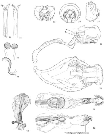
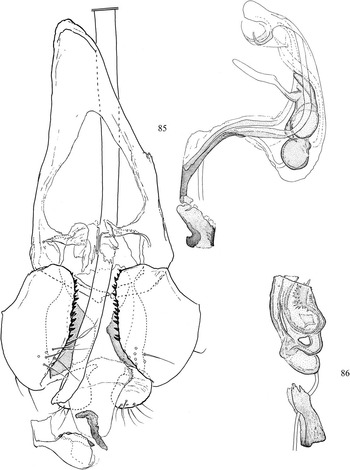
(B) The schineri group is composed following members of the former Hexomyza: H. cecidogena (Hering, Reference Hering1927), H. salicis (Malloch, Reference Malloch1913), H. schineri (Giraud, Reference Giraud1861), H. simplicoides (Hendel, Reference Hendel1920) (Figs. 8, 51–54), H. toschiae (Spencer, 1981) and H. winnemanae (Malloch, Reference Malloch1913). While the genitalic characters originally used for diagnosis (a membranous distiphallus, ring-like basiphallus) do not readily approximate the actual morphology of these species, it does mostly appear to encapsulate a tight lineage of north temperate species now orphaned, being without a parent genus. These species are provided with the new genus name Euhexomyza in the following section. The holotype of H. toschiae (host unknown) (Fig. 85) has been examined, and it is the one species that clearly belongs outside of this group – the male genitalia are very different and the clypeus is very broad and truncate, also recombining it as Ophiomyia toschiae new combination.
(C) The remaining unplaced species of Hexomyza not belonging to either the schineri or sarothamni groups are: H. abutilonicaulis (Spencer, Reference Spencer1977), H. coprosmae Spencer, Reference Spencer1976, H. gymnosporivora (Spencer, 1963), H. paederiae (Sasakawa, Reference Sasakawa1954), and H. websteri (Malloch, Reference Malloch1913). Of these, H. coprosmae and H. websteri have affinities to those “core” species in the schineri group and are also placed in the new genus Euhexomyza below, but the remaining three species are a heterogenous assemblage. Ophiomyia abutilonicaulis new combination is recombined in Ophiomyia because it has an anteriorly truncate clypeus, and while relationships with other Ophiomyia are uncertain, the elongate, asymmetrical arms of the basiphallus and the internally spinulose, bladder-like compartments of the distiphallus are found in many “typical” species (Figs. 76–80). This is the only included species from the Oriental Region (Pakistan), and it forms galls on Malvaceae (Abutilon Miller).
The Afrotropical species (South Africa) reared from Maytenus buxifolius (Richard) Grisebach (Celastraceae) is here treated as Melanagromyza gymnosporivora, its original generic combination. This robust species is relatively large (2.3 mm), and like those taxa in the schineri group, it has a subshiny dark brown body, a high lunule that forms a flat plate with the face and a relatively pronounced orbital plate (although narrower and shiny), but it is readily placed in Melanagromyza because there are two posteromedial bristles on the mid tibia, the supra-alar is well developed, the eye is partially setulose (setae minute and sparse), and most importantly, the basiphallus is small, U-shaped and symmetrical, and the distiphallus has one pair of tubules flanking the mesophallus (Fig. 86); the epandrium is not preserved with the original dissection. The clypeus is not readily visible, but it is relatively broad with the anterior margin possibly straight.
Requests to examine the holotype male of Melanagromyza paederiae, housed in the Osaka Museum of Natural History, Japan, were not returned. Label data for the type are as follows: Japan. Shikoku: Ehime Pref., Matsuyama, 11.v.1953, T. Yano. This species is here recombined as Ophiomyia paederiae new combination until the holotype can be examined, as the illustrations in Sasakawa (Reference Sasakawa1954) reveal the phallus to be similar to that of some other Ophiomyia, while not exhibiting the genitalic synapomorphies of other genera of Agromyzinae.
Melanagromyza Hendel
Melanagromyza Hendel, Reference Hendel1920: 114. Type species: Agromyza aeneoventris Fallén, 1823, by original designation. Frick Reference Frick1952: 375; Spencer Reference Spencer1969: 64; Spencer and Steyskal Reference Spencer and Steyskal1986: 18.
Limnoagromyza Malloch, Reference Malloch1920: 147. Type species: L. diantherae Malloch, Reference Malloch1920, by original designation. Synonymised in Frick (Reference Frick1952).
In comparison to similar agromyzine genera, Melanagromyza species often have a distinct metallic reflection on the abdomen and sometimes the thorax, which can be blue, green, coppery, red, or a combination of colours. The mid tibia of Melanagromyza also usually has two posteromedial bristles, less commonly one or three. There is usually only one bristle in Ophiomyia (there are none in species of the former Kleinschmidtimyia), uncommonly one in Tropicomyia and none in Euhexomyza (although mid tibia sometimes with minute medial bristle). The fore tibia also sometimes has one posteromedial bristle, which is found in many Japanagromyza, but Japanagromyza is usually also characterised by one pair of prescutellar acrostichals, bare eyes, and a flat, smooth frons (Lonsdale Reference Lonsdale2013).
The metallic shine is also uncommonly seen in Ophiomyia, but the colour is often restricted to a faint reflection on the abdomen. Also similar to some Ophiomyia, a minority of Melanagromyza also has a slightly truncated clypeus, but as discussed above, the anterior margin is usually without corners and the margins grade into a curve, the entire body is distinctly metallic, and the eye is often conspicuously pilose dorsally (Fig. 10). At this point it cannot be entirely ruled out that the similar clypeus in these few taxa is due to shared ancestry, but the large suite of derived characters also found in these Melanagromyza suggest that it is independently acquired.
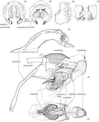
With regards to the male genitalia (Figs. 44–50), all examined species of Melanagromyza have (i) a symmetrical basiphallus (asymmetrical in most species of other agromyzine genera), which is either U-shaped or ring-like, and often very short, (ii) the epandrium sometimes has a small posterodistal spine behind the surstylus, and (iii) the ventrobasal surface of the distiphallus has one pair of thin tubules flanking the mesophallus. Preliminary results suggest that the short, ring-shaped to U-shaped basiphallus is most definitive, as are the paired tubules of the distiphallus, however, these can be shortened and difficult to locate in some, such as the African M. provecta (Meijere, 1910). The latter is possibly absent in some non-Nearctic species, many of which have not been personally examined, but it has been found in all dissected non-Nearctic species thus far (note that even if large and conspicuous, the tubules on the distiphallus are uncommonly depicted in published illustrations).
Melanagromyza aeneoventris (Fallén), male external genitalia; 44. posterior; 45. anterior; 46. left lateral; 47. ventral; 48–50. M. dianthereae (Malloch), male internal genitalia; 48. hypandrium, ventral; 49. hypandrial complex, left lateral; 50. phallus and proepiphallus, ventral.

Lastly, most representatives of the Ophiomyia genus group have one pair of dark lobes extending from the lateral margin of the proepiphallus, often obscuring the phallophorus from view laterally, but these are often only present in a reduced state in some Melanagromyza. More detailed analysis of non-Nearctic species will be necessary to determine whether this is a useful diagnostic character at the genus level. Despite these putative genitalic synapomorphies, the genus is often readily diagnosed externally through a combination of dorsally pilose eyes, a dominant metallic shine, and an absence of characters otherwise definitive of other Agromyzinae. With these characters, only a handful of species of Melanagromyza will still require genitalic dissection for generic placement.
Euhexomyza new genus
Type species . Melanagromyza simplicoides Hendel, Reference Hendel1920, by present designation.
Etymology. The generic name (feminine) compounds eu- (Gr. “true”) with the previous genus name applied to the included species, Hexomyza.
Diagnosis. Colour dark brown, including halter; without metallic shine (most Melanagromyza and some Ophiomyia) (Fig. 1a–b). Lunule with height nearly equal to width (also seen in some Melanagromyza and Japanagromyza). Clypeus broadly rounded (anteriorly truncate in all Ophiomyia and a few Melanagromyza). Orbital plate and parafacial pronounced (orbital plate shallow in Japanagromyza and most Ophiomyia), forming dark ring around eye. Prescutellar acrostichal seta absent (present in Agromyza and some Japanagromyza). Postsutural supra-alar thin, sometimes setula-like, and never larger than intra post-alar (the bristle immediately in front of anterolateral corner of scutellum), not longer and sometimes thickened or subequal to major notal setae. Scutellum sometimes with small additional setae. Tibiae without posteromedial bristles, but mid tibia sometimes with minute medial bristle (often present in remainder of Ophiomyia genus group, at least on mid tibia). Distiphallus symmetrical in ventral view, with base broadly rounded and apex pointed with medial section unpigmented; in lateral view with parallel dorsal bands nearly joined apically (Figs. 61–62). Phallophorus with ventral pouch.
Euhexomyza simplicoides (Hendel), female genitalia; 51. segment 10 and cerci, ventral; 52. segment 10 and cerci, dorsal; 53. spermathecae and end of ejaculatory duct; 54. ventral receptacle; 55–62. E. simplicoides, male genitalia; 55. external genitalia, ventral; 56. external genitalia, anterior; 57. external genitalia, left lateral; 58. hypandrium, epiphallus, and phallapodeme, left lateral; 59. hypandrium, epiphallus, and phallapodeme, ventral; 60. ejaculatory apodeme; 61. phallus, ventral; 62. phallus, left lateral.

Description. Chaetotaxy: At least four fronto-orbitals, usually with two ors and two ori, with posterior ori reclinate (i.e., appearing as ors); orbital setulae erect, inclinate, and sometimes slightly reclinate, usually 1/3–1/2 length of fronto-orbitals, and in at least one scattered row. Vibrissa relatively small and thin, sometimes only slightly larger than genal bristles. Ocellar and postvertical bristles usually not much longer than width of ocellar tubercle. Six to eight scattered rows of acrostichal setulae; prescutellar acrostichal bristle absent, although Spencer (Reference Spencer1966) mentions that some E. cecidogena (as E. salicis – see Spencer (Reference Spencer1969: 80) and Spencer (Reference Spencer1976: 36)) have one pair of prescutellar bristles, although this has not been confirmed with the material available. Two dorsocentrals near posterior margin of scutum, with anterior (second) bristle rarely near level of postsutural supra-alar. Scutellum with one well developed pair of anterolateral and apical bristles, and sometimes with small additional setae laterally (and less frequently apically). At least one well developed anepisternal and at least two katepisternal bristles. Tibiae without posteromedial bristles; mid tibia sometimes with minute medial bristle (barely stronger than surrounding setulae). Colour: Almost entirely dark brown, including halter and calypter margin and setae; without metallic shine. Head: Arista pubescent; first flagellomere small and ovate. Lunule flat and high, semicircular with apex pointed to slightly truncated, with height nearly equal to width; not bent inwards (as seen in most of Ophiomyia genus group aside from some Melanagromyza). Ocellar triangle reaching posterior ori. Face with shallow medial carina, almost flat medially; face relatively wide with width over two times height; antennae slightly separated at base. Buccal cavity broad with anterior margin relatively straight; no epistoma; clypeus broadly rounded (some E. simplicoides with one pair of small anterolateral lobes level with buccal cavity, not the raised body of the clypeus proper). Orbital plate and parafacial moderately projecting and swollen, forming dark ring around eye. Male terminalia: Surstylus fused to epandrium, curved inwards, with apical setae and inner-marginal to medial tubercle-like setae; inner surface grading into halves of stout, bare, plate-like subepandrial sclerites (=sternite 10), with halves tapered to a point dorsomedially and connected via a short membrane (Figs. 55–56). Cerci well developed, normally setose but with additional distomedial setae on inner surface (Figs. 55–57). Hypandrium with apex curved ventrally and ending in short apodeme; inner lobe of hypandrium with outstanding setae or well-developed sockets, sometimes atrophied and partially membranous but with thin band extending to base of long, rod-like phallapodeme (Figs. 58, 59). Metepiphallus with flat base continuous with lateral extensions enclosing base of phallus, sometimes with pointed ventral process; proepiphallus small, slightly curved in cross section and with one pair of similarly sized flanking plates. Phallophorus tube-like with margins weakly sclerotised and ill defined; with well-sclerotised pouch developed from distoventral margin (Figs. 61, 62) (missing in the two tentatively allied species). Basiphallus only well defined and irregularly sclerotised dorsomedially and dorsoapically. Mesophallus small, sac-like, with subbasal insertion of ejaculatory duct, and tapered apically to ventromedial insertion point into distiphallus. Distiphallus with base broad, internally spinulose and extending far past base of mesophallus, partially enclosing it; relatively flat with short ventromedial process and longer dorsomedial processes forming transverse pocket. Ejaculatory apodeme well developed and asymmetrical with dark medial rib on flat fan-shaped blade that narrows to short stem with single small laterobasal process; pileus ejaculatorius transverse and thin with ends broad and upturned (Fig. 60). Female terminalia: oviscape dark, heavily sclerotised, typical of family; T10 and S10 thin and relatively elongate with base tapered and apex setose (Figs. 51, 52). Cerci small and subcylindrical with apical and medial setae and one pair of apical peg-like setae. Ventral receptacle sinuate with slightly expanded apical chamber with ventral recess (Fig. 54). Spermatheca (one pair) dark and dome shaped with base (at duct insertion) slightly invaginated with margin spirally grooved (Fig. 53). The posterior larval spiracles have usually three pores (eight in E. coprosmae and three to four in E. websteri) on a raised stalk.
Natural history. All species of Euhexomyza are known to form oval twig galls on Salicaceae (Populus Linnaeus and Salix Linnaeus) (Fig. 2) as larvae, with adult habits largely unknown. Euhexomyza copsosmae and E. websteri form similar galls on Rubiaceae and Fabaceae, respectively.
Species recombined as Euhexomyza (alphabetical order)
Note that descriptions and a species key are not included here. Species-level boundaries and host preferences are ill defined and overlapping, and necessitate additional attention that is outside the scope of the present study. For details on these species-level difficulties, see Spencer (Reference Spencer1966, Reference Spencer1969, Reference Spencer1990) and Spencer and Steyskal (Reference Spencer and Steyskal1986).
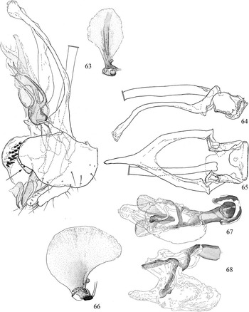
Two species presently included in Euhexomyza require especial examination as they share certain affinities with those Salicaceae-feeding species that make up the “core” of the new genus, but share none of the synapomorphies characteristic of the newly defined Ophiomyia and Melanagromyza. Species-level revisions are required to more accurately place these species and determine if the shared characters are homoplastic or due to shared ancestry. These species are E. coprosmae and E. websteri. Euhexomyza coprosmae from New Zealand is a strong geographic outlier, although Spencer (Reference Spencer1990) considered it to be close to the Japanese O. paederiae, which is also found on Rubiaceae and makes similar galls, and postulated the presence of undiscovered relatives in southeast Asia. Like the Saliceae-feeders, E. coprosmae most conspicuously shares the external features of a very high lunule that forms a large, flat plate with the face, a narrow gap between the antennal sockets that are not developed into a bulge ventrally, a reduced vibrissa, a bulging frons, a pronounced orbital plate that continues around the eye as a thick ring (although only about 2/5 the height of the gena), there are many long irregular orbital setulae, and no medial tibial bristles. Unlike core Euhexomyza, the supra-alar is slightly larger than the intra postalar (not subequal in size), the pv and oc are large (not small), the phallus is dissimilar and the phallophorus is without a ventral pouch (Figs. 67, 68).
Euhexomyza cecidogena (Hering), male genitalia, ventrolateral [arrow indicating ventral pouch of phallophorus]; 64–68. E. coprosmae (Spencer), male genitalia; 64. hypandrium, epiphallus, and phallapodeme, left lateral; 65. hypandrium, epiphallus, and phallapodeme, ventral; 66. ejaculatory apodeme; 67. phallus, ventral; 68. phallus, left lateral.

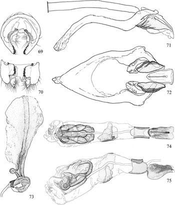
Euhexomyza websteri is the least reliably placed species in the genus, having only the high, flat lunule, a narrow gap between the antennal sockets that continues onto a flat face ventrally, a bulging frons and a high orbital plate with dense, irregular setulae (although the plate is directed more inwards than dorsally), and no medial tibial bristles. The male terminalia (nontype male dissected) (Figs. 69–75) also has none of the features characteristic of the Salicaceae-feeders aside from the short, asymmetrical basiphallus, which is also found elsewhere in the subfamily.
Euhexomyza websteri (Malloch), male genitalia; 69. external genitalia, anterior; 70. external genitalia, ventral; 71. hypandrium, epiphallus, and phallapodeme, left lateral; 72. hypandrium and epiphallus, ventral; 73. ejaculatory apodeme; 74. phallus, ventral; 75. phallus, left lateral.

Euhexomyza cecidogena (Hering) new combination
(Fig. 63)
Melanagromyza cecidogena Hering, Reference Hering1927: 325. Hendel Reference Hendel1931: 162.
Hexomyza cecidogena. Spencer, Reference Spencer1966: 39 [as synonym of Agromyza salicis Malloch], 1969: 13 [reinstated status], 1976: 36, 1990: 81; Wen and Dong Reference Wen and Dong1995: 77.
Distribution: Palaearctic: Europe, China, Japan.
Host: Salix species.
Euhexomyza coprosmae (Spencer) new combination
Hexomyza coprosmae Spencer, Reference Spencer1976: 162. Spencer Reference Spencer1990: 233.
Distribution: Australian: New Zealand.
Host: Rubiaceae – Coprosma propinqua Cunningham.
Euhexomyza salicis (Malloch) new combination
Agromyza salicis Malloch, Reference Malloch1913: 314. Shewell Reference Shewell1953: 464.
Melanagromyza salicis. Frick Reference Frick1952: 379, Frick Reference Frick1959: 365.
Hexomyza salicis. Spencer Reference Spencer1966: 39, 1990: 81; Spencer and Steyskal Reference Spencer and Steyskal1986: 36.
Distribution: Nearctic: Canada (British Columbia), United States of America (Massachussets).
Host: Salix species.
Euhexomyza schineri (Giraud) new combination
Agromyza schineri Giraud, Reference Giraud1861: 484. Malloch Reference Malloch1913: 327; Spencer Reference Spencer1966: 42 [lectotype designation].
Melanagromyza schineri. Hendel Reference Hendel1920: 128, 1931: 174; Frick Reference Frick1952: 379; Sasakawa Reference Sasakawa1961: 349.
Hexomyza schineri. Spencer Reference Spencer1966: 42, 1969: 81, Spencer Reference Spencer1980: 29, 1990: 81; Spencer and Steyskal Reference Spencer and Steyskal1986: 36; Gil-Ortiz et al. Reference Gil-Ortiz, Sánchez and Martinez2012: 275 [Spain].
Distribution: Nearctic: Canada (Alberta, British Columbia, Manitoba, New Brunswick, Ontario), United States of America (California, Colorado, Massachusetts, New Mexico, Washington); Palaearctic: western Europe.
Host: Populus species; records from Salix species in Sasakawa (Reference Sasakawa1961) are considered dubious by Spencer and Steyskal (Reference Spencer and Steyskal1986).
Comments: The holotype male was not examined; it is housed in the NMW and was collected in Austria on the banks of the Danube River.
Euhexomyza simplicoides (Hendel) new combination
Melanagromyza simplicoides Hendel, Reference Hendel1920: 128. Hendel Reference Hendel1931: 178. Spencer Reference Spencer1966: 44 [lectotype designation].
Melanagromyza kirgizica Rohdendorf-Holmanová, Reference Rohdendorf-Holmanová1959: 695. Synonymised in Spencer (Reference Spencer1966).
Hexomyza simplicoides. Spencer Reference Spencer1966: 44, 1969: 79 [potential presence in Canada], 1980: 31, 1990: 81; Spencer and Steyskal Reference Spencer and Steyskal1986: 35.
Distribution: Nearctic: United States of America (California, New York, Ohio). Palaearctic: Europe, Russia (Kirghiz).
Host: Salix; doubtful records from Populus (Spencer Reference Spencer1966).
Comments: The Melanagromyza simplicoides lectotype male housed in the NMW was not examined; it was collected on the 18 of June in Austria and is labelled “typus”. The Melanagromyza kirgizica Rohdendorf-Holmanová holotype (sex unknown), also not examined, is from Frunze, Russia; Spencer (Reference Spencer1966) noted that the type series is deposited in the E. Rohdendorf Collection, Prague, Czech Republic.
Euhexomyza websteri (Malloch) new combination
Agromyza schineri. Misidentification. Aldrich, in Amundsen Reference Amundsen1912: 730.
Agromyza websteri Malloch, Reference Malloch1913: 325. Shewell Reference Shewell1953: 465.
Melanagromyza websteri. Hendel, Reference Hendel1931: 180; Frick Reference Frick1952: 380; Sasakawa Reference Sasakawa1961: 353.
Hexomyza websteri. Spencer and Steyskal Reference Spencer and Steyskal1986: 36; Spencer Reference Spencer1990: 115.
Distribution: Nearctic: United States of America (introduced – Washington); Palaearctic: Japan.
Host: Fabaceae – Wistaria Sprengel species.
Euhexomyza winnemanae (Malloch) new combination
Agromyza winnemanae Malloch, Reference Malloch1913: 314. Shewell Reference Shewell1953: 465.
Melanagromyza winnemanae. Frick Reference Frick1952: 380, 1959: 367.
Hexomyza albicula Spencer, Reference Spencer1969: 79. Synonymised in Spencer and Steyskal (Reference Spencer and Steyskal1986).
Hexomyza winnemanae. Spencer and Steyskal Reference Spencer and Steyskal1986: 250; Spencer Reference Spencer1990: 81.
Distribution: NE: Canada (Ontario), United States of America (Maryland).
Host: Salix.
Tropicomyia Spencer
Tropicomyia Spencer, Reference Spencer1973: 180. Type species: Melanagromyza flacourthiae Séguy, 1951, by original designation. Spencer Reference Spencer1990: 391; Ipe and Ipe Reference Ipe and Ipe2004: 1311.
Epidermidomyia Ipe, Reference Ipe1986: 103. Nomen nudum.
Epidermomyia Ipe and Ipe, Reference Ipe and Ipe2004: 1312. Type species: Agromyza atomella Malloch, 1914, by original designation. New synonym.
Species presently included in Tropicomyia are entirely Old World tropical in distribution with many species exhibiting an unusually high degree of polyphagy. Species are found on all dicot subclasses, at least two superorders of monocots, ferns, and the coniferous genus Gnetum Linnaeus (Gnetaceae) (Spencer Reference Spencer1990).
Following examination of the type species, several additional described species (Tropicomyia crotonella (Spencer, Reference Spencer1964), T. vigneae (Séguy, 1951), and T. polyphyta (Kleinschmidt, 1961)), and a number of potentially undescribed species housed in the NMWC, it is apparent that there is no convenient way to characterise the genus as it is currently outlined. Generally speaking, species consist of small, entirely brown flies that often mine leaves in the epidermal or upper cell layers and usually require male dissection for confident identification. Generic placement can sometimes be made by determining an absence of characters otherwise definitive of the remaining Agromyzinae, but this is prone to error, particularly for such physically diminutive taxa. As would be expected from such a vague description, Tropicomyia is suspected to be nonmonophyletic, even by the author of the genus – see discussion in Spencer (Reference Spencer1990: 404) – who noted that additional new genus groups may be needed to accommodate several species.
Inclusion of taxa in Tropicomyia by authors appears to have been based on a combination of morphological nonagreement with other more well-defined genera and larval leaf-mining habits, the latter of which authors would have used to exclude it from other taxa incorrectly assumed to be exclusively stem-boring. Spencer (Reference Spencer1986) used a more intricate combination of characters to differentiate the genus from the highly similar Kleinschmidtimyia (here treated as a synonym of Ophiomyia), whose species have similar mines, but these have a distinct phallus and a slightly differing series of larval morphological and behavioural characters.
While redefinition of Tropicomyia is outside of the scope of the present study and requires additional investigation into agromyzine relationships, it does appear as though most species can be confidently placed in a clade containing the type species (T. flacourthiae) that Spencer (Reference Spencer1976) referred to as Tropicomyia sensu strictus. These species often have a shallow, purely epidermal leaf mine, “serrated” larval teeth, anterior larval spiracles on long stalks (also found in O. pisi), posterior spiracles with three pores on a protuberance (also frequently found elsewhere), and a costa ending at R4+5, but many species differ in one or more of these characters, confounding confident diagnosis. Of these characters, authors have placed an emphasis on the unusual epidermal leaf mine, but even this is inconsistent within Tropicomyia sensu strictus, and the difficulties associated with the use of larval behaviour in agromyzid classification are hopefully convincingly explained above. Representatives examined here also appear have two dorsocentrals, a very shallow lunule (often angled inwards and hidden within the ptilinal suture, as in most Ophiomyia), four fronto-orbitals (only the anterior bristle in strongly incurved), a rounded clypeus, a shallow gena, a well-developed supra-alar and a small intra post-alar, no tibial bristles aside from a ventroapical bristle and uncommonly a single posteromedial bristle on the mid leg, a brown halter, a brown calypter margin and setae, a flat face, small body size (~1.2–2.3 mm), and a penultimate section of vein M that often appears to be no longer than vein dm-cu.
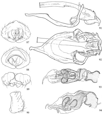
What will likely prove to most confidently define Tropicomyia, however, will be features of the male genitalia (Figs. 87–94), which confidently encapsulates a group of species larger than Spencer’s Tropicomyia sensu strictus. This lineage appears to be defined by the following: basiphallus with relatively short dorsobasal surface that extends into one pair of very broad lateral plates that often cover part of the distiphallus laterally; distiphallus forming a broad, often flat and internally spinulose sac that may be produced posterodorsally over the mesophallus; mesophallus (insertion ventromedial or ventrobasal) continuing ventromedially into one pair of clear, unified tubules disrupted by a single, short ventromedial process. This reliance on genitalic features is inconvenient for diagnosis, so it is hoped that additional characters can be found, particularly because numerous species are known only from females.
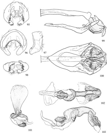
Genitalic features will likely also predominate in the definition of any additional genera erected for unrelated species in this group because of the external uniformity of these taxa, which at least includes T. crotonella (Figs. 95–103), a miner in a lower leaf layer. The terminalia of T. crotalariae (Hering, 1957) (not examined) also excludes this species from Tropicomyia, and while the illustration in Spencer (Reference Spencer1990) is reminiscent of Ophiomyia, very little else can be inferred from the drawing.
With regards to higher classification, male genitalic structures readily place this taxon in the Ophiomyia genus group, and the molecular analyses of Scheffer et al. (Reference Scheffer, Winkler and Wiegmann2007) recovered Tropicomyia theae as the sister group to Euhexomyza schineri+Ophiomyia “A”. Morphology partially suggests a sister group relationship with Melanagromyza, however, as both groups have one pair of spines developing from the posteroventral margin of the epandrium, and a short, ring to U-shaped basiphallus. The latter character is of particular importance to the generic definition of Melanagromyza (discussed above), but the additional distolaterally produced plates emerging from this structure in Tropicomyia are unique.
Synonymy of Epidermomyia Ipe and Ipe with Tropicomyia
With the partial redefinition of Tropicomyia above and recognition of the core lineage of taxa including the type species M. flacourthiae, there is enough evidence to reduce one last genus to synonymy: Epidermomyia Ipe and Ipe, Reference Ipe and Ipe2004. This is not to be confused with Epidermidomyia Ipe, Reference Ipe1986, which was a nomem nudum published in the abstracts of the First International Congress of Dipterology (Pape and Thompson Reference Pape and Thompson2012). Agromyza atomella was selected as the type species for Epidermomyia, and a total of 10 species were included in the genus. These 10 species strangely included the type species of Tropicomyia, despite treating Tropicomyia as a valid genus in the same publication. In their own words, the “erection of a new genus for the dimunitive upper surface cuticle miners are justifiable and essential to mainly accommodate the Tropicomyia s.s. of Spencer” – that is, they decided to provide a name for a genus that had already been named. Epidermomyia new synonymy is here formally included as a junior synonym of Tropicomyia. The following species treated as Epidermomyia are here returned to Tropicomyia: T. atomella; T. flacourthiae [misspelled “flacourtiae” in Ipe and Ipe (Reference Ipe and Ipe2004)]; T. polyphaga (Spencer, 1961); T. theae; and T. vigneae.
The following species were also treated as Epidermomyia, but justification for placement in that genus was never provided. These species are returned to their position in Melanagromyza – the generic combination held before recombination by Ipe and Ipe (Reference Ipe and Ipe2004) – until further evidence can reliably support alternate placement. The generic placement of Melanagromyza sanctijohani Garg, Reference Garg1971 is at present uncertain, but its treatment as Epidermomyia appears to have been solely based on termination of the costa at R4+5. Unfortunately the genitalic illustration does little to reveal its generic affinities, so the more cautious approach will be to return the species to Melanagromyza, following Sasakawa (Reference Sasakawa1977). Sasakawa (Reference Sasakawa1977) is also followed in treating Melanagromyza latifrons Garg, Reference Garg1971 as a junior synonym of M. conspicua Spencer, as the type has not been examined and little can be determined from the genitalic illustration in Garg (Reference Garg1971). The placement of Melanagromyza eravensis Sing and Ipe, 1973 and M. padmanabhi Sing and Ipe, 1973 in Tropicomyia are also uncertain, as they are known from one or two female adults, and as such, are divorced from the essential male genitalic and (possibly) biological data needed for placement in Tropicomyia. Melanagromyza thekadi Singh and Ipe, Reference Singh and Ipe1973 is also known from a single female, but the larva makes “upper epidermal mines on its host” (Singh and Ipe Reference Singh and Ipe1973).
Material examined
Euhexomyza cecidogena (Hering). GERMANY. Syntype [circular label with blue margin], Type [red label], Berlin-Frohnau, 1.v.1927, “N. 306i”, O.andM. Hering, Galle on: Salix aurita [holotype, Fig. 63] (1♂, BMNH). Mecklenburg, Neuer [illegible] Zweiggalle an Salix, 15.vi.1960, rehderiana [illegible], (1♀, CNC). ITALY. Fano, 20.v.1935, Ex cec. Salic, F. Venturi (1♂, BMNH).
Euhexomyza coprosmae (Spencer). NEW ZEALAND. South Island: Kohatu, Begg’s Corner, Motueka Valley, ex Coprosma propinqua, coll. 15.viii.1968, em. 10.x.1969, J.S. Dugdale [paratype] (1♂ 1♀, NZAC), ibid, except em. unknown [paratypes, Figs. 62–68] (1♂ 1♀, NZAC).
Euhexomyza salicis (Malloch). United States of America. Massachusetts: Reading, 16.v.1908, New York State Collection, from Willow, E.P. Felt [holotype] (1♂, USNM).
Euhexomyza schineri (Giraud). CANADA. Ontario (117♂♀, CNC).
Euhexomyza simplicoides (Hendel). CANADA. Alberta: Cherry Point, F.I.S., 20.vi.1962, ex Salix sp. galls (1♂ 3♀, CNC), MB: Mine Dock, Em. 24.vi.1969, ex Salix sp. galls (1♂, CNC), Ontario: Mantourwadge [sic], 28.vi.1962, ex Salix sp. galls (1♂, CNC), Manitouwadge, 8.vi.1964, ex Salix sp. galls, F.I.S. (2♂ 1♀, CNC). SPAIN. Algeciras, “9.6.1965”, ex Salix pedicillata, “24.4.55”, K.A. Spencer (Figs. 51–54) (1♀, CNC). Los Barrios, Algeciras, “30.5.55”, Salix pedicillata “25.4.55”, K.A. Spencer (Figs. 55–62) (1♂, CNC).
Euhexomyza websteri (Malloch). United States of America. California: San Diego, Wistaria buds from Japan, E.O. Amundsen (Figs. 69–75) (1♂, USNM), Washington: Seattle, F.M. Rhoder, galls on Wistaria fr. Japan, issued 21.i.1913, Type No. 15587 [holotype] (1♀, USNM), “ex galls”/“on pink Wistaria”, Japan, B.B. Whitney, “No. 745 coll” [paratype] (1♂, USNM).
Euhexomyza winnemanae (Malloch). CANADA. Ontario: Ottawa, 7.iv.1948, ex gall on Salix sp., G.E. Shewell [holotype, Hexomyza albicula Spencer] (1♂, CNC), Dryden, 12–13.vi.1960, Kelton and Whitney [paratype, H. albicula Spencer] (1♀, CNC). United States of America. Maryland: Plummers Island, 27.vi.1909, W.L. McAtee [holotype Agromyza winnemanae Malloch] (1♀, USNM).
Melanagromyza gymnosporivora Spencer. SOUTH AFRICA. Hout Bay (C.P.), galls twigs Gymnosporia buxifolia, coll. 26.xi.1961, em. 10.xii.1961, K.A. Spencer [holotype, Fig. 86] (1♂, BMNH).
Ophiomyia abutilonicaulis (Spencer). PAKISTAN. Mir pur Khas, 8.i.1976, maggot in galls Abutilon sp., F.I.B.C, Abu -1/76-16 [holotype, Figs. 76–80] (1♂, BMNH), Hyderibad, 6–8.iii.1980, ex stem Abutilon indicum (1♂, BMNH).
Ophiomyia abutilonicaulis (Spencer), male genitalia; 76. external genitalia, ventral; 77. hypandrium and epiphallus, ventral; 78. ejaculatory apodeme; 79. phallus, ventral; 80. phallus, left lateral; 81–84. O. sarothamni (Hendel), male genitalia; 81. external genitalia, ventral; 82. ejaculatory apodeme; 83. phallus, ventral; 84. phallus, left lateral.

Ophiomyia aloes Spencer. SOUTH AFRICA. Pretoria, vi.1942, H.K. Munro., Ac.P.4717, mine Aloe petricola, 6.42 [paratype, Figs. 41–43] (1♂, BMNH), Mine an: Aloe petricola, ether fixed, Süd-Africa, paratype [red label], No type status, det. C.A. Couch 1983 (1♂, BMNH).
Ophiomyia arabica (Deeming). SAUDI ARABIA. Jazan, Gizan, 6.ii.2012, J.C. Deeming (1♂, NMWC), Riyadh, Rhader Karim, 19.ii.2012, J.C. Deeming (Figs. 4, 26–32) (1♀, NMWC).
Ophiomyia centaurae (Spencer). ITALY. Rnme [sic], Centaurea, Acc. 163D, solstitialis, 3.iv.1960, reared from head, L. Andres [holotype] (1♂, USNM).
Ophiomyia dianellae (Kleinschmidt). AUSTRALIA. Australian Capital Territory: Black Mtn., light trap, 20.xi.1964, I.F.B. Common (1♂, CNC).
Ophiomyia longilingua (Hendel). FRANCE. “Type”, Mine an Knautia arvensis, Hendel, Verson b. Caen, 4903, 5.4.1943, Dr. H. Buhr, Nr., Melanagromyza knautia m. Type. Det. Hering, 1943, O. longilingua, K.A. Spencer, det. 1964 [Holotype knautiae Hering] (1♀, BMNH). GREECE. “Korinth”, “50000.iv” [Holotype longilingua Hering, Fig. 5a–c] (1♂, ZMHU).
Ophiomyia orbiculata (Hendel): AUSTRIA. Ober Osterreich, Ennsdorf, 23.vi1962, Hering, Fang an: Centaurea scabiosa (2♀ [same pin], CNC). GERMANY. [illegible]-Görlitz, 15.vi.1952, Hering No. 58, F. Achill. Mitt. (1♂, CNC), Biehain, Lausitz, Hering (1♀, CNC).
Ophiomyia phaseoli (Tryson). CHINA. Hong Kong, Tai Lung, N.T., P.Y.SO, 12-9-65, C.I.E. Coll., No. A884, larvae feeding on long bean (1♀, CNC). NIGERIA. Ile Ife, 2.i.1970, on bean, C.I.E. A4342 (2♂[same pin], CNC). TAIWAN. Taipeh, Formosa, spring 1959, Ex Glycine sojae [K.A. Spencer] (1♂ 1♀[with puparia], CNC). United States of America. Hawaii: Oahu, Waimanala, 19.i.1971, ex snap bean, G. Funasati coll. (1♀, CNC).
Ophiomyia pisi (Kleinschmidt). AUSTRALIA. South Australia: 23 km SW by S of Etadunna H.S. B/ville Track, SA (Green Vly.), 16.ix.1972, Z. Liepa (Figs. 33–40) (1♂, CNC).
Ophiomyia rostrata (Hendel). SPAIN. Ibiza, Portinatx, 6–13.v.1983, J.C. Deeming (1♀, BMNH).
Ophiomyia rotata Spencer. THAILAND. Bangkok, 15.xi.1985, K.A. Spencer, Ja-tu-jak Park opp. Central plaza (Figs. 4, 18–24) (2♂, BMNH).
Ophiomyia sarothamni (Spencer). ITALY. Sicily: Mt. Etna, Sinofsi, Em. 27.iv.1964, gall Spartium junceum 8/4, K.A. Spencer (1♀, CNC), Mt. Etna, Sinofsi, em. 18.iv. 1964, ex gall Spartium junceum 8.4, K.A. Spencer (Figs. 81–84) (1♂, CNC).
Ophiomyia simplex (Loew). GERMANY. Berlin, from C. Schirmer (1♂ ♀, USNM). United States of America. California: Lakeside, 4.iv.1944, Asparagus (1♂ 1♀, USNM), DC: “DC” (1♂, USNM), Illinois: Chicago, A.L. Melander (1♂, USNM), IO: Pleasant Valley, 29.vii.1931, H.M. Harris (1♀, USNM), GA: Tifton, 24.ix.1896, A.L. Melander (1? [head and abdomen missing], USNM), Maryland: Cabin John, 10.v.1897 (2♂, USNM), Chillum, “3-6-14”, collected on Asparagus (1♂, USNM), Colesville, 20.viii.1975, Malaise trap, W.W. Wirth (1♂, USNM), College Park, “6-29”, C.T. Greene (2♂, USNM), College Park, “8/10/14”, collected on potato, W.H. White (1♂, USNM), College Park, “6-29”, C.T. Greene (1♀, USNM), MI: E Lansing, 1.vi.1929, on asparagus, R.W. Pettit (2♂ 1♀, USNM), Grand Rapids, 13.iv.1931, C.W. Sabrosky (1♂ 1♀, USNM), New York: Ithaca, A.L. Melander (2♂ 2♀, USNM).
Ophiomyia toschiae (Spencer). United States of America. California. California: Kern Co., Wofford Heights, 8 mi W, 29.iv.1964, G.A. Toschi [holotype, Fig. 85] (1♂, CAS).
Ophiomyia toschiae (Spencer), genitalia (ventral view) with phallus inset at top right (right lateral view); 86. Melanagromyza gymnosporivora Spencer, phallus, left lateral.

Ophiomyia wikstroemiae Kleinschmidt. AUSTRALIA. New South Wales: Kangaroo Valley, 5.ix.1948, E.F. Reik (1♂ 1♀, CNC).
Tropicomyia crotonella (Spencer). ETHIOPIA. Addis Ababa, Little Akaki River, 2300 m, E.M. Hering, mine an Croton macrostachys, 27.x.1959 [paratype, Figs. 95–103] (1♂, CNC), 15.xi.1959 [paratypes] (1♂ 1♀[same pin], CNC), 16.xi.1959 [paratype] (1♀, CNC).
Tropicomyia crotonella (Spencer), male genitalia; 95. external genitalia, posterior; 96. external genitalia, anterior; 97. external genitalia, left lateral; 98. external genitalia, ventral; 99. hypandrium, epiphallus, and phallapodeme, left lateral; 100. hypandrium, epiphallus, and phallapodeme, ventral; 101. ejaculatory apodeme; 102. phallus, ventral; 103. phallus, left lateral.

Tropicomyia flacourthiae (Séguy). CAPE VERDE ISLAND. São Antao: Paúl, on coffee, xii.1989, C. Diaz (Figs. 87–94) (5♂ 4♀, NMWC).
Tropicomyia flacourthiae (Séguy), male genitalia; 87. external genitalia, posterior; 88. external genitalia, anterior; 89. external genitalia, ventral; 90. external genitalia, left lateral; 91. hypandrium, epiphallus, and phallapodeme, left lateral; 92. hypandrium, epiphallus, and phallapodeme, ventral; 93. phallus, ventral; 94. phallus, left lateral.

Tropicomyia polyphyta (Kleinschmidt). AUSTRALIA. Queensland: Brisbane, on Erythrina sp., K.A. Spencer (1♀, CNC).
Tropicomyia vigneae (Séguy). ETHIOPIA. Addis Ababa, Little Akaki River, 2300 m, 25.x.1959, E.M. Hering, Mine an Glycine javanica [paratype Melanagromyza dolichi Sehgal] (1♀, CNC), Addis Ababa, Little Akaki River, 2300 m, 27.x.1959, E.M. Hering, Mine an Glycine javanica [paratype M. jonesi Spencer] (1♂, CNC). INDIA. Nankum, 22.xii.1963, “Ex leaf mines on Dolicos lablab Linn.”, V.H. Sehgal [paratype M. dolichi] (1♂, CNC).
Acknowledgements
The following curators are thanked for providing some of the material examined here: K. Goodger (BMNH); A. Kirk-Spriggs (National Museum Bloemfontein, South Africa); A. Plant and J. Deeming (NMWC); A. Norrbom (USNM); and J. Ziegler (ZMHU). Discussions on agromyzid characteristics, phylogenetics, and nomenclature were generously shared by M. v.Tschirnhaus, who also provided generous comments on the text and references, despite disagreeing with some of the author’s findings. Comments on the manuscript were also provided by B.J. Sinclair, S. Gaimari, and an anonymous reviewer, who are thanked for their time and insights. Photographs of Euhexomyza schineri adults and galls (Figs. 1, 2) were provided by C. Eisemann; the photo of Ophiomyia aloes (Fig. 3) was taken by J. Read; photographs of O. longilingua (Figs. 5a–c) were taken by C. Boudreault and H. Goulet. C. Boudreault and S. Laplante provided the abstract translation.