Introduction
Computed tomography (CT) is the principal imaging modality in radiation therapy. Reference van Elmpt and Landry1 It provides the necessary information required for the treatment planning system (TPS), such as patient CT images for target volumes and organs at risk delineation, and Hounsfield unit (HU) that are converted into physical densities or relative electronic densities from a CT number-relative electron density (CT-RED) calibration curve for dose calculation. Reference van der Heyden, Öllers, Ritter, Verhaegen and van Elmpt2,Reference Vergalasova, McKenna, Yue and Reyhan3 Thus, the quality and fidelity of the treatment plans depend on both image quality and calculation accuracy of HU. Reference Davis, Palmer, Pani and Nisbet4,Reference Chen, Noid and Tai5 Consequently, to provide high image quality, CT image acquisition protocols should be varied according to patient morphology and region of interest (ROI). Reference Li, Yu and Anastasio6–Reference El Mansouri, Choukri, Talbi and Hakam9 However, these variations can affect the HU values. Reference Davis, Palmer, Pani and Nisbet4,Reference Nhila, Talbi, El, Katib and El Chakir10,Reference Zurl, Tiefling, Winkler, Kindl and Kapp11 In this context, Zurl et al. concluded that the use of different CT protocols leads to variations of up to 20% in HU values. Reference Zurl, Tiefling, Winkler, Kindl and Kapp11 A number of studies have quantified the dose change in the radiation therapy planning process due to HU variation. Reference Saini, Pandey, Kumar, Singh and Pasricha12–Reference Cozzi, Fogliata, Buffa and Bieri14 One of these reports to the International Atomic Energy Agency (IAEA) has found that a variation of ± 60 HU can result in ± 1% deviation of the calculated dose for the 6 MV photon beam crossing 5 cm of bone equivalent material. Therefore, a number of recommended tolerances of HU values used in the CT-RED calibration curve are available in the literature. Reference Davis, Palmer and Nisbet15–Reference Bissonnette, Balter and Dong18 For example, two of the most recent references, one is from the Institute of Physics and Engineering in Medicine (IPEM), which quotes a change of ± 30 HU in soft tissue to limit the dose change to ± 1%, and with a dose change of ± 2% in lung and bone corresponded to a HU change of ± 50 and ± 150 HU, respectively. Reference Patel, Steven and Antony17 The second which is the most widely adopted in recent studies concluded that a dose change of about 1% corresponded to a change of ± 20 HU in soft tissue and ± 50 HU in bone, as well as in air. Reference Davis, Palmer and Nisbet15
For this reason, several studies have investigated the effect of acquisition parameters, Reference Nhila, Talbi, El, Katib and El Chakir10,Reference Fang, Mazur, Mutic and Khan13,Reference Das, Cheng, Cao and Johnstone19,Reference Ebert, Lambert and Greer20 type of scanner Reference Chung, Mossahebi and Gopal21,Reference Cropp, Seslija, Tso and Thakur22 and type of phantom Reference Koniarova23,Reference Hasani, Farhood and Ghorbani24 on the HU variation. The most influential parameters are the voltage tube Reference Saini, Pandey, Kumar, Singh and Pasricha12,Reference Zheng, Al-Hayek and Cummins25,Reference Afifi, Abdelrazek, Deiab, Abd El-Hafez and El-Farrash26 and the reconstruction filters, whereas, for this last, the HU change depends on the brand of scanner. Reference Vergalasova, McKenna, Yue and Reyhan3,Reference Zurl, Tiefling, Winkler, Kindl and Kapp11,Reference Davis, Muscat and Palmer27 For this purpose, the setting of reconstruction filters is recommended during the commissioning phase. Reference Lillicrap28 In the literature, some studies have provided methodologies to evaluate the effect of reconstruction filters on the number of HU. Reference Vergalasova, McKenna, Yue and Reyhan3,Reference Davis, Palmer, Pani and Nisbet4,Reference Zurl, Tiefling, Winkler, Kindl and Kapp11,Reference Davis, Muscat and Palmer27 Most of these studies are performed with the most commonly used tube voltage in clinical routine, which is 120 kVp. Reference van der Heyden, Öllers, Ritter, Verhaegen and van Elmpt2 Furthermore, these studies are conducted with various scanner brands such as Toshiba, Siemens, GE and Philips. However, none of these studies have evaluated the Hitachi Supria scanner algorithms.
In our previous study, the effect of CT acquisition protocols on HUs for this brand of scanner has been investigated. Reference Nhila, Talbi, El, Katib and El Chakir10 It was concluded that changing the reconstruction filter from body to head and using beam hardening correction (BHC) significantly affects HU in dense bone. Moreover, this study was conducted with a single energy (120 kVp). However, in clinical workflows, dosimetric scans may be performed with other tube voltage settings. Nevertheless, there is a lack of knowledge regarding the effect of reconstruction filters on the HU when selecting voltages higher or lower than 120 kVp.
The purpose of this work is to evaluate the effect of Hitachi Supria scanner reconstruction filters on HU and to see how this effect varies as a function of kVp. This can help the clinicians to quantify any possible change in HU when varying both reconstruction filters and tube voltage.
Materials and Methods
The measurements were carried out on a Hitachi Supria scanner 16-slice. The gantry opening was 75 cm in diameter. The tube voltage settings were 80 kVp, 100 kVp, 120 kVp, and 140 kVp. The tube current could be adjusted from 10 to 400 mA. This scanner has an automatic exposure control and modulates tube current called Intelli EC and a dose reduction function that applies an iterative reconstruction technology called Intelli IP. It achieves both low dose and high image quality by reducing image noise and artefacts.
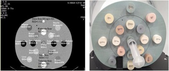
The scans of a computerised imaging reference system M062 phantom were acquired. The distribution of the tissue equivalent inserts in the phantom as well as their relative electron densities (REDs) is shown in Figure 1.
The distribution of tissue equivalent inserts in the phantom and their REDs (a) CIRS M062 and (b) CT image of CIRS M062.

As a first step, the acquisitions were done using the four tube voltage settings while keeping the other acquisition parameters constant (300 mAs, 500 FOV, 2·5mm slices, 1·065 × 0·625 collimation). As a second step, the raw data were reconstructed using different reconstruction algorithms. The reconstruction filters studied are listed in Table 1. The mean HUs of each insert were obtained through the central slice of the phantom using a ROI of 10 mm diameter; the ROI must be spaced far from the extremities of the inserts to avoid statistical fluctuations.
Main reconstruction filters of Hitachi Supria 16-slice scanner

Results
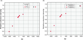
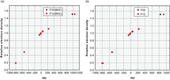
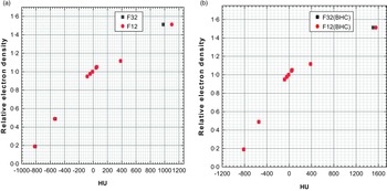
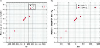
Figures 2, 3, 4 and 5 show the HU variations of the CT-RED calibration curves when comparing the reconstructions filter of body (F32) against head (F12) with tube voltage settings of 80 kVp, 100 kVp, 120 kVp and 140 kVp, respectively. All these figures show that the HU values coincide for all materials except for the RED equal to 1·51, which explains that there is no significant difference between the reconstruction filters, except for the dense bone. In addition, Figures 2 and 5 show a significant difference in the HU values of dense bone when changing the reconstruction filters without BHC: up to 159 HU for 80 kVp (Figure 2a) and 92 HU for140 kVp (Figure 5a). Nevertheless, when selecting the BHC, this variation decreases strongly for 80 kVp to become 51 HU (fig2B) and decreases slightly for 140 kVp to become 74 HU (Figure 5b), but it is still out of tolerance which is ±50 HU.
CT-RED calibration curves of F12 versus F32 at 80 kVp (a) without BHC and (b) with BHC.

CT-RED calibration curves of F12 versus F32 at 140 kVp (a) without BHC and (b) with BHC.

CT-RED calibration curves of F12 versus F32 at 100 kVp (a) without BHC and (b) with BHC.

CT-RED calibration curves of F12 versus F32 at 120 kVp (a) without BHC and (b) with BHC.

In contrast, Figures 3 and 4 show the effect of the reconstruction filters without and with BHC on the HU variation for voltage setting of 100 kVp and 120 kVp, respectively. The results still show that changing the filters has a significant impact on the HU values in the dense bone, 121 HU and 98 HU differences between the filters without BHC for tube voltage setting of 100 kVp (Figure 3a) and 120 kVp (Figure 4a), respectively. However, in this case, when selecting the BHC the impact of the filters decreases and the HU differences become within the tolerances indicated for the dense bone (Figures 3b and 4b).
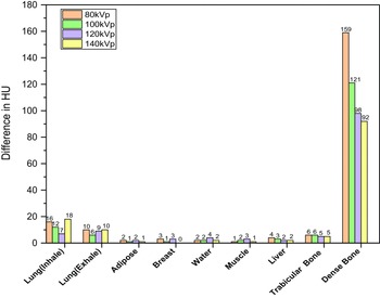
Figure 6 shows the difference between the F12 and F32 reconstruction filters without BHC; according to the tube voltage setting, the difference was greater for the materials at the extremes of the HU scale (the lowest and highest densities). However, it was still within the tolerances for lung. Hence, for dense bone, the variation in HU values was increased when the tube voltage was decreased from 140 kVp to 80 kVp.
HU variation between F32 and F12 without BHC according to different kVp settings.

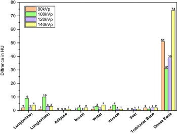
Figure 7 shows the variation of HU of substitute materials between F12 and F32 with BHC according to the tube voltage settings. In this case, the greatest variation appeared in the tube voltage extremes, which are 140 kVp and 80 kVp.
HU variation between F32 and F12 with BHC according to different kVp settings.

Discussion
In the present study, we aimed to evaluate the effect of reconstruction filters on HU variation and investigate the differences based on kVp settings.
According to the results, we concluded that the reconstruction filters have no significant effect on the HU change except in high-density materials; the degree of this variation depends on the selected tube voltage. Thus, for dense bone, the difference in HU between F12 and F32 increased as energy decreased from 140 kVp to 80 kVp. This result is consistent with the study of Sarah et al. Reference Kirwin, Langmack and Nightingale29 On a Siemens Emotion Duo scanner, a range of head and body reconstruction algorithms were tested with varying energies of 80 kV, 110 kV and 130kV. They concluded that the maximum difference between reconstructions filters increases when decreasing energy.
However, this no longer applies when selecting the BHC. In addition, the selection of BHC reduces the effect of the reconstruction filters on the HU, and only the differences between the filters when selecting the extreme energies (80 kVp and 140 kVp) become out of tolerance (±50 HU) for the dense bone. For other materials, we had a minimal effect of about 2–3 HU, as reported in Figure 7.
This can be explained by the change in energy spectrum resulting from the BHC algorithm, which aims to transform polychromatic attenuation data into the monochromatic equivalent before image reconstruction. Reference Ketcham and Hanna30
Therefore, changes in HU depend on the BHC algorithm of the scanner brand. Reference Zheng, Al-Hayek and Cummins25 In the literature, the effect of these algorithms is not well described, although the extent of the variation is not clearly indicated. For instance, on a Toshiba Aquilion scanner, Zurl et al. Reference Zurl, Tiefling, Winkler, Kindl and Kapp11 concluded that the use of a beam-hardening filter resulted in a dose difference of 0·6% in response to a density change of about 5%. Nevertheless, this study does not precisely indicate the HU changes of each substitute material.
The common point between the effects of the reconstruction filters as a function of all tube voltage settings is that only HU values of dense bone were significantly affected. Similarly, a tolerable effect was observed for the lung and negligible for the soft tissue. In this context, several studies have investigated the effect of reconstruction filters on HU using different types of scanners. One of them was conducted by Anne et al. Reference Davis, Muscat and Palmer27 on three brands of scanner: GE (Chicago, USA), Toshiba (now Canon, Tochigi Prefecture, Japan) and Siemens (Erlangen, Germany). They found that the degree of HU change depended strongly on the selected reconstruction filter, with some resulting in little or no change. The largest HU changes were observed for reconstruction filters on GE (Chicago, USA) and Toshiba (now Canon, Tochigi Prefecture, Japan) CT scanners, even for soft tissue. In contrast, Siemens showed insignificant variations in HU.
Typically, radiotherapy centres insert the calibration curves in TPS taking into account the effect of kVp change, but few centres take into account the effect of reconstruction filters on HU. It seems that this may be caused by the lack of studies in this context.
However, a significant number of studies have recently been published, which evaluate the effect of reconstruction filters from different scanner brands on HU variations. Reference van der Heyden, Öllers, Ritter, Verhaegen and van Elmpt2,Reference Vergalasova, McKenna, Yue and Reyhan3,Reference Ebert, Lambert and Greer20,Reference Davis, Muscat and Palmer27 These studies can help physicists to understand the performance of their scanner in HU calculation. Thus, a change in kVp during a dosimetric scan can be associated with a change in the acquisition parameters, including the reconstruction filter. However, these earlier studies only examined the effect of filters for a single energy. Therefore, it was important to investigate the effect of reconstruction filters on HU values according to the selected energy, which was the objective of this work.
The decision to implement a new calibration curve or not is based on the effect of HU variations on the dose calculation. For example, third generation algorithms such as Monte Carlo take into account small variations in the electron density of the medium and include them in dose calculations. Reference Saini, Pandey, Kumar, Singh and Pasricha12
Therefore, with the advent of new techniques like intensity-modulated radiation therapy, the tolerances of variations in dose calculation have become more stringent. Consequently, each centre must establish its own HU tolerances according to its technical platform (scanner, phantom, TPS, accelerator, etc.) and treatment techniques.
Conclusion
The purpose of this work is to evaluate the effect of Hitachi Supria scanner reconstruction filters on HU and to see how this effect varies as a function of kVp. This can help the clinicians quantify any possible change in HU when varying both reconstruction filters and tube voltage.