Introduction
The genus Llimoniella was introduced for two lichenicolous fungi growing on terricolous lichens of gypsum soils (Hafellner & Navarro-Rosinés Reference Hafellner and Navarro-Rosinés1993). Up to now, thirteen taxa have been recognized within the genus (Diederich & Etayo Reference Diederich and Etayo2000; Diederich et al. Reference Diederich, Ertz and Etayo2010). The original concept of Llimoniella has recently been enlarged to include a group of taxa, referred to as the L. phaeophysciae group, which does not exhibit the K+ violet, purplish or green pigments in the epihymenium and excipulum but are otherwise typical of Llimoniella (Diederich et al. Reference Diederich, Ertz and Etayo2010). During the course of recent work on lichens and lichenicolous fungi in Alaska (e.g., Spribille et al. Reference Spribille, Pérez-Ortega, Tønsberg and Schirokauer2010), we found an undescribed species of lichenicolous fungus growing on the soralia of Ramboldia cinnabarina that matches the concept of the Llimoniella phaeophysciae group. Here we describe the new taxon and discuss its affinity to Llimoniella and related genera within the Helotiales.
Material and Methods
Specimens were examined using a Leica S8 stereomicrocope. Hand-cut sections of ascomata were studied in distilled water, lactophenol cotton blue and KOH. The amyloid reactions were tested in Lugol's iodine solution (Panreac 251774) directly (I) and after pre-treatment with KOH (K/I). Pigment reactions were observed under KOH and c. 40% nitric acid. Habit photographs were taken using a Leica S8 stereomicrocope fitted with a Leica EC3 digital camera. Microscopic pictures were taken using a Zeiss AX10 microscope fitted with ‘Nomarski’ differential interference contrast and an AxioCam digital camera. All measurements were made in water. Ascospore size measurements are indicated as (minimum−) x−σx–x+σx (−maximum), with the average together with ± standard deviation in italics; values are approximated to the nearest 0·5 µm.
Taxonomy
Llimoniella cinnabarinae Pérez-Ortega, Etayo & T. Sprib. sp. nov
MycoBank No.: MB 519894
Llimoniella species, in thallis Ramboldiae cinnabarinae vigens, ascomatibus atris, 180–300 µm diam., excipulo et epihymenio brunneo, K+ brunneo-aurantiaco; hymenio non insperso; ascis subcylindricis ad clavatis, 8-sporis; ascosporis stricte ellipsoideis, (15 − )16 − 18(−20) × 3–4(−5) µm.
Typus: USA, Alaska, Matanuska-Susitna Borough, Denali National Park, Tokositna River below terminus of Tokositna Glacier, west bank of river, 62°38·947′N, 150°47·590′W, on Ramboldia cinnabarina, on young Picea, 264 m elev., 18 August 2008, T. Spribille 27868 with C. Printzen, P. Nelson & J. Walton (NY— holotypus).
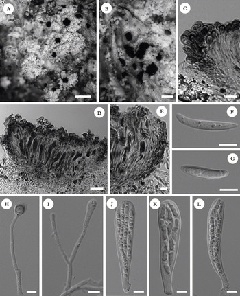
(Fig. 1A–L)
Llimoniella cinnabarinae (holotype, Spribille 27868, NY). A & B, habit; C & E, details of excipulum; D, longitudinal section of an ascoma; F & G, ascospores; H, paraphysis showing a swollen apex and a pigmented apical cell; I, young and branched paraphysis; J–L, asci. C, D & E: in lactophenol blue; F, G, H, I, J, K & L: in water after KOH pretreatment (differential interference contrast). Scale bars: A = 500 µm; B = 250 µm; C & E = 15 µm; D = 30 µm; F– L = 5 µm.

Ascomata usually aggregated on the host thallus (soralia), immersed in the substratum to slightly superficial; dark brown to black, matt or slightly shiny, disc usually faintly lighter, closed when young but soon with exposed hymenium, irregularly rounded to elongated, up to 300 µm wide at the widest point; margin thin, prominent, especially when young, without hairs; disc concave to applanate. Exciple thin, composed of radiating to interwoven hyphae, with thick walls and usually with an enlarged and subglobose terminal cell, with an orange to light brown pigment in the upper part, disappearing in the lower part, K+ darker brown, N+ brighter orange, 17–35 µm thick in the upper part, 16–25 µm in the hyaline, lowest part; not stipitate at the base. Subhymenium colourless. Hymenium hyaline to light yellowish, not inspersed, 55–70 µm tall, I−, K/I−. Epihymenium with an orange, medium brown pigment, K+ dark brown, N+ brighter orange. Paraphyses hyaline, septate, 1·5–2·5 µm thick, thicker at the tips, up to 5–6 µm, with pigmented caps, sparingly branched. Asci clavate to subcylindrical, 35–55 × 8–12 µm, wall apically not thickened, I−, K/I−, 8-spored. Ascospores hyaline, narrowly ellipsoid to fusiform, usually slightly curved, non-septate, (15 − )16–18(−20) × 3–4(−5) µm; wall smooth, without perispore.
Conidiomata unknown.
Host. On the thallus, mostly on the soralia of Ramboldia cinnabarina.
Distribution. The species is so far known only from two localities in Alaska (USA). The host is very common in this region and the species can be expected to be more common than these two records would seem to indicate.
Remarks. Our new taxon is characterized by its immersed to superficial ascomata, thin exciple, paraphyses with swollen apices and pigmented caps, brownish orange excipular and epihymenial pigments that react K+ dark brown and N+ lighter orange, non-thickened ascus apex and non-septate ascospores, which are c 16–18 × 3–4 µm. This description falls within the new, enlarged concept of Llimoniella (Diederich et al. Reference Diederich, Ertz and Etayo2010). The genus was originally conceived to include species with black, superficial apothecia, with a slightly concave to convex disc, upper exciple composed of thick, radiating cells forming a margin externally lacking hairs, a combination of characteristic pigments that usually react K+ violet or purplish, thin-walled, non-amyloid asci and simple to septate, straight to sigmoid, hyaline ascospores (Hafellner & Navarro-Rosinés Reference Hafellner and Navarro-Rosinés1993; Diederich & Etayo Reference Diederich and Etayo2000). The description and inclusion of a group of species without such pigments, namely the L. phaeophysciae group, has broadened the concept of Llimoniella (Diederich et al. Reference Diederich, Ertz and Etayo2010). Within this group, the most similar species to the one we describe here is L. heppiae (Nav.-Ros., Hladun & Llimona) Diederich & Ertz. This species, growing on the terricolous lichen Heppia despreauxii, was originally described in Gelatinopsis (Navarro-Rosinés et al. Reference Navarro-Rosinés, Hladun and Llimona2008), and only later combined into Llimoniella (Diederich et al. Reference Diederich, Ertz and Etayo2010). Llimoniella cinnabarinae shares with L. heppiae the apically swollen paraphyses with a distinct pigment cap, a character exclusive to these two species, as well as a brownish orange pigment. Llimoniella cinnabarinae differs from L. heppiae in ascospore shape and size, with ascospores irregularly ellipsoid, straight to slightly curved and 10·5–13 × 4·5–5·5 µm in L. heppiae, and narrowly ellipsoid to fusiform, usually curved, and 16–18(−20) × 3–4 µm in L. cinnabarinae. Both species also differ in their ascus shape, which is apically applanate in L. heppiae, but not in L. cinnabarinae, and in their different hosts.
The new species is also reminiscent of two other genera in Helotiales: Gelatinopsis Rambold & Triebel Reference Rambold and Triebel1990 and Rhymbocarpus Zopf 1896 (Rambold & Triebel Reference Rambold and Triebel1990; Diederich & Etayo Reference Diederich and Etayo2000). Gelatinopsis was described to include two fungi that were lichenicolous and fungicolous, respectively (Rambold & Triebel Reference Rambold and Triebel1990). Further incorporations to the genus have shown that fungicolous species differ from lichenicolous species in the presence of a thicker apical ascus wall and, at least in the type, strongly convex and immarginate apothecia (Baral & Marson Reference Baral, Marson and Consiglio2001). This fact led Diederich et al. (Reference Diederich, Ertz and Etayo2010) to combine all previously described lichenicolous Gelatinopsis species into Llimoniella or Rhymbocarpus. Rhymbocarpus is probably closely related to Llimoniella, but differs from it in that the ascomata are immersed and almost perithecioid when young, with a greenish, K+ olivaceous pigment present in the epihymenium, and an excipulum composed of elongate cells that are sometimes filiform in the apex or prolonged in the form of external, excipular hairs (Diederich & Etayo Reference Diederich and Etayo2000). In L. cinnabarinae the hymenium is exposed from an early stage, not appearing perithecioid, and it does not contain a greenish, K+ olivaceous pigment in the epihymenium. Llimoniella cinnabarinae also possesses an exciple composed of radiating to entangled hyphae that are usually swollen in the apex, not filiform as in Rhymbocarpus.
Spirographa cf. fusisporella was found growing together with L. cinnabarinae in the holotype. However, S. fusisporella is a very different species easily distinguishable from Llimoniella by its sigmoid and twisted ascospores.
Additional specimen examined. USA: Alaska: Klondike Gold Rush National Historic Park, Chilkoot trail just above Sheep Camp, 59°39·930′N, 135°15·912′W, on Ramboldia cinnabarina, 378 m elev., 2008, Pérez-Ortega 1440 et al. (hb. Pérez-Ortega).
SPO is supported by the program JAE-Doc (Consejo Superior de Investigaciones Científicas). We thank Dr J. Wierzchos (CSIC, Spain) for his kind help with microscopy questions. Support for fieldwork in Klondike Gold Rush National Historical Park was provided by the U.S. National Park Service Natural Preservation Program and the Southeast Alaska Inventory and Monitoring Program, with special thanks to Dave Schirokauer. Fieldwork in Denali National Park was made possible by Peter Nelson, James Walton and Carl Roland and is gratefully acknowledged.