INTRODUCTION
The protozoon parasite Histomonas meleagridis is the causative agent of histomonosis, also known as blackhead disease or infectious enterohepatitis, of gallinaceous birds (Tyzzer, Reference Tyzzer1920). The complete ban of all available licensed drugs effective against histomonosis raised an urgent demand for intensive research on the development of new effective strategies against the disease (Hess et al. Reference Hess, Liebhart, Bilic and Ganas2015).
The establishment of a monoeukaryotic culture of H. meleagridis, derived from one singular cell of a virulent histomonad strain (Hess et al. Reference Hess, Liebhart, Grabensteiner and Singh2008; Liebhart et al. Reference Liebhart, Windisch and Hess2010) via micromanipulation and the attenuation of the parasite by long-term passaging was described recently (Hess et al. Reference Hess, Kolbe, Grabensteiner and Prosl2006, Reference Hess, Liebhart, Grabensteiner and Singh2008). Subsequently, the protective capability of this attenuated clonal strain could be demonstrated in various trials (Liebhart et al. Reference Liebhart, Ganas, Sulejmanovic and Hess2017). During ongoing research on this attenuated parasite a deviation from the phenotype generally expressed by H. meleagridis in other in vitro cultures was observed.
So far, the parasite has been described as a highly pleomorphic protozoon that occurs in various forms depending on the site of its dwelling (Tyzzer, Reference Tyzzer1919, Reference Tyzzer1920; Bishop, Reference Bishop1938). Soon after its discovery an aflagellated tissue form, 6–20 µm in size, that assumes an amoeboid shape while invading the tissue, and a more spherical vegetative form within the tissue were reported (Tyzzer, Reference Tyzzer1919, Reference Tyzzer1920; Lee et al. Reference Lee, Long, Millard and Bradley1969). When residing in the avian caecal lumen, the parasite is flagellated and rounded but still expresses the capacity for amoebic movement by forming pseudopodia. This form has a smaller size of 3–16 µm and is comparable to the form usually seen in in vitro settings (Honigberg and Bennett, Reference Honigberg and Bennett1971; McDougald and Reid, Reference McDougald and Reid1978; Mielewczik et al. Reference Mielewczik, Mehlhorn, Al-Quraishy, Grabensteiner and Hess2008; Munsch et al. Reference Munsch, Lotfi, Hafez, Al-Quraishy and Mehlhorn2009). However, some discrepancies regarding the protozoon's morphology can be found in the existing literature as several authors based their morphological descriptions on cultures of both H. meleagridis and the related flagellate Histomonas wenrichi also assigned as Parahistomonas wenrichi (Tyzzer, Reference Tyzzer1919, Reference Tyzzer1920; Wenrich, Reference Wenrich1943; Lund, Reference Lund1963). Furthermore, investigations were somewhat complicated by the presence of morphologically similar parasites such as Blastocystis spp. or Tetratrichomonas gallinarum in cultures established from feces (Delappe, Reference Delappe1952; Harrison et al. Reference Harrison, Hansen, DeVolt, Holst and Tromba1954).
Earlier studies on long-time in vitro propagation of H. meleagridis were performed by Lund et al. (Reference Lund, Augustine and Ellis1966, Reference Lund, Augustine and Chute1967). Although those authors also reported a loss of virulence of the parasite after long-term passaging they based this on a selection process between various strains present within the cultures used in their cultivation experiments and denied that H. meleagridis could be attenuated (Lund et al. Reference Lund, Augustine and Ellis1966). This theory, however, was refuted by demonstrating an attenuation process in parasite cultures derived from a single cell (Hess et al. Reference Hess, Liebhart, Grabensteiner and Singh2008). Furthermore, Lund et al. (Reference Lund, Augustine and Chute1967) could not detect any significant changes in morphology despite having cultivated parasites for over 7 years.
Therefore, the objective of this study was to conduct detailed investigations on the morphology of both attenuated and virulent parasites derived from previously established clonal monoxenic strains of H. meleagridis. Additionally, the influence of attenuation on growth behaviour and tenacity were determined. Comparing the attenuated form with its virulent clone, the aim was to gain further insights into important characteristics of the parasite which are of value for the ongoing research on the formulation and application of a H. meleagridis live vaccine.
MATERIALS AND METHODS
Cultures
All experiments were carried out using the monoxenic clonal culture H. meleagridis/turkey/Austria/2922-C6/04, established by micromanipulation after isolation from a naturally infected turkey (Hess et al. Reference Hess, Kolbe, Grabensteiner and Prosl2006). Cultures had been previously transferred to a monoxenic state by replacing the original fecal bacteria with the laboratory strain Escherichia coli DH5α (Invitrogen, Vienna, Austria) (Ganas et al. Reference Ganas, Liebhart, Glösmann, Hess and Hess2012). Histomonads of high [>290x (HP)] and low [<50x (LP)] in vitro passages of this strain had been cryopreserved at −150 °C. After thawing, cells were cultivated in sterile Falcon tubes (Sarstedt, Wiener Neudorf, Austria) in Medium 199 containing Earle's salts, L-glutamine, 25 mm HEPES and L-amino acids (Gibco™, Invitrogen, Vienna, Austria), 15% heat-inactivated fetal calf serum (Biochrom GmbH, Berlin, Germany) and 0·22% sterilized rice starch (Carl Roth, Karlsruhe, Germany). Cultures were maintained at an incubation temperature of 40 °C.
Passaging of the cultures was performed every 48–72 h by transferring 1 mL of cultured cells into 9 mL of fresh medium. For the experiments cells were propagated in sterile T25 tissue culture flasks (Sarstedt, Wiener Neudorf, Austria) by transferring 1 mL of cultured parasites together with 1 mL of overnight E. coli DH5α culture into 28 mL of fresh medium and incubating them for 48–72 h. Cell count and vitality of the parasites were determined using a Neubauer counting chamber and Trypan Blue dye (GIBCO™, Invitrogen).
For the tenacity assays the content of the tissue culture flasks was transferred to Falcon tubes and after determination of the cell count, centrifuged at 300 g for 5 min. The supernatant was removed and the cell pellet re-suspended in the calculated amount of fresh culture medium to establish a cell count of 1 × 10⁶ cells mL−1.
Morphological analysis
The first part of the comparative morphological analysis of histomonad cells was performed with cells derived from 48 to 72 h old HP and LP cultures under standard cultivation conditions. After extracting samples to study the parasites' morphology, these cultures were supplemented with an antibiotic mixture (50 µg mL−1 doripenem, 500 µg mL−1 neomycin and 300 µg mL−1 rifampicin) for 24 h to examine the stressful effect of the eradication of co-cultivated E. coli DH5α on cell morphology. This protocol of antibiotic treatment is in conformity with that used for the corresponding tenacity assay later on. Absence of live bacteria in the culture medium was confirmed by transferring aliquots of the cultures onto Columbia Agar supplemented with 5% sheep blood (COS) (BioMérieux, Vienna, Austria) and Coliform agar (CF) (Merck, Vienna, Austria) and incubation for 24 h at 37 °C.
Light microscopy
Comparison of live HP and LP histomonad cells was performed on an Olympus BX53 microscope (Olympus Austria GmbH, Vienna, Austria) using the Olympus cellSens 1·5 digital imaging software (Olympus Austria GmbH, Vienna, Austria). Aliquots of 15 µL were taken from the tissue culture flasks and transferred to glass slides for examination. Photographs of the samples were taken immediately after preparation of the slide to avoid a possible influence of the environmental changes on cell morphology. Afterwards, the images were evaluated using the cellSens software. Three times 300 cells per culture type were evaluated during three separate runs of the experiment by tracing the cell contour either manually in case of the irregular cells or via a spherical template adjustable in size for the round parasites. Based on this, cell types were categorized into two morphological groups, irregularly shaped and completely spherical. In addition, the whole cell area was used as a marker for cell size, in order to compare round and irregularly-shaped cell forms.
Trichrome stain
Permanent smears of the different cultures were prepared by polyvinyl alcohol fixation (PVA Gold Standard Fixative, Polysciences Europe GmbH, Hirschberg an der Bergstrasse, Germany) and stained, using a modification of Gomori's trichrome stain established by Wheatley (Reference Wheatley1951). Wheatley's Trichrome Blue (Polysciences Europe GmbH, Hirschberg an der Bergstrasse, Germany) staining was performed according to the manufacturer's instructions and cells were viewed by light microscopy under oil immersion.
Electron microscopy
To assess the cell morphology on an ultrastructural level, transmission electron microscopy (TEM) and scanning electron microscopy (SEM) were performed on cell suspensions from cultures before and after antibiotic treatment.
Transmission electron microscopy
For TEM, parasitic cells were fixed in 5% glutaraldehyde (Merck, Darmstadt, Germany) in 0·1 m phosphate buffer (Sigma-Aldrich, Vienna, Austria), pH 7·2, at 4 °C for 3 h. A cell pellet was generated by centrifugation of the culture and fixative mix at 300 g for 5 min. This cell pellet was then washed in 0·1 m phosphate buffer resuspended in 1% osmium tetroxide (Merck, Darmstadt, Germany) at 4 °C for 1 h. The cells were coated in agarose to form a dense pellet, which was then trimmed into 1–2 mm cubes using a razor blade. Dehydration of the samples occurred in an alcohol gradient series and propylene oxide (Merck, Darmstadt, Germany) followed by embedding in glycid ether (Serva, Heidelberg, Germany). Samples were cut on a Leica Ultramicrotome (Leica Ultracut S, Vienna, Austria) to obtain ultrathin sections and stained with uranyl acetate (Sigma-Aldrich, Vienna, Austria) and lead citrate (Merck, Darmstadt, Germany). The cells were examined with a Zeiss TEM 900 electron microscope (Carl Zeiss, Oberkochen, Germany).
Scanning electron microscopy
For SEM, histomonad cultures were incubated overnight on poly-L-lysine coated glass cover slips to allow adherence of the cells, which were then fixed in 2·5% glutaraldehyde in 0·1 m cacodylate buffer, pH 7·2 for 2 h at 4 °C. Post-fixation occurred in 1% OsO4 in cacodylate buffer for 15 min followed by dehydration in a graded series of ethanol. The slides were critical point-dried in a Bal-TEC CPD030 (Leica Microsystems GmbH, Wetzlar, Germany), mounted and sputter-coated with gold in a Polaron SC7640 (Quorum Technologies Ltd., Newhaven, UK). The samples were examined in a JEOL JSM-5800 scanning electron microscope (Jeol Ltd., Tokyo, Japan).
Growth behaviour
Tissue culture flasks containing 28 mL of fresh culture medium and 1 mL of overnight E. coli DH5α culture were inoculated with 1 mL fresh culture medium containing 3 × 10⁵ histomonads, either from HP or LP cultures and incubated at 40 °C. Cell counts were monitored over 8 days. Growth curves were generated during three separate runs of the experiment, from the mean cell counts of three individual tissue culture flasks at every time point using a Neubauer counting chamber. Only vital cells, unstained by Trypan blue dye (GIBCO™, Invitrogen) were counted to evaluate the growth pattern of the parasites. Furthermore, parasite morphology was monitored throughout the growth phases by light microscopy. To exclude a possible influence of differing bacterial numbers within the cultures, aliquots of cultures of both passages were examined for comparable bacterial cell counts on COS and CF plates.
Tenacity
To evaluate cell tenacity the assessment of parasite survival and stress response under a change of culture conditions was performed in 1·5 mL Eppendorf tubes with 1 mL of histomonad culture containing 1 × 10⁶ cells. With exception of the temperature assay, all experiments were performed at an incubation temperature of 40 °C. All experiments were performed three times, all of them in triplicate.
Temperature
Test tubes were incubated at 40, 22 and 4 °C to mimic an incubation optimum, room temperature and a severe drop in temperature, respectively. The cell counts of vital histomonads from HP and LP cultures were measured after an incubation time of 12 and 24 h, respectively. After 24 h the cultures were re-incubated at 40 °C and examined for histomonad growth over 72 h to confirm the absence of vital cells.
Reduction of bacteria
The number of vital H. meleagridis cells in the absence of live bacteria was assessed after 24 h of incubation with a combination of antibiotics (50 µg mL−1 doripenem, 500 µg mL−1 neomycin and 300 µg mL−1 rifampicin). Dilution series of the cultures were transferred to COS and CF plates before and after antibiotic treatment to monitor antibacterial influence and to confirm the absence of live bacteria.
Change in pH level
Cell vitality and morphology was monitored over a time period of 24 h after Histomonas cells were confronted with a deviation of the pH level from the near neutral standard cultivation conditions to a pH level of either 8·5 or 4·5, by adding hypochlorite or acetic acid, respectively. The PH levels were adjusted by titration of acid or lye to sterile culture medium and assessed by pH meter and pH test strips (MColorpHast™, Merck, Vienna, Austria). Cultures containing parasites and bacteria were supplemented with the previously established amount of lye and acid, resulting in concentrations of 0·03% hypochlorite (Sigma-Aldrich, Vienna, Austria) and 0·17% acetic acid (Sigma-Aldrich, Vienna, Austria). At the time no vital cells could be detected anymore in the cultures, the test tubes were centrifuged for 5 min at 300 g and the supernatant was removed. The cell pellet was washed with Medium 199 and then re-suspended in 1 mL of fresh standard cultivation medium enriched with E. coli DH5α. Cultures were examined for histomonad growth over a period of 72 h.
Statistical analysis
Statistical evaluation was performed using the chi-squared (χ2) test of independence and t-test for unpaired samples in SPSS and Microsoft Excel. Differences were considered statistically significant when the P value was ⩽0·05.
RESULTS
Morphology and ultrastructure
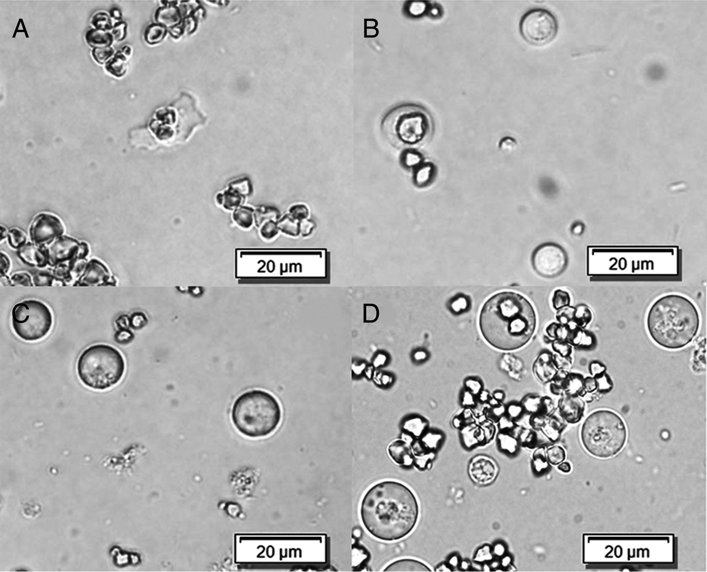
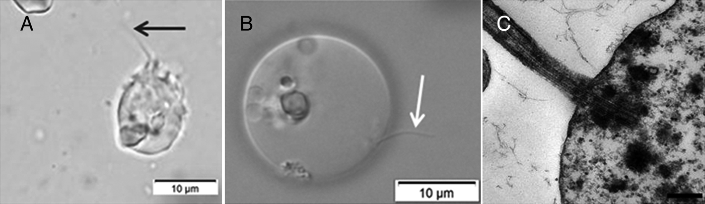
Cultures of the attenuated parasite consisted of about 94% irregularly shaped amoeboid cells, whereas only 6% of the measured cell population assumed a completely rounded morphology. These amoebic cells often appeared flat and thinly spread out on the slides with a highly irregular contour (Fig. 1A). In LP cultures, almost 70% of the cells appeared spherical (Fig. 1B) and the remaining 30% showed small irregularities by forming pseudopodia. The very distinct amoeboid cell shape, typical for the attenuated histomonads, could not be found in cultures with virulent parasites. Killing of bacteria, by adding of antimicrobials for 24 h, led to the formation of up to 100% spherical cells (Fig. 1C and D) in both the virulent and avirulent cultures (Supplementary Fig. 1). The size varied widely within both HP (43–529 µm2, with a mean value of 150 µm2) and LP (17–518 µm2, with a mean value of 140 µm2). Despite the wide range in size, on average, parasitic cells of the long-term in vitro propagated cultures were significantly larger. The spherical parasites in the cultures supplemented with antibiotics also exhibited a wide variation in size (45–624 µm2, with mean values of 147 µm2 for HP and 162 µm2 for LP) with no significant difference between the cells of HP and LP (Supplementary Fig. 2). Parasites with one or sometimes two flagella could be found in all cultures, before and after treatment (Fig. 2).
Light micrographs of H. meleagridis. (A, B) Untreated cells from cultures after 48 h of incubation at 40 °C in unchanged standard cultivation medium showing either an (A) amoeboid morphology in the HP sample or a (B) rounded cell form in the LP sample. (C, D) Spherical morphology of parasites from cultures of (C) HP and (D) LP after 24 h of treatment with antibiotics.

Expression of the flagellum (arrows) by HP parasites. (A) Oil immersion light micrograph of an amoeboid parasite under standard cultivation conditions. (B) Phase contrast micrograph of a cell with typical spherical morphology after 24 h antibiotic treatment. (C) Protrusion of the flagellum depicted in TEM micrograph. The scale represents 250 nm (Magnification × 30 000).

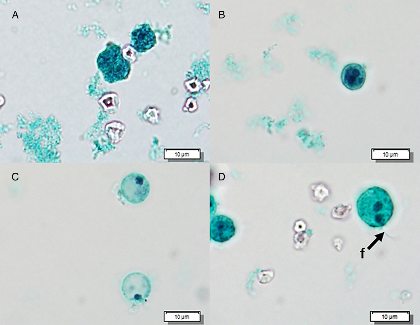
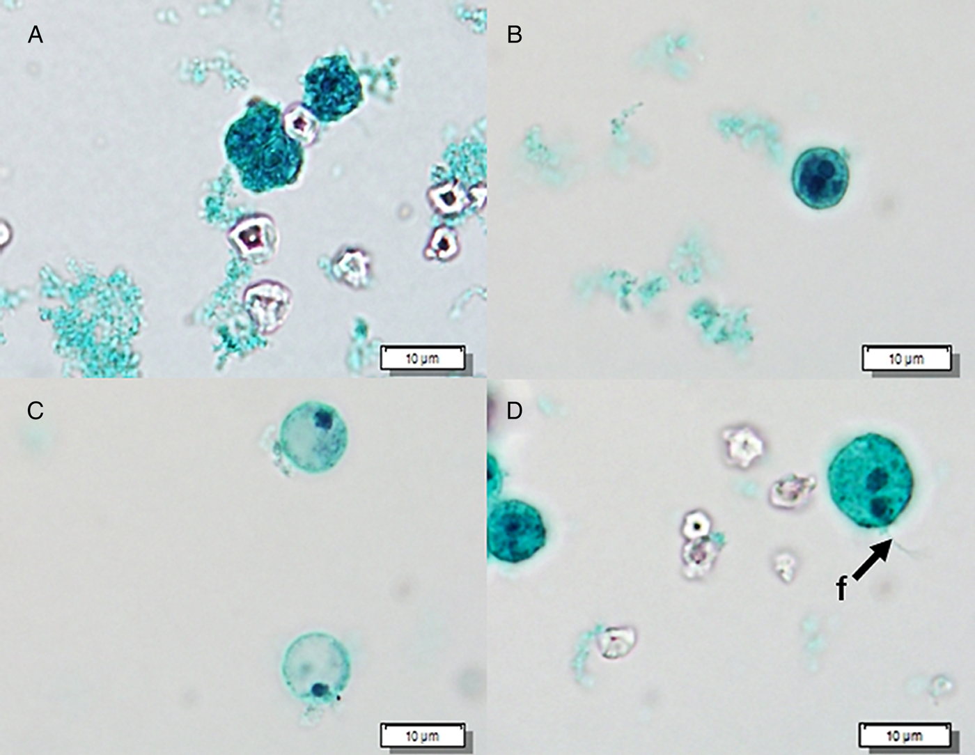
A more detailed visualization of cell morphology was achieved by evaluating smears stained by trichrome blue (Fig. 3), revealing differences in staining characteristics between parasites before and after antibiotic treatment, whereas untreated parasites stained mostly very intensely (Fig. 3A and B), the round forms, noticed after bacterial eradication, often appeared largely unstained and empty, lacking the usually visible inclusions of rice starch (Fig. 3C). Bi-nucleated cells (Fig. 3D), sometimes expressing two flagella, could be revealed in all cultures, also after antibiotic treatment.
Light micrographs of H. meleagridis after PVA fixation and trichrome staining obtained from cultures incubated at 40 °C for 48 h in unchanged standard culture medium. Parasites showed either (A) amoeboid morphology in the HP sample or (B) a rounded cell form in the LP sample. Inside the cells the darker stained nucleus and ingested rice starch granules are visible, surrounded by intensely stained cytoplasm. (C) Spherical cells of the high passage after antibiotic treatment with darker stained nuclei and otherwise relatively transparent cytoplasm. (D) Bi-nucleated spherical cell of the low passage culture after antibiotic treatment with a visible flagellum (f).
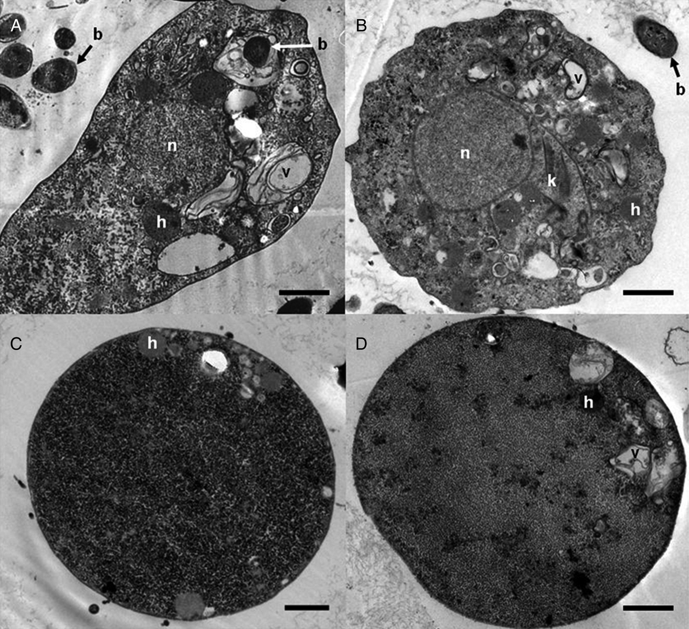
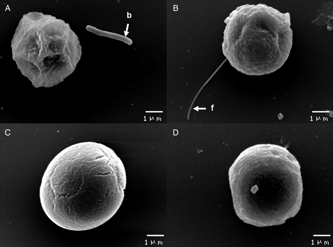
Changes in morphology after long-term cultivation and the response to the antibiotic treatment were also obvious by electron microscopy (Figs. 4 and 5). In the TEM micrographs, both the HP (Fig. 4A) and LP (Fig. 4B) forms of the parasite contained the typical organelles such as nucleus, Golgi apparatus, hydrogenosomes and multiple food vacuoles along with numerous glycogen granules. Histomonads from the cultures that underwent antibiotic treatment differed by appearing very densely packed with glycogen granules (Fig. 4C and D). In the investigated sections, organelles could, if at all, only be observed as a dense cluster at the cell periphery. All membrane irregularities, as seen in the untreated parasites, had smoothed out to form a completely even surface. Flagellated parasites could be seen in all cultures before and after antibiotic treatment.
TEM micrographs of fixed H. meleagridis cells from (A) HP and (B) LP cultures under standard cultivation conditions. Note the elongated amoeboid shape of the attenuated parasite. Virulent parasites showed a round shape and sometimes slightly wavy surface. The nucleus (n), kinetic apparatus (k) as well as numerous hydrogenosomes (h) and food vacuoles (v) can be viewed. The surrounding cytoplasm appeared to be filled with glycogen granules. Co-cultivated E. coli DH5α (b) can be found in the surroundings and inside food vacuoles after ingestion by the parasite. Cells from both the (C) HP and (D) LP culture assumed a spherical shape after antibiotic treatment for 24 h. In the cell periphery only few organelles such as hydrogenosomes and food vacuoles could be depicted. Otherwise, the cytoplasm appeared densely packed with glycogen particles. The spherical cells are surrounded by a smooth membrane, lacking irregularities such as pseudopodia. The scale represents 1000 nm (Magnification: A- C × 7000; D × 4400).

SEM micrographs of H. meleagridis from (A) HP and (B) LP cultures under standard cultivation conditions. Multiple indentations of the cell surface amounting to an amoeboid morphology in the (A) attenuated parasite could be contrasted to a more rounded shape in the (B) virulent histomonads. Note the flagellum (f) adhering to the glass cover slip and co-cultivated E. coli DH5α (b). Cells of the (C) attenuated and (D) virulent form after antibiotic treatment for 24 h appeared spherical and exhibited a smoothed out surface texture.

Similar results could be obtained by SEM regarding cell shape before and after antibiotic treatment (Fig. 5A–D). In the treated histomonads, a change towards a smoother surface texture of the cells could be observed (Fig. 5C and D).
Growth
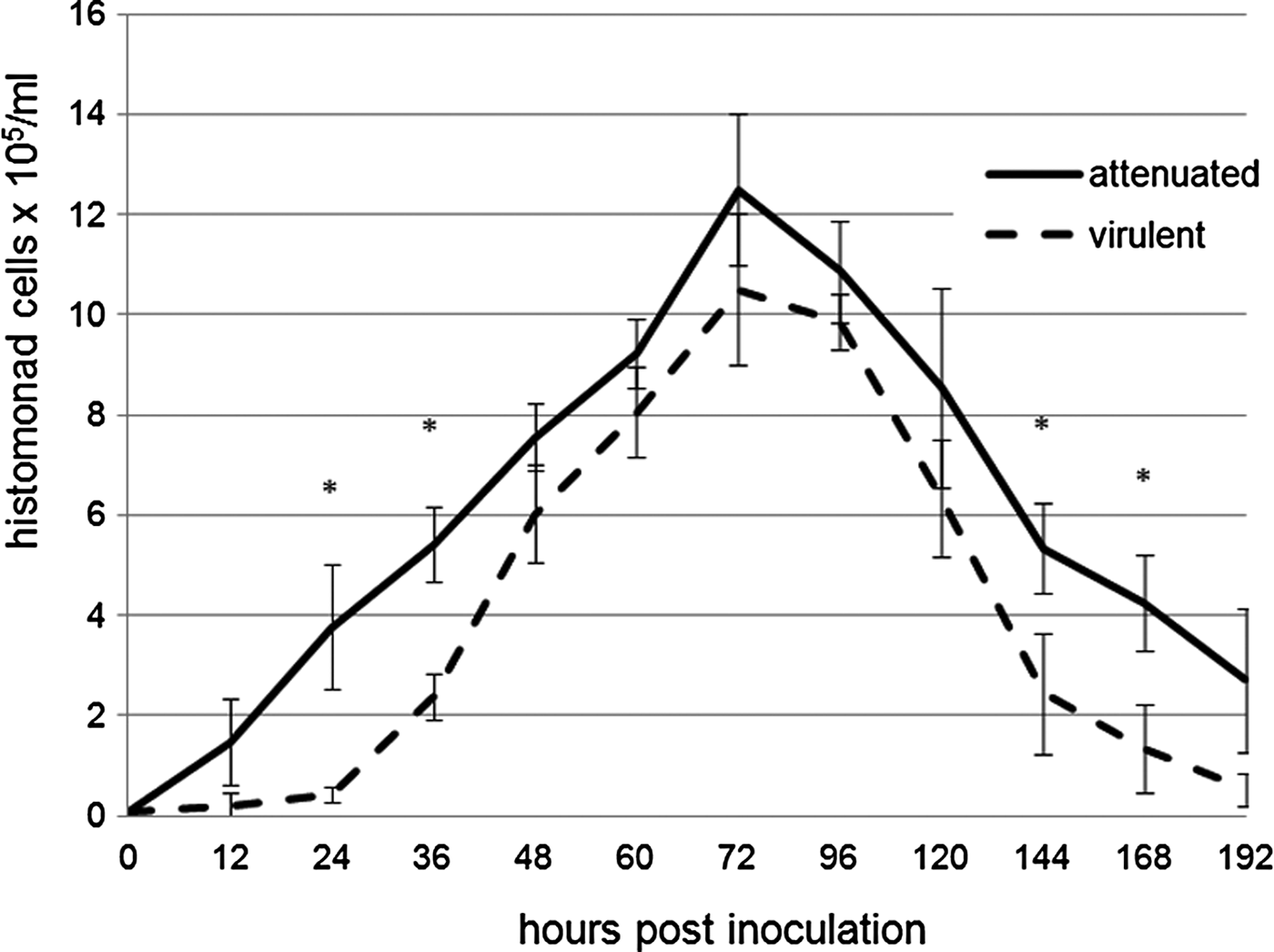
Growth of H. meleagridis in vitro under standard cultivation conditions is shown in Fig. 6. In general, all cultures reached a peak of growth level at 72 h, after which the number of histomonads began to decline again. Under identical cultivation conditions and an equal monoxenic setting (~5 × 10⁸ bacterial cells mL−1 of E. coli DH5α) higher cell counts were noted in HP cultures in each individual run of the experiment. However, combining the values of the individual experiments for statistical analysis, the difference could not be proven to be statistically significant. Nevertheless, LP parasites differed by having a significantly longer lag-phase during the first 48 h after passaging and a steeper decline of cell numbers from day 5 onwards. The above described differences in morphology between HP and LP cultures persisted throughout the growth phases (data not shown).
Growth curves of attenuated and virulent histomonads under standard culture conditions generated over 8 days. Significantly (*) higher cell counts could be documented for the HP clone at multiple time points post inoculation (p.i.). A certain lag phase within the first 24 h, visible for the cultures containing the virulent parasites, was not exhibited by the attenuated strain. Moreover, during the decline of parasites after a peak of growth at 72 h p.i. the HP parasites were able to maintain significantly higher cell counts.

Tenacity
Temperature
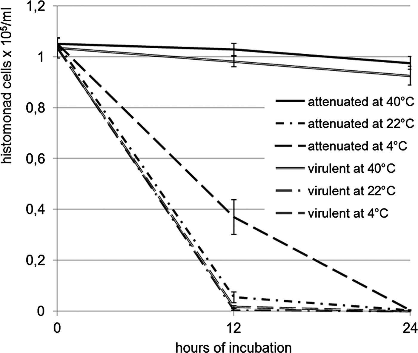
Incubation at lowered cultivation temperatures of 22 and 4 °C led to a rapid decline in cell count within 24 h, in contrast to cultures that had been kept at an incubation optimum of 40 °C (Fig. 7). Again, an increase in spherical forms within the cultures could be observed. Comparing the effect of temperature changes on the two different forms of the parasite at 22 °C, significantly higher cell numbers were revealed for the attenuated strain. When incubated at 4 °C the HP cultures were also able to maintain higher cell counts over 12 h when compared with the same culture at 22 °C and LP cultures at both temperatures. After 24 h no vital parasites of the virulent strain could be detected anymore, whereas the cultures with the attenuated clone still contained a low number of vital cells. This result was confirmed by re-incubating the cultures at 40 °C with successful re-growth of parasites only in the HP cultures.
Cell counts of vital histomonads in HP and LP cultures in standard cultivation medium at incubation temperatures of 22 and 4 °C. Cultures incubated at 40 °C served as a negative control. After 24 h, a low number of vital cells could still be detected in HP cultures at both temperatures, whereas only dead cells could be found in LP cultures.

Reduction of bacteria
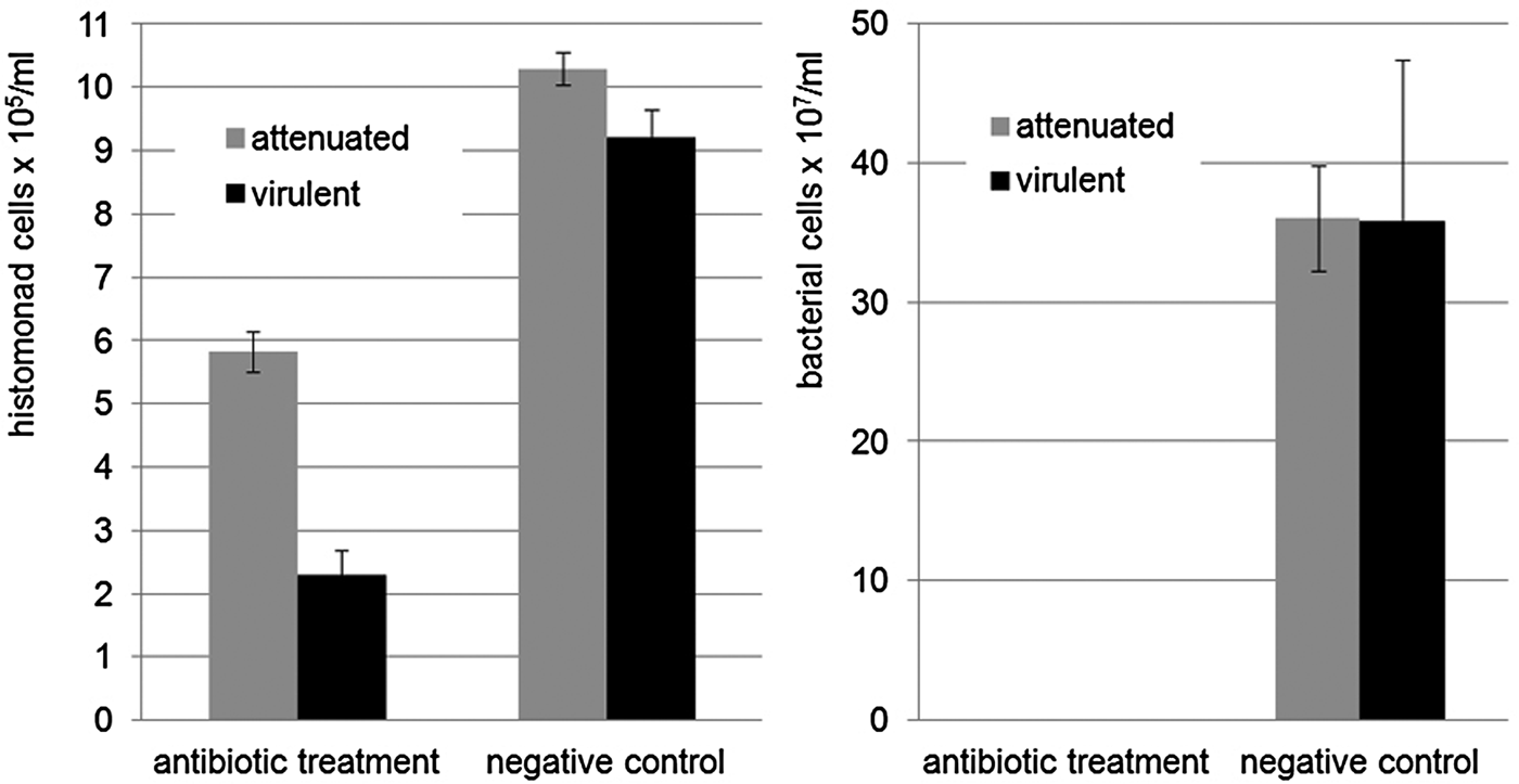
Elimination of vital E. coli DH5α from the culture medium could be achieved within the 24 h time span of the experiment, verified by the absence of bacterial growth on COS and CF plates. Both the attenuated and virulent parasites reacted to this by assuming a spherical shape and a certain decline in vital cells was detected. However, the decrease in parasite numbers was significantly less pronounced in HP cultures containing the attenuated parasites (Fig. 8).
Quantification of parasites and bacteria in HP and LP cultures in a standard cultivation medium after 24 h of antibiotic treatment (doripenem 50 µg mL−1, neomycin 500 µg mL−1, rifampicin 300 µg mL−1).

Change in pH level
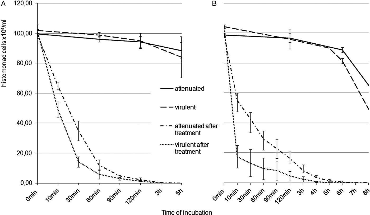
Both, alkalization (Fig. 9A) and acidification (Fig. 9B) of culture medium led to a shift towards the completely spherical cell form. The drastic change of the pH resulted in a steep decline of parasite numbers in both HP and LP within 60 min after addition of the respective chemical but had a more severe impact on cell numbers of the virulent parasite. The attenuated parasites were able to survive longer by 2–3 h with an overall survival time of 5 h in the alkaline pH (8·5) and 8 h in the acidic pH (4·5). The absence of vital cells at the respective time points was confirmed by negative re-cultivation results.
Influence of pH change on the vitality of histomonads. (A) LP cultures in a medium supplemented with hypochlorite (0·03%) appeared more sensitive to the alkaline pH level (8·5) than the HP cultures, reflected in a more rapid decline in vital cells within the first 90 min and an overall shorter survival time by 2–3 h. (B) Similar results could be observed in the medium acidified by acetic acid (0·18%) to a pH level of 4·5.

DISCUSSION
Various morphological forms of H. meleagridis inside the host have been described, the flagellated lumen dwelling form and two aflagellated types of cells, the amoeboid invasive and the spherical tissue form (Tyzzer, Reference Tyzzer1919, Reference Tyzzer1920). In vitro cultivated parasites were reported similar to the lumen dwelling form (Bishop, Reference Bishop1938; Lund et al. Reference Lund, Augustine and Chute1967; Schuster, Reference Schuster1968; Honigberg and Bennett, Reference Honigberg and Bennett1971). However, descriptions of cultivated parasites vary widely in the existing literature. In the more recent studies, a rounded flagellated type and an amoeboid unflagellated form were frequently described, although parasites with a different history of in vitro passages and culture conditions were investigated (Mielewczik et al. Reference Mielewczik, Mehlhorn, Al-Quraishy, Grabensteiner and Hess2008; Munsch et al. Reference Munsch, Lotfi, Hafez, Al-Quraishy and Mehlhorn2009; Zaragatzki et al. Reference Zaragatzki, Hess, Grabensteiner, Abdel-Ghaffar, Al-Rasheid and Mehlhorn2010a).
In the present study, however, it could be demonstrated that long-term in vitro cultivation under a well-defined and optimal cultivation regime caused the cells to deviate from a rounded morphology, as seen in the virulent parasites, towards a distinctly amoeboid phenotype without losing the flagellum. This form is similar to that seen by Bishop (Reference Bishop1938) who had established cultures from infected liver tissue and cultivated them for 9–10 months without further studies in vivo. Those parasites showed the ability to assume a similar amoebic morphology when observed on slides heated to 37 °C, whereas parasites viewed without the aid of a plate warmer, scarcely showed any amoeboid movement. A similar phenomenon was observed by Honigberg and Bennett (Reference Honigberg and Bennett1971). However, the attenuated parasites used in the present investigation expressed a highly amoeboid phenotype and were capable to maintain it for an extended period of time even when studied at room temperature.
This flagellated amoeboid form, by offering a large surface area, could in a way be the most effective shape to utilize the full range of offered food sources in vitro, rice starch, bacteria and nutrients of the cultivation medium, while the aflagellated amoeboid tissue form only feeds on lysed host tissue by pinocytosis (Lee et al. Reference Lee, Long, Millard and Bradley1969). It can also be hypothesized that the continued expression of the flagellum of amoeboid parasites offers an additional advantage regarding motility in the liquid medium, which is supported by a higher growth rate of HP histomonads in the actual study. Therefore, it can be concluded that the parasite adapted its phenotype to the environmental conditions which was also observed in other long-term in vitro passaged strains of the parasite (data not shown). However, the results contradict observations reported by Lund et al. (Reference Lund, Augustine and Chute1967) who did not notice any significant changes in the parasite's morphology after long-term cultivation despite certain adaptations to the cell physiology. Such parasites were passaged over 1000 times and a loss of virulence could be demonstrated. However, frequent variations made to the cultivation conditions, as described by the authors, might have prevented a distinctive and pronounced morphological adaptation. In comparison, the strain presently investigated was attenuated applying a consistent cultivation protocol for over 290 passages (Hess et al. Reference Hess, Liebhart, Grabensteiner and Singh2008). In addition, improvements in the cultivation procedures for H. meleagridis, e.g. addition of rice starch and changes in composition of the medium, which have been established since the experiments in 1967, might have further contributed to these obvious differences (Dwyer, Reference Dwyer1970; Van der Heijden et al. Reference Van der Heijden, McDougald and Landman2005; Hauck et al. Reference Hauck, Armstrong and McDougald2010).
In the tenacity experiments, H. meleagridis responded to a confrontation with agonistic stimuli by assuming a strictly spherical morphology. It could be argued that the rounded shape presents a defence mechanism due to the fact that it minimizes the exposed surface area. These forms could reflect cyst-like stages, which were previously described (BonDurant and Wakenell, Reference BonDurant, Wakenell and Kreier1994; Mielewczik et al. Reference Mielewczik, Mehlhorn, Al-Quraishy, Grabensteiner and Hess2008; Munsch et al. Reference Munsch, Lotfi, Hafez, Al-Quraishy and Mehlhorn2009; Zaragatzki et al. Reference Zaragatzki, Hess, Grabensteiner, Abdel-Ghaffar, Al-Rasheid and Mehlhorn2010a, Reference Zaragatzki, Mehlhorn, Abdel-Ghaffar, Al-Rasheid, Grabensteiner and Hessb). Similarly, such a shift towards a spherical morphology was also observed in other flagellates of the family Trichomonadidae under unfavourable environmental conditions (Friedhoff et al. Reference Friedhoff, Kuhnigk and Müller1991; Granger et al. Reference Granger, Warwood, Benchimol and De Souza2000; Pereira-Neves et al. Reference Pereira-Neves, Ribeiro and Benchimol2003). Recent reports suggest that a cyst stage could also be of importance in the transmission of H. meleagridis' closest relative, Dientamoeba fragilis (Munasinghe et al. Reference Munasinghe, Vella, Ellis, Windsor and Stark2013; Munasinghe, Reference Munasinghe2016; Stark et al. Reference Stark, Garcia, Barratt, Phillips, Roberts, Marriott, Harkness and Ellis2014).
The findings of the present study are also in agreement with the reports of Zaragatzki et al. (Reference Zaragatzki, Hess, Grabensteiner, Abdel-Ghaffar, Al-Rasheid and Mehlhorn2010a, Reference Zaragatzki, Mehlhorn, Abdel-Ghaffar, Al-Rasheid, Grabensteiner and Hessb), who described a very similar response of H. meleagridis to stress, including the rounding of the cells and a reduction in food vacuoles by TEM. SEM micrographs utilized in the actual study supported the morphological changes. Furthermore, from TEM and the trichrome stained smears an intracellular accumulation of glycogen granules could be concluded; whereas in TEM these histomonads appeared densely packed with glycogen granules, almost empty cells were noticed in the trichrome stained smears as glycogen remains unstained by this method (Zeibig, Reference Zeibig2004). Increased storage of glycogen could be interpreted as a further sign of the formation of a defensive form as it was also described for the encystation process of a similar protozoon, Entamoeba histolytica (Osada, Reference Osada1959). Furthermore, a significantly higher amount of stored glycogen, when compared to that of the highly motile invasive stage of H. meleagridis, has been described for the vegetative stage of the parasite that resides immotile in the host tissue (Lee et al. Reference Lee, Long, Millard and Bradley1969). The fact that a less active stage of the parasite would contain more glycogen further supports the theory that these spherical cells resemble a resting stage. However, with the flagellum still present under all conditions applied in the in vitro assays and without noticeable changes in membrane structure or visible formation of a cyst wall, the induction of a true encystation process could not be demonstrated within the presented experiments.
As a parasite dependant on the presence of suitable microbiota (Franker and Doll, Reference Franker and Doll1964), H. meleagridis could so far not be successfully cultivated under axenic conditions (Hauck et al. Reference Hauck, Armstrong and McDougald2010; Ganas et al. Reference Ganas, Liebhart, Glösmann, Hess and Hess2012). Based upon very limited knowledge, it is a general believe that the co-cultivated bacteria are necessary to produce an anaerobic environment favourable for H. meleagridis and that they might act as a nutrient source since bacteria are taken up by cultured protozoa (Munsch et al. Reference Munsch, Lotfi, Hafez, Al-Quraishy and Mehlhorn2009; Ganas et al. Reference Ganas, Liebhart, Glösmann, Hess and Hess2012). This strong dependence was also noticed in the actual study following antibiotic treatment, undertaken for the morphological assay and tenacity testing. However, within the course of 24 h the absence of live bacteria was much better tolerated by the attenuated parasites.
In culture, H. meleagridis is very sensitive to changes in temperature (DeVolt and Davis, Reference DeVolt and Davis1936; Bishop, Reference Bishop1938; Gerhold et al. Reference Gerhold, Lollis, Beckstead and McDougald2010). The findings of the present experiment are very much in accordance with those of previous experiments reported by Bishop (Reference Bishop1938) who observed the death of nearly all protozoa within 24 h in cultures incubated either at room temperature or at 5 °C. In the present study, however, vital cells could still be detected in all HP cultures after 24 h independent of the incubation temperature. Apparently, the attenuated parasites were able to handle the change of temperature somewhat better. In related parasites such as Monocercomonas sp. or T. foetus, changes in incubation temperature can be used to induce pseudo-cyst formation (Granger et al. Reference Granger, Warwood, Benchimol and De Souza2000; Borges et al. Reference Borges, Gottardi, Stuepp, Larré, Vieira, Tasca and Carli2007). Hence, it could be assumed that lowering the temperature to 4 °C in the culture medium also triggered a sort of survival mechanism in H. meleagridis. Induction of encystation or pseudo-cyst formation in other protozoa coincides with the down regulation of the parasites' metabolism, as has been described for example for Entamoeba invadens (Jeelani et al. Reference Jeelani, Sato, Husain, Escueta-de Cadiz, Sugimoto, Soga, Suematsu and Nozaki2012). Similarly, H. meleagridis had a higher survival rate at 4 °C in comparison with 22 °C indicating that lowering its metabolic activity could support survival at critical temperatures. This seems another indicator for the parasites' adaptation during long-term in vitro propagation, as this phenomenon was not observed in cultures containing virulent histomonads.
Overall, it could be demonstrated that the parasite underwent a substantial adaptation process as a result of long-term (>290x) in vitro passaging. This is characterized by a shift in phenotype regarding its morphology, growth behaviour and tenacity when compared to its virulent low passage clone. A defence mechanism of H. meleagridis was observed in both forms of the parasite, resembling a previously reported cyst-like stage. The formation of a true cyst form, however, could not be observed in these in vitro settings.
The obtained results support the development of a vaccination strategy based upon long-term cultivated parasites.
SUPPLEMENTARY MATERIAL
The supplementary material for this article can be found at https://doi.org/10.1017/S0031182017000646.
ACKNOWLEDGEMENTS
All ELMI samples were examined at the Institute of Pathology and Forensic Veterinary Medicine, Department of Pathobiology, Veterinary University, Vienna. Special thanks to Dr Michael Szostak from the Institute of Bacteriology for the substantial help with the preparation of samples and SEM imaging and Mag. Nora Dinhopl from the Institute of Pathology and Forensic Veterinary Medicine for her support in sample preparation and TEM imaging.