Introduction
Sperm cryopreservation is a valuable broodstock management method for stock enhancement and, hence, for the conservation of endangered fish species. However, the current sperm cryopreservation protocols for small endangered fish species have not yet been standardized or even attempted (Tiersch Reference Tiersch2001; Yang & Tiersch, Reference Yang and Tiersch2009), because small semen volume and short duration of spawning period limit the performance of a systematic study for sperm cryopreservation. Furthermore, in most fish species, most hatched larvae are lost and only a small fraction survive to sexual maturity.
Cryopreservation of spermatogonia may provide a valid alternative to cryopreservation of sperm because functional sperm can be obtained via germ-line chimeras produced by spermatogonial transplantation (Okutsu et al., Reference Okutsu, Shikina, Kanno, Takeuchi and Yoshizaki2007) and in vitro spermatogenesis (Sakai, Reference Sakai2002). Furthermore, cryopreservation of spermatogonia other than sperm can be beneficial for the preservation of endangered fish species as this alternative expands the option of cell types for preservation and also enables the utilization of non-spawning fish and even juveniles as genetic resources.
Honmoroko Gnathopogon caerulescens is a small cyprinid (≈10 cm body length and ≈10 g body weight) that is endemic to ancient Lake Biwa in Japan. Although this fish has been an important target of the lake fisheries over a long period of time, the natural stock has rapidly decreased in recent decades and now this fish is listed as a critically endangered species in the Red List of Japan (Ministry of the Environment, Japan, 2015). A very short spawning period (July to March; Nakamura, Reference Nakamura and Nakamura1969) and a large loss of the juvenile population of this fish during the winter (Maki, Reference Maki1968) make the collection of sperm for cryopreservation difficult. Hence, we have recently established an in vitro spermatogenesis system to produce functional sperm from spermatogonia for this fish (Higaki et al., Reference Higaki, Shimada, Kawamoto, Todo, Kawasaki, Tooyama, Fujioka, Sakai and Takada2017).
In our previous study, testicular cells were dissociated enzymatically before cryopreservation (Higaki et al., Reference Higaki, Shimada, Kawamoto, Todo, Kawasaki, Tooyama, Fujioka, Sakai and Takada2017). This method may cause inevitable cell loss due to the multiple washing steps by centrifugation that are required to obtain a single-cell suspension for cryopreservation. Therefore, in this study, we developed a cryopreservation technique for whole testis that diminished the loss of spermatogonia during cryopreservation.
Vitrification is a process by which liquids solidify without ice crystal formation (Luyet & Hodapp, Reference Luyet and Hodapp1938), requiring only suitable cryoprotective solutions and a rapid cooling rate, which is generally archived by immersion into liquid nitrogen (Amorim et al., Reference Amorim, Curaba, Van Langendonckt, Dolmans and Donnez2011; Saragusty & Arav, Reference Saragusty and Arav2011). Vitrification can be performed quick and easy, and no special equipment to manage the cooling rate is needed. As special equipment is difficult to acquire at remote sites where this endemic species lives, we chose vitrification as the cryopreservation method. The production of gametes from vitrified whole testes through germ-line chimeras has been recently reported for medaka (Oryzias latipes; Seki et al., Reference Seki, Kusano, Lee, Iwasaki, Yagisawa, Ishida, Hiratsuka, Sasado, Naruse and Yoshizaki2017). To the best of our knowledge, however, this report is the first to describe the successful production of functional sperm from vitrified whole testis via in vitro spermatogenesis in fish.
Materials and Methods
Fish and preparation of testis
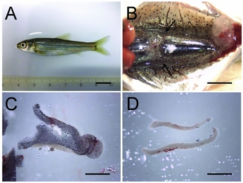
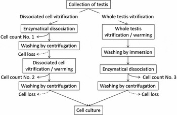
Juvenile male G. caerulescens (7–10 months of age, approximately 6 cm in total length) used in this experiment were artificially raised from embryos at the Laboratory of Cell Engineering, Department of Pharmaceutical Sciences, Ritsumeikan University, under a natural light-dark cycle at 24 ± 2°C. The fish were euthanized by immersion in ice-cold water and the surface was disinfected with 70% (v/v) ethanol; subsequently, their testes were aseptically dissected out. High cooling and warming rates are crucial for successful vitrification (Yavin & Arav, Reference Yavin and Arav2007), hence adherent fatty and non-testicular tissues were removed from the testes (Fig. 1) to increase the cooling and warming rates by minimizing the sample volume. The trimmed testes (approximately 10 mm in length and 1 mm in width) were transferred into sterile phosphate-buffered saline (PBS) supplemented with 0.5% (v/v) commercial bleach. After a 2-min immersion, the testes were washed three times in sterile PBS for 2 min each and used for histological examination and two types of cryopreservation protocols: dissociated cell vitrification and whole testis vitrification (Fig. 2).
Flow chart of the two vitrification protocols for testicular cell cryopreservation in honmoroko Gnathopogon caerulescens. In the vitrification protocol for dissociated testicular cells, a dissected testis was enzymatically digested, washed by centrifugation, and vitrified in cryo-vial with a cryomedium for human ES/iPS cells (RCHEFM001: ReproCELL, Kanagawa, Japan) by direct immersion in liquid nitrogen. Vitrified dissociated cells were warmed with 10% (v/v) fetal bovine serum in Leibovitz's L-15 medium and washed by centrifugation. In the vitrification protocol for whole testis, a dissected testis was vitrified on a strip-shaped nylon mesh by direct immersion in liquid nitrogen after serial exposures to self-made pretreatment solution (PS) and vitrification solution (VS). Vitrified whole testis was warmed and washed by sequential immersion into thawing and washing solutions and enzymatically digested. Cell count nos. 1–3 indicate the timing of the cell counts and cell viability assays performed during this study.

Testis preparation for whole testis vitrification in juvenile honmoroko Gnathopogon caerulescens. Seven- to 10-month-old juvenile fish (approximately 6 cm in total length) were euthanized by immersion in ice-cold water (A). Following surface disinfection with 70% (v/v) ethanol, a ventral midline incision was made from the urogenital opening to just beneath the jaw. The intestine was cut at the posterior end and removed anteriorly to expose the testes (B). Aseptically dissected testes were transferred into sterile phosphate-buffered saline (C). Fatty and non-testicular tissues attached to the testes were removed (D). Arrows in (B) indicate the testes. Bar in (A) = 1 cm. Bars in (B), (C), and (D) = 2 mm.

To prepare unfertilized eggs for artificial insemination assays, mature female G. caerulescens (> 1 year of age) were purchased during their spawning season from a local fish farmer 1 day before use. In total, 39 juvenile male and six mature female fish were used in this study. All experiments were carried out according to the guidelines of Ritsumeikan University.
Histological examination of testis
To examine the developmental stages of spermatogenic cells, testes obtained from 10 juveniles were fixed with 4% (w/v) paraformaldehyde in PBS supplemented with 10% (v/v) acetic acid overnight at 4°C. The fixed testes were dehydrated in ethanol and xylene, and embedded in paraffin. Some of the sections (5-μm thickness) were deparaffinized and stained with hematoxylin/eosin for examination of the germ cell stages. The corresponding adjacent sections were stained immunohistochemically with antibodies against the Vasa protein (a germ cell marker (Raz, Reference Raz2000)) to confirm the developmental stage of the germ cells. In brief, deparaffinized sections were immersed in heated (98°C) antigen retrieval solution (Immunosaver; Nissin EM, Tokyo, Japan) for 45 min. The tissue sections were then incubated overnight at 4°C with diluted polyclonal rabbit anti-Vasa homologue protein (GTX128306: GeneTex, Irvine, CA, USA; diluted 1:1500) in PBS containing 4% (v/v) normal goat serum. The sections were then incubated with Alexa Fluor 488-conjugated goat anti-rabbit IgG (Invitrogen, Carlsbad, CA, USA; diluted 1:500) in PBS for 45 min at room temperature. Cell nuclei were counterstained with 4′,6′-diamidino-2-phenylindole (DAPI; Dojindo Laboratory, Kumamoto, Japan) for 20 min. Negative controls lacking the primary antibody were also examined. Images were obtained using an inverted microscope (IX70, Olympus, Japan) at magnification ×400.
Vitrification of dissociated testicular cells
To compare and validate the efficiency of whole testis vitrification, vitrification of dissociated testicular cells were performed (Higaki et al., Reference Higaki, Shimada, Kawamoto, Todo, Kawasaki, Tooyama, Fujioka, Sakai and Takada2017) using the commercially available DAP213 solution (2 M DMSO, 1 M acetamide, and 3 M propylene glycol) (RCHEFM001: ReproCELL, Kanagawa, Japan) (Fujioka et al., Reference Fujioka, Yasuchika, Nakamura, Nakatsuji and Suemori2004). In short, each testis was minced with microscissors and digested in 500 µl Leibovitz's L-15 medium containing 500 U/ml collagenase (17104–019, Life Technologies) for 30 min at 28°C under constant shaking (140 rpm) with gentle pipetting every 10 min. The cell suspension was washed by centrifugation at 120 g for 5 min with 5 ml HEPES-buffered Leibovitz's L-15 medium containing 1% (w/v) bovine serum albumin (BSA). After discarding the supernatant, the pelleted cells were resuspended in 200 μl DAP213 solution, transferred into a 1.8 ml CryoTube vial, and cooled rapidly by plunging the vial into liquid nitrogen within 15 s.
For warming, the cryopreserved vial was removed from the liquid nitrogen and the cells were warmed quickly by the adding 1 ml prewarmed (37°C) Leibovitz's L-15 medium supplemented with 10% fetal bovine serum (FBS) within 15 s. The cell suspension was diluted with 9 ml medium (37°C) and washed by centrifugation at 120 g for 3 min. The experiment was repeated eight times using one testis for each replicate.
Vitrification of whole testis
Whole testis was vitrified according to the previously described procedures for the cryopreservation of zebrafish primordial germ cells by vitrification of whole embryos (Higaki et al., Reference Higaki, Kawakami, Eto, Yamaha, Nagano, Katagiri, Takada and Takahashi2013) with modifications. Briefly, the pretreatment solution (PS) consisted of Leibovitz's L-15 medium supplemented with 2 M ethylene glycol (EG), 1 M dimethyl sulfoxide (DMSO), and 0.35% BSA. The vitrification solution (VS) consisted of Leibovitz's L-15 medium supplemented with 3 M EG, 2 M DMSO, 0.5 M sucrose, and 0.7% BSA. In total, 1 ml of PS and VS were each prepared in individual wells of a 4-well dish (176740, Thermo Fisher Scientific, Waltham, MA, USA). For vitrification, each testis was serially exposed to PS for 20 or 30 min and to VS for 5, 10 or 20 min, and then loaded onto a strip-shaped (approximately 5 mm × 30 mm) nylon mesh (100-μm pore size, NYTAL 13XX-100: SEFAR, Thal, Switzerland) using fine-tipped forceps. The testis-loaded nylon mesh was then plunged into liquid nitrogen directly. The nylon mesh was transferred into a CryoTube vial and stored in liquid nitrogen.
For warming, the cryopreserved nylon mesh was removed from the vial in liquid nitrogen and immersed in 10 ml thawing solution (TS), which consisted of Leibovitz's L-15 medium supplemented with 0.7% BSA and 0.5 M sucrose. After a 4-min immersion, the testis detached from the nylon mesh were transferred into 1 ml of a 1:1 mixture of TS and testicular cell culture medium (TCCM) (Sakai, Reference Sakai2006) supplemented with 10% FBS (TCCM–FBS) and kept there for 5 min. The testis was then washed twice in 1 ml fresh TCCM–FBS for 5 min each and dissociated into single cells as described above. All cooling and warming procedures were performed in an air-conditioned room set at 25°C. Fish embryo extract, which was contained in TCCM, was prepared from common carp (Cyprinus carpio) embryos as described previously (Westerfield, Reference Westerfield and Westerfield2007). In each exposure protocol for PS/VS, the experiment was repeated four times using one testis for each replicate.
Viability analysis of testicular cells
The viability of testicular cells was assessed by the intactness of the plasma membrane using trypan blue exclusion test when cell counts were obtained. Briefly, dissociated testicular cells were stained with 0.25% (w/v) trypan blue (Kanto Chemical, Tokyo, Japan) for 3 min at room temperature. The numbers of trypan blue-negative and trypan blue-positive cells were counted using a hematocytometer, and the percentages of membrane-intact cells were calculated according to the following formula:
percentage of membrane-intact cells = number of trypan blue-negative cells/number of total cells.
Cell culture of vitrified whole testis
Testicular cells dissociated from vitrified whole testes that were cryopreserved following serial exposures to PS for 20 min and VS for 10 min were cultured under adhesion and suspension conditions as described previously (Higaki et al., Reference Higaki, Shimada, Kawamoto, Todo, Kawasaki, Tooyama, Fujioka, Sakai and Takada2017). Briefly, dissociated testicular cells were resuspended in TCCM–FBS supplemented with 3% (v/v) common carp serum, 100 ng/ml epidermal growth factor (EGF, Peprotech, Rocky Hill, NJ, USA), 100 ng/ml basic fibroblast growth factor (bFGF, Peprotech), 100 ng/ml insulin-like growth factor-I (IGF-I, Peprotech), 10 μM forskolin (Sigma-Aldrich), 0.1 mM β-mercaptoethanol (Sigma-Aldrich), 10 IU/ml human chorionic gonadotropin (HCG, ASKA Pharmaceutical, Tokyo, Japan), 10 IU/ml pregnant mare's serum gonadotropin (PMSG, ASKA Pharmaceutical), 50 ng/ml 11-ketotestosterone (11-KT, Cosmo Bio, Tokyo, Japan), 100 ng/ml 17β-estradiol (E2, Wako Pure Chemicals, Osaka, Japan), and 10 ng/ml 17α,20β-dihydroxy-4-pregnen-3-one (DHP, Toronto Research Chemicals, North York, NO, Canada). One pair of testes was plated into four 35-mm plastic tissue culture dishes (3000-035, AGC Techno Glass, Shizuoka, Japan) coated with 0.1% (w/v) gelatin for adhesion culture, or one pair of testes was plated onto one plastic non-treated Petri dish (1000-035, AGC Techno Glass) for suspension culture. Testicular cells were cultured at 18°C in humidified air, and the medium was changed every 3 or 4 days. Adhesion culture was used for assessing germ cell differentiation, because the morphological changes of adherent germ cells were easily discernible. While suspension culture was suitable for flow cytometric analyses and fertilization assays, because efficient sperm production was achieved by suspension culture rather than adhesion (Higaki et al., Reference Higaki, Shimada, Kawamoto, Todo, Kawasaki, Tooyama, Fujioka, Sakai and Takada2017). Adhesion and suspension culture experiments were repeated two and six times, respectively, using two testes for each replicate.
Flow cytometry analysis of testicular cells to confirm spermatozoa presence
To assess meiotic progression in cell culture, testicular cells cultured under suspension conditions were dissociated into single cells, fixed with methanol and stained with propidium iodide (PI), and the DNA content of nuclei was measured by flow cytometry. Briefly, culture medium containing suspended testicular cells was centrifuged at 800 g for 7 min, and the pelleted cells were washed with PBS twice by centrifugation. The cells were dissociated by treatment with PBS containing 0.06% (w/v) trypsin and 1.32 mM ethylene diamine tetraacetic acid for 30 min at room temperature. The trypsin was neutralized by the addition of Leibovitz's L-15 medium supplemented with 10% FBS, and the suspended cells were washed again with PBS by centrifugation. The cells were fixed with 70% (v/v) ice-cold ethanol for 30 min. The cells were then washed with PBS by centrifugation and treated with 1 mg/ml RNase A (Nakalai Tesque, Kyoto, Japan) and 50 μg/ml PI (Wako Pure Chemicals, Osaka, Japan) for 15 min in the dark. The stained cells were filtered through a 40-μm nylon mesh to remove cellular debris and cell clumps, and analysed by flow cytometry. Flow cytometry was performed on a FACSCalibur machine (BD Biosciences, San Jose, CA, USA) using CellQuest software (BD Biosciences). The experiment was repeated three times using suspension-cultured testicular cells. As a control, non-cultured testicular cells obtained from five juvenile fish of the same age were combined and analysed.
In vitro fertilization assay
To validate the fertilization and developmental competence of sperm differentiated in vitro, the sperm were subjected to in vitro fertilization assays. Briefly, the medium containing sperm was collected from each culture dish, and sperm were concentrated by centrifugation at 800 g for 7 min. The majority of the supernatant was discarded, and the cell pellet was resuspended in the remaining medium (<100 μl). To avoid female effects, the sperm suspension was divided into two aliquots and added to two dishes each containing a half batch of unfertilized eggs, which were obtained from two different HCG-primed female G. caerulescens, and the dish was swirled for 1 min to mix the sperm and eggs. Dechlorinated tap water was added gradually with shaking, and the eggs were incubated at 23°C. Embryonic development was assessed at the somite (24 h after insemination) and hatching stages (5 days after insemination). The experiment was repeated three times using suspension-cultured testicular cells obtained from one culture dish (containing two testes) and two females for each replicate.
Statistical analysis
Data were expressed as the mean ± standard deviation. Percentages of membrane-intact cells were transformed using the arcsine transformation (arcsin √%) before analysis to normalize the variance. Data were analysed by one-way analysis of variance (ANOVA) followed by the Tukey–Kramer post hoc test. The SPSS for Windows computer program (version 12.0: SPSS Inc., IL, USA) was used for statistical analyses. Differences were considered statistically significant at P < 0.05.
Results and Discussion
Successful cryopreservation of whole testis/testicular tissue by the conventional slow freezing and vitrification methods were reported in a few fish species, such as slow freezing for testicular tissue in Siberian sturgeon (Acipenser baerii; Pšenička et al., Reference Pšenička, Saito, Rodina and Dzyuba2016), slow freezing for whole testis in rainbow trout (Oncorhynchus mykiss; Lee et al., Reference Lee, Iwasaki, Shikina and Yoshizaki2013), and vitrification for whole testis in medaka (Seki et al., Reference Seki, Kusano, Lee, Iwasaki, Yagisawa, Ishida, Hiratsuka, Sasado, Naruse and Yoshizaki2017). Although functional sperm was obtained from the cryopreserved testis through germ-line chimeras (Lee et al., Reference Lee, Iwasaki, Shikina and Yoshizaki2013; Seki et al., Reference Seki, Kusano, Lee, Iwasaki, Yagisawa, Ishida, Hiratsuka, Sasado, Naruse and Yoshizaki2017), we successfully produced functional sperm via in vitro spermatogenesis from vitrified whole testis in fish.
Histological examination of testis
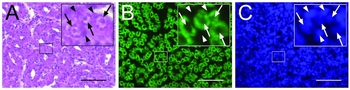
As shown in Fig. 3, all Vasa-positive spermatogenic cells in juvenile testis had the typical appearance of spermatogonia, with a few compact nucleoli and a relatively large nucleus (Leal et al., Reference Leal, Cardoso, Nóbrega, Batlouni, Bogerd, França and Schulz2009).
Testicular histology of juvenile honmoroko Gnathopogon caerulescens. Testicular sections were stained with hematoxylin and eosin (A). Corresponding adjacent sections were immunohistochemically stained with antibodies against the Vasa protein (B). Nuclei were counterstained with 4´,6´-diamidino-2-phenylindole (C). Insets in each panel represent high-magnification images of the selected areas. Arrows and arrowheads indicate the nuclei of germ and somatic cells, respectively. Note the specific expression of Vasa protein in germ cells (green in B). Bars = 50 μm.

Vitrification of dissociated testicular cells
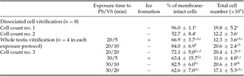
As shown in Table 1, both the percentage of membrane-intact cells and the total number of dissociated testicular cells decreased from the pre-vitrification step (cell count no. 1) to the post-vitrification step (cell count no. 2; P < 0.05). As a result, the live cell recovery rate was calculated to be 33.8% (live cell number in cell count nos. 1 and 2 were calculated to be 19.0 × 10⁵ cells and 6.4 × 10⁵ cells, respectively) using the conventional vitrification protocol for dissociated testicular cells.
Ice formation during cooling and percentages of membrane-intact cells recovered from vitrified whole testes of juvenile honmoroko Gnathopogon caerulescens

PS and VS represent pretreatment solution and vitrification solution, respectively. ‘+’ and ‘−’ indicate that ice formation was observed in the whole, in part of, and none of the testes during cooling, respectively. Membrane intactness of the cryopreserved cells was determined by trypan blue dye exclusion.
a,b,c,d,e Values (means ± standard deviation (SD) of four replicates) with different superscripts differ significantly within the same row (P < 0.05).
Vitrification of whole testis
Under the tested conditions, the highest percentage of membrane-intact cells (84%) with the largest total cell number (20.6 × 10⁵ cells) was obtained from testis that was vitrified following serial exposures to PS for 20 min and VS for 10 min (Table 1).
Ice formation was observed in parts of the testis sample that was rapidly cooled after a 5-min immersion in VS, regardless of the time of exposure to PS (Table 1). Although the percentages (67 and 63%) of membrane-intact cells of ice-formed testes are not low in general, these numbers are lower than that of the testis vitrified under the best conditions (84%; P < 0.05). In addition, the total cell numbers obtained from these ice-formed testes were approximately 60% of that of the testes vitrified under the best condition. This may result from both severe plasma membrane damage caused by ice crystal formation (Mazur, Reference Mazur1984; Fowler & Toner, Reference Fowler and Toner2005) and cell lysis caused by warming and/or cell dissociation.
With a relatively prolonged exposure to PS and VS (PS/VS for 20/10, 20/20, 30/10, and 30/20 min), rapidly cooled testes were found to be translucent in liquid nitrogen (Table 1 and Fig. 4 A), indicating that observable ice formation was prevented during the cooling process (Scheffen, Reference Scheffen1986). After cryogenic storage, vitrified whole testes had a well preserved morphology, even after warming (Fig. 4 B). This finding also suggests that vitrified whole testis was not subjected to severe cryoinjuries, which can lead to extracellular matrix damage and disruption of tissue morphological integrity (Brockbank & Taylor, Reference Brockbank, Taylor and Baust2006). While, relatively low percentages of membrane-intact cells with large total cell numbers from testes vitrified after a 20-min exposure to VS (PS/VS for 20/20 and 30/20 min) might be the result of the cytotoxic effects of the cryoprotectants used. Cryoprotectant toxicity can be reduced by cooling the PS and VS during immersion of testis.
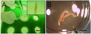
Vitrified whole testis of juvenile honmoroko Gnathopogon caerulescens. Trimmed whole testis were loaded onto a strip-shaped nylon mesh after serial exposures to pretreatment solution (PS) and vitrification solution (VS) for 20 and 10 min, respectively, and then rapidly cooled by direct immersion in liquid nitrogen (A). Vitrified whole testis was quickly warmed by immersion in thawing solution (TS) followed by immersion in a 1:1 mixture of TS and TCCM–FBS and washed in fresh TCCM–FBS (B). Bar in (A) = 1 cm. Bar in (B) = 2 mm.

Live cell recovery rates of vitrified whole testis were difficult to determine under the experimental conditions studied because the cell numbers of whole intact testis could not be counted. However, although a direct comparison was difficult, more than 2.5 times the number of live cells was recovered from a vitrified testis than the conventional vitrification protocol for dissociated testicular cells (17.3 × 10⁵ cells and 6.4 × 10⁵ cells, respectively).
In vitro differentiation of cryopreserved spermatogonia
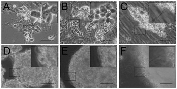
Hereafter, we used vitrified whole testes that were cryopreserved following serial exposures to PS for 20 min and VS for 10 min, because we obtained the largest total cell number with the highest percentage of membrane-intact cells from these testes. The morphologies of the cells, which were cultured under adhesion or suspension conditions using vitrified whole testes are shown in Fig. 5. Under both cell culture conditions, cryopreserved spermatogonia differentiated into flagellated sperm, as reported previously (Higaki et al., Reference Higaki, Shimada, Kawamoto, Todo, Kawasaki, Tooyama, Fujioka, Sakai and Takada2017). During the first week, most observable spermatogenic cells were spermatogonia. Spermatocytes emerged during the second week, and flagellated motile sperm was first observed during the third week (Videos S1 and S2).
Cell cultures prepared from vitrified juvenile whole testis in honmoroko Gnathopogon caerulescens. Vitrified-warmed whole testis was enzymatically digested and cultured under adhesion (A–C) and suspension conditions (D–F) at 18°C in air. As a spermatogenic cells, spermatogonia was dominantly observed in cell cultures at the first week (A, D). At the second week, many spermatocytes were observed (B, E). Flagellated sperm first emerged at the third week (C, F). Insets in (A), (B), and (C) represent spermatogonia, spermatocytes, and flagellated sperm, respectively. Bars = 50 μm.

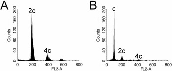
Flow cytometric analyses of the DNA content of cells cultured for 30 days in suspension showed a clear haploid peak, indicating sperm and spermatids, in addition to diploid and tetraploid peaks. These peaks were not observed in freshly dissociated juvenile testicular cells (Fig. 6). These results suggest that cryopreserved spermatogonia underwent successful meiosis in vitro after cryopreservation.
Flow cytometric analyses of DNA content in pre- and post-culture testicular cells of juvenile honmoroko Gnathopogon caerulescens. Pre-culture testicular cells prepared from juvenile fish showed only diploid and tetraploid peaks (A). The cell culture prepared from vitrified whole testis showed clear haploid peak in addition to diploid and tetraploid peaks at day 30 (B). FL2-A indicates the total fluorescence of propidium iodide; c, 2c, and 4c represent haploid, diploid, and tetraploid DNA content, respectively.

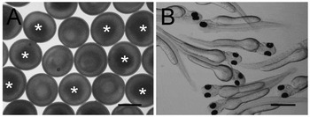
As shown in Fig. 7, many morphologically normal fertilized embryos and hatched larvae were obtained through in vitro fertilization with in vitro-differentiated sperm from vitrified whole testes (Table 2). The fertility rate was 100% (n = 6). The average fertilization rate of the present study of 16.7% tended to be higher than that of a previous study using a conventional vitrification protocol for dissociated testicular cells (4.8 ± 2.3%; range 0.9–18.3%; Higaki et al., Reference Higaki, Shimada, Kawamoto, Todo, Kawasaki, Tooyama, Fujioka, Sakai and Takada2017), although sperm were obtained from one-fifth of the number of testicular cells in the previous study (testicular cells prepared from one and five juveniles were cultured in the present and previous studies, respectively, for artificial insemination using eggs from one female). In G. caerulescens, there are no reports of gynogenesis under normal rearing conditions; additionally, artificially induced haploid gynogenote fish cannot survive until hatching (Fujioka, Reference Fujioka1993). These findings suggest that sperm differentiated from vitrified spermatogonia possess normal fertilization and developmental competence.
Morphology of embryos and hatched larvae produced with in vitro-differentiated sperm in honmoroko Gnathopogon caerulescens. Eggs were fertilized with in vitro-differentiated sperm from vitrified juvenile whole testis of G. caerulescens (A). Resultant fertilized embryos developed normally and hatched out at around 4 days post insemination (B). Asterisks in (A) indicate unfertilized eggs. Bars = 1 mm.

Number of eggs successfully fertilized by in vitro-differentiated sperm from vitrified whole testes of juvenile honmoroko Gnathopogon caerulescens

Testicular cells obtained from vitrified whole testes, that were cryopreserved following serial exposures to PS for 20 min and VS for 10 min, were cultured under suspension condition for 30 days and differentiated sperm were subjected to in vitro fertilization assays. Embryonic development at the somite and hatching stages was assessed 24 h and 5 days after insemination, respectively. Total percentages were the means ± standard deviation (SD).
In conclusion, juvenile whole testis was successfully cryopreserved by vitrification in the critically endangered cyprinid G. caerulescens. The survival rate of the cells recovered from vitrified whole testis was high (84%). More than 2.5 times live cells were estimated to be obtained from a vitrified whole testis (17.3×10⁵ cells; 84% of the 20.6×10⁵ cells) compared with the conventional vitrification protocol for dissociated testicular cells (6.4×10⁵ cells; 52.7% of the 12.2×10⁵ cells). Cryopreserved spermatogonia dissociated from the vitrified whole testis differentiated into functional sperm in vitro. These results indicated that the vitrification of juvenile whole testis provides an approach for the preservation of male genetic material in endangered fish species.
Conflict of interest
The authors declare that they have no conflicts of interests.
Acknowledgements
The authors thank Syuichi Shimomura (Kusatsu Honmoroko Seisan Kumiai) and Tetsuo Yamamoto (Yamasho Honmoroko Youshoku Koubou), and Taemon Yamamoto (Yamamoto Yougyojo) for providing honmoroko G. caerulescens and carp serum. We also thank Takefumi Yamamoto and Yasuhiro Mori (Shiga University of Medical Science) for their excellent technical support. This work was funded in part by a Grant-in-Aid for challenging Exploratory Research (23651248 and 15K14440 to T.T.) and Scientific Research on Priority Area (21028021 to T.T.) from the Ministry of Education, Culture, Sports, Science and Technology (MEXT) of Japan. This study was also supported by NIG Collaborative Research Program (2010-A41, 2011-A36, 2012-A28, 2013-A47, 2014-A45, and 2016-A36 to T.T.), Ritsumeikan Global Innovation Research Organization (R-GIRO) program (to T.T.), Center of Innovation Trial Program from Japan Science and Technology Agency, JST (to T.T.), and Science Research Promotion Fund from the Promotion and Mutual Aid Corporation for Private Schools of Japan (to S.H.).
Supporting information
Additional Supporting Information is available online at the publisher's website.
Video S1.
Video S2.
Supplementary material
To view supplementary material for this article, please visit https://doi.org/10.1017/S0967199417000430