Introduction
Neuroimaging studies have provided evidence for subtle structural brain abnormalities in patients with bipolar disorder (BD). Most of these studies reported on cerebral volume and density as indices of structural neuropathology. In particular, smaller volumes of prefrontal cortical grey matter, corpus callosum and cerebral white matter, and larger volumes of the lateral ventricles and striatum were found in BD patients relative to healthy controls (Emsell & McDonald, Reference Emsell and McDonald2009; Savitz & Drevets, Reference Savitz and Drevets2009). The volume of the amygdala appears larger in BD adults, but smaller in BD children and adolescents (Savitz & Drevets, Reference Savitz and Drevets2009). However, an absence of volumetric abnormalities of the total brain, grey and white matter has also been described (Emsell & McDonald, Reference Emsell and McDonald2009). In addition, a recent meta-analysis (Selvaraj et al. Reference Selvaraj, Arnone, Job, Stanfield, Farrow, Nugent, Scherk, Gruber, Chen, Sachdev, Dickstein, Malhi, Ha, Ha, Phillips and McIntosh2012) of voxel-based morphometry studies demonstrated a smaller regional grey matter volume in frontal and temporal regions but did not include two studies that noted larger regional volumes in these (Adler et al. Reference Adler, Delbello, Jarvis, Levine, Adams and Strakowski2007; Bearden et al. Reference Bearden, Thompson, Dalwani, Hayashi, Lee, Nicoletti, Trakhtenbroit, Glahn, Brambilla, Sassi, Mallinger, Frank, Kupfer and Soares2007) and parietal regions (Adler et al. Reference Adler, Delbello, Jarvis, Levine, Adams and Strakowski2007). Measures of brain volume in BD patients are possibly influenced by lithium use, familial load, mood status, illness duration, age, heterogeneity of subject groups and variability in imaging methodology, probably contributing to the inconsistencies across studies (Emsell & McDonald, Reference Emsell and McDonald2009; Savitz & Drevets, Reference Savitz and Drevets2009; Hajek et al. Reference Hajek, Kopecek, Hoschl and Alda2012).
The heritability of BD is estimated to be around 85% (McGuffin et al. Reference McGuffin, Rijsdijk, Andrew, Sham, Katz and Cardno2003). Twin studies have shown an association between the genetic liability to develop BD and smaller cerebral white matter volume (Kieseppa et al. Reference Kieseppa, van Erp, Haukka, Partonen, Cannon, Poutanen, Kaprio and Lonnqvist2003; van der Schot et al. Reference van der Schot, Vonk, Brans, van Haren, Koolschijn, Nuboer, Schnack, van Baal, Boomsma, Nolen, Hulshoff Pol and Kahn2009), implicating that genes involved in the risk to develop BD are related to smaller white matter volume. In contrast, environmental factors related to BD were found to be significantly associated with smaller cortical grey matter volume (van der Schot et al. Reference van der Schot, Vonk, Brans, van Haren, Koolschijn, Nuboer, Schnack, van Baal, Boomsma, Nolen, Hulshoff Pol and Kahn2009; Hulshoff Pol et al. Reference Hulshoff Pol, van Baal, Schnack, Brans, van der Schot, Brouwer, van Haren, Lepage, Collins, Evans, Boomsma, Nolen and Kahn2012). This suggests that environmental factors unique for each individual influence directly (or indirectly through a causal relationship) both BD and grey matter volume.
Cortical volume is the ‘product’ of surface area and thickness (Panizzon et al. Reference Panizzon, Fennema-Notestine, Eyler, Jernigan, Prom-Wormley, Neale, Jacobson, Lyons, Grant, Franz, Xian, Tsuang, Fischl, Seidman, Dale and Kremen2009). The cortex has a columnar organization (Mountcastle, Reference Mountcastle1997). The number of cells in ontogenetic columns that run perpendicular to the surface of the brain influence cortical thickness while the size of the surface is suggested to be dependent of the number of columns (Rakic, Reference Rakic1988, Reference Rakic1995). Although these measures are inherently biologically correlated, they appear to be genetically distinct but may singly or both drive cortical volume (Rakic, Reference Rakic1988, Reference Rakic1995; Pakkenberg & Gundersen, Reference Pakkenberg and Gundersen1997; Jansen & Andermann, Reference Jansen and Andermann2005; Fornito et al. Reference Fornito, Yucel, Wood, Adamson, Velakoulis, Saling, McGorry and Pantelis2008; Im et al. Reference Im, Lee, Lyttelton, Kim, Evans and Kim2008; Pontious et al. Reference Pontious, Kowalczyk, Englund and Hevner2008; Panizzon et al. Reference Panizzon, Fennema-Notestine, Eyler, Jernigan, Prom-Wormley, Neale, Jacobson, Lyons, Grant, Franz, Xian, Tsuang, Fischl, Seidman, Dale and Kremen2009; Rakic et al. Reference Rakic, Ayoub, Breunig and Dominguez2009; Winkler et al. Reference Winkler, Kochunov, Blangero, Almasy, Zilles, Fox, Duggirala and Glahn2010; Eyler et al. Reference Eyler, Prom-Wormley, Panizzon, Kaup, Fennema-Notestine, Neale, Jernigan, Fischl, Franz, Lyons, Grant, Stevens, Pacheco, Perry, Schmitt, Seidman, Thermenos, Tsuang, Chen, Thompson, Jak, Dale and Kremen2011). Consequently, the genetic contribution to brain volume in healthy individuals (Baare et al. Reference Baare, Hulshoff Pol, Boomsma, Posthuma, de Geus, Schnack, van Haren, van Oel and Kahn2001; Thompson et al. Reference Thompson, Cannon, Narr, van Eep, Poutanen, Huttunen, Lonnqvist, Standertskjold-Nordenstam, Kaprio, Khaledy, Dail, Zoumalan and Toga2001; Peper et al. Reference Peper, Brouwer, Boomsma, Kahn and Hulshoff Pol2007) and BD patients (Noga et al. Reference Noga, Vladar and Torrey2001; van der Schot et al. Reference van der Schot, Vonk, Brans, van Haren, Koolschijn, Nuboer, Schnack, van Baal, Boomsma, Nolen, Hulshoff Pol and Kahn2009; Hulshoff Pol et al. Reference Hulshoff Pol, van Baal, Schnack, Brans, van der Schot, Brouwer, van Haren, Lepage, Collins, Evans, Boomsma, Nolen and Kahn2012) is probably dependent on the genetic influences on cortical surface area and/or cortical thickness.
An association between liability to develop BD and cortical surface area has not yet been established nor have case–control studies found evidence for cortical surface area abnormalities in BD (Fornito et al. Reference Fornito, Yucel, Wood, Bechdolf, Carter, Adamson, Velakoulis, Saling, McGorry and Pantelis2009; Rimol et al. Reference Rimol, Nesvag, Hagler, Bergmann, Fennema-Notestine, Hartberg, Haukvik, Lange, Pung, Server, Melle, Andreassen, Agartz and Dale2012) whereas cortical thickness in BD has been investigated (Lyoo et al. Reference Lyoo, Sung, Dager, Friedman, Lee, Kim, Kim, Dunner and Renshaw2006; Rimol et al. Reference Rimol, Hartberg, Nesvag, Fennema-Notestine, Hagler, Pung, Jennings, Haukvik, Lange, Nakstad, Melle, Andreassen, Dale and Agartz2010; Foland-Ross et al. Reference Foland-Ross, Thompson, Sugar, Madsen, Shen, Penfold, Ahlf, Rasser, Fischer, Yang, Townsend, Bookheimer and Altshuler2011; Hulshoff Pol et al. Reference Hulshoff Pol, van Baal, Schnack, Brans, van der Schot, Brouwer, van Haren, Lepage, Collins, Evans, Boomsma, Nolen and Kahn2012). Quantification of genetic and environmental contributions to cortical surface area and cortical thickness independently in BD may further illuminate the association between grey matter volume deficits and the risk to develop BD (McDonald et al. Reference McDonald, Bullmore, Sham, Chitnis, Wickham, Bramon and Murray2004; van der Schot et al. Reference van der Schot, Vonk, Brans, van Haren, Koolschijn, Nuboer, Schnack, van Baal, Boomsma, Nolen, Hulshoff Pol and Kahn2009). Previously, we reported on whether patients with schizophrenia and patients with BD showed overlapping abnormalities in cortical thickness (Hulshoff Pol et al. Reference Hulshoff Pol, van Baal, Schnack, Brans, van der Schot, Brouwer, van Haren, Lepage, Collins, Evans, Boomsma, Nolen and Kahn2012). In the present twin study, using the same BD twin sample as Hulshoff Pol et al. (Reference Hulshoff Pol, van Baal, Schnack, Brans, van der Schot, Brouwer, van Haren, Lepage, Collins, Evans, Boomsma, Nolen and Kahn2012), we set out to quantify the additive genetic, common and unique environmental contributions to the association between BD liability and cortical measures in 39 cortical regions of interest (ROIs) per hemisphere. In addition, we specifically explored the degree to which associations between regional cortical volume and BD liability reflect the associations of cortical surface area and cortical thickness with BD liability.
Method
Subjects
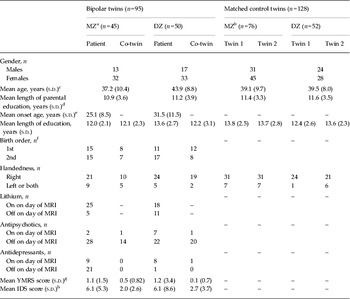
A total of 118 monozygotic (MZ) and dizygotic (DZ) twin pairs underwent magnetic resonance imaging (MRI) (van der Schot et al. Reference van der Schot, Vonk, Brans, van Haren, Koolschijn, Nuboer, Schnack, van Baal, Boomsma, Nolen, Hulshoff Pol and Kahn2009, Reference van der Schot, Vonk, Brouwer, van Baal, Brans, van Haren, Schnack, Boomsma, Nolen, Hulshoff Pol and Kahn2010). Of this group, 13 subjects were excluded from analysis due to bad image quality. Consequently, 44 twin pairs affected with BD (nine MZ concordant, 12 MZ discordant; four DZ concordant, 19 DZ discordant) and seven twins from incomplete pairs (three MZ co-twins from discordant pairs; two DZ patients and two DZ co-twins from incomplete discordant pairs), and 61 matched control twin pairs (37 MZ, 24 DZ) plus two MZ and four DZ healthy control twins from incomplete pairs were included in the analyses. All twins were raised together, except for one control pair where twins were separated at 12 years of age when both parents died. Subjects were between 18 and 60 years of age at the time of enrolment in the study. Demographic information is presented in Table 1. Clinical diagnosis of Axis I psychiatric disorders and Axis II personality disorders was confirmed using the Structured Clinical Interview for DSM-IV (SCID) (First et al. Reference First, Spitzer, Gibbon and Williams1997) and the Structured Interview For DSM-IV Personality (SIDP) (Pfohl et al. Reference Pfohl, Blum and Zimmerman1997), respectively, and through available medical records. Patients were also interviewed on their medication history. The twin pairs had no history of drug or alcohol dependency for the last 6 months prior to inclusion in the study, for this was an exclusion criterion. Moreover, none had severe medical illness, verified with a medical history inventory. The current mood state of BD patients was assessed using the Young Mania Rating Scale (YMRS) (Young et al. Reference Young, Biggs, Ziegler and Meyer1978) and the Inventory for Depressive Symptomatology (IDS) (Beck et al. Reference Beck, Ward, Mendelson, Mock and Erbaugh1961). Upon inclusion, all patients were euthymic with a YMRS score of 4 or less and an IDS score of 12 or less, except for nine BD patients who were mildly to severely depressed (IDS scores of 14, 14, 14, 15, 17 20, 22 and 38, respectively) or hypomanic (YMRS score of 17). Healthy control pairs were matched to the bipolar pairs for zygosity, gender, age and parental education. Control pairs had no history of Axis I or II disorders according to DSM-IV criteria (confirmed with the SCID and SIDP, respectively) and no history of severe medical illness. Furthermore, they had no first-degree relative with a history of a major Axis I psychiatric disorder (DSM-IV). Family histories of all twins were obtained via the Family Interview Genetic Studies (Nurnberger et al. Reference Nurnberger, Blehar, Kaufmann, York-Cooler, Simpson, Harkavy-Friedman, Severe, Malaspina and Reich1994), performed with both twins of each pair. Zygosity was determined with DNA fingerprinting using high polymorphic microsatellite markers 9 to 11. The medical ethics review board of the University Medical Center Utrecht approved the study and all participants gave written informed consent after full explanation of the study aims and procedures.
Demographic characteristics of the bipolar and matched healthy control twin pairs

Data are given as number of individuals or as mean (s.d.).
MZ, Monozygotic; DZ, dizygotic; s.d., standard deviation; MRI, magnetic resonance imaging; YMRS, Young Mania Rating Scale; IDS, Inventory for Depressive Symptomatology.
a Nine MZ concordant pairs; 12 MZ discordant pairs; three MZ co-twins from incomplete pairs; four DZ concordant pairs; 19 DZ discordant pairs; two DZ patients and two DZ co-twins from incomplete pairs.
b 37 MZ healthy control pairs; two MZ healthy controls from incomplete pairs; 24 DZ healthy control pairs; four DZ healthy controls from incomplete pairs.
c DZ patient pairs were significantly older than MZ patient pairs (F 1,93 = 11.55, p < 0.05), MZ control pairs (F 1,124 = 7.95, p < 0.05) and DZ control pairs (F 1,100 = 7.30, p < 0.05).
d Years of parental education for two bipolar twin pairs could not be determined.
e Age of onset could not be determined for four bipolar patients; significant difference between MZ and DZ (F 1,53 = 5.47, p < 0.05).
f Birth order was not determined for one bipolar twin pair.
g YMRS score was not ascertained for two MZ patients, two MZ co-twins and three DZ patients.
h IDS score was not ascertained for two MZ patients, two MZ co-twins and two DZ patients.
Brain imaging
For information on previously described MRI acquisition and pre-processing protocols, please refer to the Supplementary Method, available online.
Cortical surface and cortical thickness estimates were obtained using the CLASP (Constrained Laplacian Anatomic Segmentation Using Proximity) algorithm in a custom implementation of CIVET, developed at the McConnell Brain Imaging Centre of the Montreal Neurological Institute (MacDonald et al. Reference MacDonald, Kabani, Avis and Evans2000; Kabani et al. Reference Kabani, Le, MacDonald and Evans2001; Kim et al. Reference Kim, Singh, Lee, Lerch, Ad-Dab'bagh, MacDonald, Lee, Kim and Evans2005). The grey/white/cerebrospinal fluid (CSF) segments from our own segmentation algorithm, as described earlier, were used as inputs for the original CLASP algorithm. A three-dimensional surface consisting of the 81 920 polygons and 40 962 vertices was fitted to the grey/white matter intersection, thereby creating the inner surface of the cortex. The outer surface of the cortex was then constructed by expanding the inner surface to fit the grey matter/CSF intersection (MacDonald et al. Reference MacDonald, Kabani, Avis and Evans2000; Kim et al. Reference Kim, Singh, Lee, Lerch, Ad-Dab'bagh, MacDonald, Lee, Kim and Evans2005). The surfaces are modelled as nets of polygons (triangles) with vertices being the points where the polygons meet (i.e. the angular points of the nets). The surfaces of the subjects (Lyttelton et al. Reference Lyttelton, Boucher, Robbins and Evans2007) were registered to an average surface, allowing for comparison of cortical thickness locally. Each polygon vertex on the outer surface had its counterpart on the inner surface; cortical thickness was defined as the distance between these inner and outer surface polygons. Subsequently, automated anatomical labelling was executed to create parcellations of 39 bilateral brain regions that encompass these vertices (Tzourio-Mazoyer et al. Reference Tzourio-Mazoyer, Landeau, Papathanassiou, Crivello, Etard, Delcroix, Mazoyer and Joliot2002). Cortical surface area was estimated from the mid-surface, positioned halfway between the outer and inner surfaces. Cortical volume was calculated via multiplication of cortical surface area and cortical thickness.
Model fitting
Twin designs allow for quantification of the influence of genes and environment on measured traits. Comparison of correlations within MZ and DZ twins provides a basis on which the degree of genetic and environmental influences on such traits can be judged. A larger correlation between traits in MZ twins than in DZ twins suggests that genetic factors play a role, because MZ twins are genetically identical whereas DZ twins only share on average 50% of their segregating genes. When there are no differences between MZ and DZ correlations, but there is phenotypic resemblance for the trait, then environment shared by twins is more likely influencing the trait (Boomsma et al. Reference Boomsma, Busjahn and Peltonen2002).
To estimate the extent of the contribution of genetic and unique environmental factors to the association between cortical measures and BD liability, a bivariate liability threshold model was chosen. This model was implemented in structural equation modelling software OpenMx (Kenny et al. Reference Kenny, Andric, Boker, Neale, Wilde and Small2009), running under the statistical programming environment R (2008; http://www.r-project.org/). Prior to model fitting, linear regression on gender, age and handedness (and lithium use for BD patients only, measured as on/off on day of MRI) was performed to eliminate their contribution to the variance in cortical measures. We found that handedness significantly (α < 0.05) or at trend level influenced some of the cortical measures. Therefore, correction for handedness was applied, given that doing so would only result in correction of the measures that showed influence of handedness and not of those that did not show such an influence.
A bivariate Cholesky decomposition was fitted to the data to estimate additive genetic (A) and unique environmental (E) variance components of cortical surface area, cortical thickness and cortical volume, and phenotypic, genetic and environmental overlap between BD disease liability and cortical measures (online Supplementary Fig. S1). No evidence of shared environmental influences (C) on BD has been found previously (McGuffin et al. Reference McGuffin, Rijsdijk, Andrew, Sham, Katz and Cardno2003) and in the present study we found no evidence of such influences on our cortical measures. Shared environmental factors were therefore not included in the model. Disease status was dichotomous and assumed to represent an underlying continuous liability with a mean of 0 (s.d. = 1). A patient will have a high value on the liability scale, thereby crossing a certain threshold (patient status = 1). All other individuals will have lower liability scores and will not cross the critical threshold (patient status = 0; discordant co-twin of patient or control twin pairs). The critical threshold and heritability for the underlying liability to develop BD was not based on this sample because we included approximately equal numbers of concordant, discordant and healthy twin pairs. Prevalence and heritability (the relative contribution of genetic variance to total variance) of BD were fixed to population values; prevalence was set to 1% (Regeer et al. Reference Regeer, ten Have, Rosso, Hakkaart-van, Vollebergh and Nolen2004) and heritability was set to 85% (McGuffin et al. Reference McGuffin, Rijsdijk, Andrew, Sham, Katz and Cardno2003). Importantly, we tested whether using alternative BD prevalences of 2.5% and 4% would influence the number and nature of phenotypic associations between BD and cortical measures. It did not. If anything, we may have been too conservative in setting prevalence at 1%, but feel confident in doing so, based on the available literature on BD prevalence in the Netherlands (see Regeer et al. Reference Regeer, ten Have, Rosso, Hakkaart-van, Vollebergh and Nolen2004). In order to apply the threshold model to the brain measures, the standardized residuals that were obtained for these measures via linear regression [on gender, age, handedness and lithium use (in patients only)] were rendered into five categories identical for all subjects – thereby equating them across groups – and put in the model. Thresholding was based on normality plots, with the boundaries of the ‘outer’ two categories set at –1.5 standard deviations and 1.5 standard deviations, respectively, and the other three categories that fall in between being 1 s.d. wide.
The phenotypic correlation (r ph), an index of association between phenotypes (e.g. liability to develop BD and brain measure), was based on calculations of within-twin/between-trait correlations. Heritability (h 2) and influence of unique environment (e 2) as well as disentanglement of the observed correlation between liability to develop BD and brain measures into genetic (r g) and environmental (r e) components was based on polychoric cross-twin/within-trait and cross-twin/cross-trait correlations within MZ and DZ groups (Neale & Miller, Reference Neale and Miller1997). The heritability of brain measures was thus determined within the bivariate model. The genetic correlation indicates the degree of overlap in genes influencing both phenotypes. Unique environmental correlations indicate overlap in unique environmental factors influencing both phenotypes. The phenotypic correlation is a function of the genetic and environmental correlations weighted by the square root of the heritabilities (r g × h BD × h brain) and the square root of e 2 (r e × e BD × e brain). We refer to these quantities as r ph-g and r ph-e (Toulopoulou et al. Reference Toulopoulou, Picchioni, Rijsdijk, Hua-Hall, Ettinger, Sham and Murray2007).
A saturated model in which means, variances and correlations are estimated freely served as a baseline model to which the more restrictive AE model was compared, using Akaike's information criterion. Compared with the saturated model, the AE model had the best fit in 95% of ROIs and was therefore applied to all ROIs. For relevant estimates, 95% confidence intervals were obtained (Neale & Miller, Reference Neale and Miller1997). To account for multiple testing in multiple regions, Bonferroni correction was applied. This was done by dividing the α value by the number of regions per hemisphere, which resulted in a critical level of significance of 0.05/39 = 0.0013 for each parameter (r ph, r g, r e, r ph-g, r ph-e). In the tables, significance at α < 0.05 and at α < 0.0013 is indicated.
Results
Global cortical measures
Genetic and environmental influences on global cortical measures
Heritability of all cortical measures was estimated irrespective of disease (see online Supplementary Tables S1, S2 and S3). Total cortical surface area was predominantly explained by genes (h 2: 90% and 91%, left and right hemispheres, respectively), as was total cortical grey matter volume (h 2: 88% and 86%, left and right hemispheres, respectively). Mean cortical thickness across the entire cortex was moderately influenced by genes (h 2: 51% and 48%, left and right hemispheres, respectively).
Association between liability to develop BD and global cortical measures
There were no significant phenotypic associations between liability to develop BD and total cortical surface area in either hemisphere, indicating that genetic and environmental factors influencing BD are not associated with global cortical surface area. However, unique environmental factors related to BD were associated with a thinner cortex and smaller total cortical volume of the right hemisphere.
Regional cortical measures
Genetic and environmental influences on regional cortical measures
Regionally, heritability of all cortical measures was estimated irrespective of disease. At this level, cortical surface area, cortical thickness and cortical volume were strongly influenced by unique environmental factors (see online Supplementary Tables S1, S2 and S3).
Association between liability to develop BD and regional cortical surface area
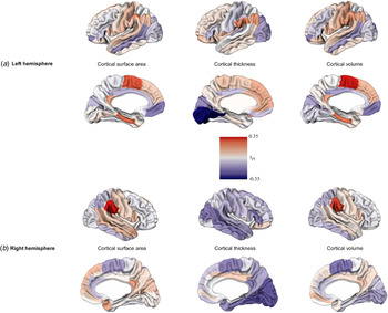
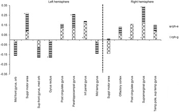
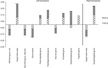
In local brain regions, liability to develop BD was associated with both larger and smaller cortices (see online Supplementary Table S1, Figs. 1 and 2). In particular, genetic liability to develop BD was significantly associated with larger cortical surface area in the left inferior parietal gyrus and right posterior cingulate and supramarginal gyri. In contrast, unique environmental factors related to BD were significantly associated with larger cortical surface in the left supplementary motor area, parahippocampal gyrus and right supramarginal gyrus, and smaller cortical surface area in the left gyrus rectus and middle temporal gyrus. This indicates that the genetic liability to develop BD appears to be primarily associated with larger regional cortical surface areas whereas unique environmental factors related to BD are associated with both larger and smaller regional cortical surface areas.
Phenotypic correlations (range r ph: 0.35 to −0.35) between bipolar disorder liability and cortical surface area, cortical thickness and cortical volume in the left (a) and right (b) hemispheres. Red indicates positive correlation, blue indicates negative correlation and white indicates no correlation.

Genetic (r ph-g) and environmental (r ph-e) contributions to the significant phenotypic correlations between liability to develop bipolar disorder and cortical surface area. * Significant contributions of r ph-g and r ph-e to the total phenotypic correlation (r ph) (p < 0.05). front, Frontal; orb, orbital; suppl, supplementary; sup, superior; med, medial; inf, inferior; pariet, parietal; temp, temporal.

Association between liability to develop BD and regional cortical thickness
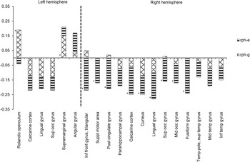
At the regional level, liability to develop BD was associated with both thicker and thinner cortices in a number of brain regions (see online Supplementary Table S2 and Figs. 1 and 3). Particularly, genetic liability to develop BD was significantly associated with thicker cortex in the left rolandic operculum and supramarginal gyrus. In contrast, unique environmental factors related to BD were significantly associated with thinner cortices in the right triangular inferior frontal gyrus, supplementary motor area, posterior cingulate gyrus, calcarine cortex, cuneus, lingual gyrus, superior occipital gyrus, middle occipital gyrus, fusiform gyrus and middle temporal gyrus. In sum, genes influencing BD appeared to be primarily associated with regionally thicker cortex whereas unique environmental factors influencing BD were more likely to be associated with thinner cortices.
Genetic (r ph-g) and environmental (r ph-e) contributions to the significant phenotypic correlations between liability to develop bipolar disorder and cortical thickness. * Significant contributions of r ph-g and r ph-e to the total phenotypic correlation (r ph) (p < 0.05). sup, Superior; occ, occipital; inf, inferior; front, frontal; suppl, supplementary; temp, temporal.

Association between liability to develop BD and regional cortical volume
Liability to develop BD was associated with both larger and smaller cortical volumes (see online Supplementary Table S3 and Figs. 1 and 4). Unique environmental factors related to BD were significantly associated with larger cortical volumes of the bilateral supramarginal gyri and left supplementary motor area and parahippocampal gyrus, and smaller cortical volume of the left gyrus rectus. There was no significant association between genetic liability to develop BD and regional cortical volume. Therefore, regionally, cortical volume appears chiefly associated with unique environmental factors, probably related to the illness itself, and not with genes influencing BD.
Genetic (r ph-g) and environmental (r ph-e) contributions to the significant phenotypic correlations between liability to develop bipolar disorder and cortical volume. * Significant contributions of r ph-g and r ph-e to the total phenotypic correlation (r ph) (p < 0.05). front, Frontal; orb, orbitofrontal; suppl, supplementary; sup, superior; med, medial; occ, occipital; inf, inferior; pariet, parietal.

The pattern of phenotypic association between BD liability and cortical volume appeared to a large extent to mimic the pattern of phenotypic association between BD liability and cortical surface area (compare online Supplementary Tables S1 and S3, and see Fig. 1), which indicates that cortical volume is primarily dependent on cortical surface area, and not cortical thickness.
In sum, across all measures, the majority of associations between BD liability and the cortex were predominantly influenced by unique environmental factors associated with BD (see online Supplementary Tables S1, S2, S3 and S4).
Discussion
To the best of our knowledge, this is the first twin study reporting on the influence of genes and environment on the association between BD and cortical surface area. We evaluated to what degree genetic and environmental factors associated with BD were associated with surface area, thickness and volume of the cortex. Furthermore, we studied the extent to which the pattern of association between BD liability and cortical volume would mimic the pattern of association of cortical surface area or cortical thickness with BD liability
Genetic and environmental influences on surface area, thickness and volume of the cortex
Total cortical surface area and cortical volume were highly heritable, and global cortical thickness was moderately heritable, irrespective of disease. In contrast, regional cortical measures were predominantly influenced by unique environmental factors. A similar reduction in heritability from global to local brain measures has been described earlier and probably reflects an increase in the degree of measurement error (Panizzon et al. Reference Panizzon, Fennema-Notestine, Eyler, Jernigan, Prom-Wormley, Neale, Jacobson, Lyons, Grant, Franz, Xian, Tsuang, Fischl, Seidman, Dale and Kremen2009; Eyler et al. Reference Eyler, Prom-Wormley, Panizzon, Kaup, Fennema-Notestine, Neale, Jernigan, Fischl, Franz, Lyons, Grant, Stevens, Pacheco, Perry, Schmitt, Seidman, Thermenos, Tsuang, Chen, Thompson, Jak, Dale and Kremen2011). In a ROI approach, the magnitude of genetic influence may be underestimated in areas that extend beyond regional boundaries determined by a brain atlas (Eyler et al. Reference Eyler, Prom-Wormley, Panizzon, Kaup, Fennema-Notestine, Neale, Jernigan, Fischl, Franz, Lyons, Grant, Stevens, Pacheco, Perry, Schmitt, Seidman, Thermenos, Tsuang, Chen, Thompson, Jak, Dale and Kremen2011).
Cortical surface area in BD
The risk of developing BD was not associated with abnormalities of global cortical surface area. However, larger regional cortical surfaces of bilateral parietal and right limbic regions were associated with the genetic risk of developing BD. Furthermore, unique environmental factors influencing BD were associated with larger surface area in left medial frontal and right parietal regions, and smaller surface area in left orbitofrontal and temporal regions. We also found a larger surface area of the left parahippocampal gyrus to be associated with unique environmental factors related to BD. Interestingly, the parahippocampus is part of the limbic system and is involved in emotion processing (Phillips et al. Reference Phillips, Drevets, Rauch and Lane2003; Emsell & McDonald, Reference Emsell and McDonald2009; Aldhafeeri et al. Reference Aldhafeeri, Mackenzie, Kay, Alghamdi and Sluming2012). Deficits in the volume and thickness of this structure have been previously observed in BD (Savitz & Drevets, Reference Savitz and Drevets2009; van der Schot et al. Reference van der Schot, Vonk, Brouwer, van Baal, Brans, van Haren, Schnack, Boomsma, Nolen, Hulshoff Pol and Kahn2010; Wang et al. Reference Wang, Lohmann, Yang, Zimmerman, Pantazopoulos, Herring, Berretta, Heckers and Konradi2011; Hulshoff Pol et al. Reference Hulshoff Pol, van Baal, Schnack, Brans, van der Schot, Brouwer, van Haren, Lepage, Collins, Evans, Boomsma, Nolen and Kahn2012; Delvecchio et al. Reference Delvecchio, Sugranyes and Frangou2013). Abnormal emotion processing appears to be a key feature of BD (Samamé et al. Reference Samamé, Martino and Strejilevich2012). Further exploration of the link between structural deficits in emotion-processing brain regions and the genetic and environmental risk for BD is therefore appropriate.
In summary, genes influencing BD are predominantly associated with regional larger cortical surfaces while unique environmental factors related to BD are associated with both larger and smaller regional cortical surfaces. It has been suggested that surface area size may be dependent on white matter volume, particularly with regard to the degree of myelination of the axons. In the maturing brain, the overlying cortex may stretch as the white matter increases (Seldon, Reference Seldon2005; Hogstrom et al. Reference Hogstrom, Westlye, Walhovd and Fjell2013). Furthermore, cortical surface area appears to be positively associated with local gyrification, indicating that greater gyrification implies larger cortical surface area (Hogstrom et al. Reference Hogstrom, Westlye, Walhovd and Fjell2013). Also, increased sulcal width and decreased sulcal depth have been suggested to relate to cortical surface shrinkage (Kochunov et al. Reference Kochunov, Mangin, Coyle, Lancaster, Thompson, Rivière, Cointepas, Regis, Schlosser, Royall, Zilles, Mazziotta, Toga and Fox2005). Perhaps the larger and smaller surface sizes we found may be relative to regional indices of white matter volume and local gyrification.
Cortical thickness in BD
At the global level, a mean thinner cortex in the right hemisphere was associated with unique environmental factors influencing BD whereas mean cortical thickness of the left hemisphere was not associated to BD liability. Our findings on regional cortical thickness are, not surprisingly, largely in line with our previous vertex-wise analyses (Hulshoff Pol et al. Reference Hulshoff Pol, van Baal, Schnack, Brans, van der Schot, Brouwer, van Haren, Lepage, Collins, Evans, Boomsma, Nolen and Kahn2012). Again, we found an association between genetic liability to develop BD and a thicker left supramarginal gyrus, and a phenotypic association between BD liability and a thinner right parahippocampus. A thinner right calcarine cortex was associated with unique environmental factors related to BD. In this study we also found that the genetic liability to develop BD was associated with a thicker left central region.
In this study, using the ROI approach, we now also found that unique environmental factors influencing BD were associated with thinner right frontal, limbic, temporal and occipital cortices. In part, these results are in line with prior findings of a voxel-based morphometry study in this cohort, where we found unique environmental factors influencing BD to be associated with lower grey matter density in right inferior frontal, cingulate, inferior temporal, lingual and limbic regions (van der Schot et al. Reference van der Schot, Vonk, Brouwer, van Baal, Brans, van Haren, Schnack, Boomsma, Nolen, Hulshoff Pol and Kahn2010). Possibly, measures of grey matter density and cortical thickness partly register the same cortical abnormalities, as has been suggested in schizophrenia (Narr et al. Reference Narr, Bilder, Toga, Woods, Rex, Szeszko, Robinson, Sevy, Gunduz-Bruce, Wang, DeLuca and Thompson2005).
Cortical volume in BD
We found that smaller total cortical volume of the right hemisphere was associated with unique environmental factors influencing BD, which is in line with the previously found overlap in unique environmental factors influencing both BD and smaller cortical grey matter volume (van der Schot et al. Reference van der Schot, Vonk, Brans, van Haren, Koolschijn, Nuboer, Schnack, van Baal, Boomsma, Nolen, Hulshoff Pol and Kahn2009). Regionally, genetic liability to develop BD was not associated with cortical volume. However, unique environmental factors influencing BD were associated with larger cortical volumes of bilateral parietal and left frontal and limbic regions, and smaller left orbitofrontal cortical volume. Only a small number of studies have examined regional brain volumes in subjects with genetic vulnerability for BD, demonstrating associations between genetic risk for BD and a larger volume of the left caudate nucleus (Noga et al. Reference Noga, Vladar and Torrey2001), and smaller volumes of the right anterior cingulate gyrus and ventral striatum (McDonald et al. Reference McDonald, Bullmore, Sham, Chitnis, Wickham, Bramon and Murray2004). Remarkably, BD liability was associated with regional cortical volume and cortical surface in many of the same brain regions in this study, in a similar fashion. Moreover, in many brain regions where cortical thickness was associated with unique environmental factors related to BD, cortical volume was not.
Thus, our results agree with previous reports that cortical surface area and not cortical thickness primarily promotes cortical volume (Pakkenberg & Gundersen, Reference Pakkenberg and Gundersen1997; Im et al. Reference Im, Lee, Lyttelton, Kim, Evans and Kim2008; Rakic, Reference Rakic2009; Winkler et al. Reference Winkler, Kochunov, Blangero, Almasy, Zilles, Fox, Duggirala and Glahn2010). Importantly, although abnormalities in cortical surface and especially cortical thickness were not always reflected in abnormalities in cortical volume, they were nevertheless present. In the same vein, Fornito et al. (Reference Fornito, Yucel, Wood, Adamson, Velakoulis, Saling, McGorry and Pantelis2008) found larger surface and thinner cortex in the anterior cingulate gyrus in schizophrenia patients compared with controls but did not find volume differences in this structure between groups. Consequently, the authors stressed the importance of moving beyond traditional measures of grey matter volume when assessing cortical abnormalities in clinical populations (Fornito et al. Reference Fornito, Yucel, Wood, Adamson, Velakoulis, Saling, McGorry and Pantelis2008). Critical evaluation of structural neuropathology should therefore include assessment of structural brain elements comprising volume and not be limited to assessment of volume only.
Importantly, the majority of phenotypic associations between liability to BD and cortical measures showed strong unique environmental involvement, indicating overlap of unique environmental factors influencing both BD and the cortex. Possibly, these unique environmental factors are illness-related and dependent on the individual's behaviour (i.e. self-generated), for example poor work performance leading to job loss. In bipolar spectrum disorders (not BD type I) and unipolar depression, it has been hypothesized that stress may in part be self-generated which, in turn, may contribute to the maintenance of symptoms (Hammen, Reference Hammen1991; Safford et al. Reference Safford, Alloy, Abramson and Crossfield2007; Bender et al. Reference Bender, Alloy, Sylvia, Urosevic and Abramson2010). However, whether this is the case here and if the cortex is affected as a result require further investigation and are merely speculation at this point.
Limitations
Certain limitations in our study should be considered. First, the issue of statistical power remains challenging to tackle in a single-twin MRI study. According to Visscher (Reference Visscher2004) and Posthuma & Boomsma (Reference Posthuma and Boomsma2000), large sample sizes and a larger number of MZ than DZ twin pairs are required to adequately estimate the magnitudes of genetic and environmental sources of variance in both univariate and bivariate designs. This is particularly relevant when heritability is expected to be moderate, as was the case with the regional cortical measures in our study. However, the heritabilities of global cortical measures were much higher and more reliably estimated within our model. Furthermore, in a number of regions the associations between BD and cortical measures were highly significant, in some cases surviving Bonferroni correction for multiple comparisons, indicating that for those regions we had sufficient power to detect an association with BD. In contrast, this may not have been true for other regions, the implication of which is that our estimate of the total number of associations may be conservative. Additionally, regional differences in gyrification or signal strength (causing signal–noise variability) may have affected (regional) heritability estimates.
Second, although MZ and DZ patient twin pairs were matched to the MZ and DZ control pairs for gender, there were more females than males in both patient and control pairs. Also, DZ patient twin pairs were significantly older than MZ patient and DZ control pairs. Therefore, cortical measures were corrected for gender and age. Third, although we corrected for lithium use in BD patients, correction for antipsychotics, antidepressants or other types of medication was not applied. This was due to unreliable reporting by patients on their use and the irregular frequency of medication use other than lithium. Fourth, the liability threshold model assumes a dichotomous disease variable, which may not fully capture the complex nature of bipolar phenomenology. Additionally, prevalence and heritability of BD were set a priori and not based on this sample. Finally, in our bivariate model assessing genetic and environmental influences on the cortex in BD, possible gene–environment interactions could not be ascertained.
Conclusion
Unique environmental factors, probably illness-related, played a prominent role in driving the majority of phenotypic associations between liability to develop BD and cortical measures. Further evaluation of their influence on the cortical mantle is recommended. Furthermore, phenotypic associations between BD liability and cortical volume followed a distribution similar to that of phenotypic associations between BD liability and cortical surface area, indicating cortical volume to be primarily driven by cortical surface area. However, cortical abnormalities are not restricted to anomalies in surface and volume but also extend to the thickness of the cortex. Assessment of genetic and environmental influences on cortical surface area and cortical thickness could enhance our understanding of the nature of disease-related structural neuropathology and may serve as a basis for judgments on the degree of disease segregation or overlap, particularly in the context of (neuro)development.
Supplementary material
For supplementary material accompanying this paper visit http://dx.doi.org/10.1017/S0033291714001251.
Acknowledgements
This work was supported by the Netherlands Organization for Scientific Research (NWO), grant no. 9120818.
Declaration of Interest
None.