Introduction
Keratomas, also known as cholesteatomas, are mainly found in the temporal bone (including the external auditory canal, middle ear, mastoid process, cerebellopontine angle and pyramid apex) and are usually associated with chronic disease of the middle ear. Keratomas are considered rare in the sinonasal regions, and extremely rare in the frontal sinus. A review of the English language literature revealed only 13 previously reported cases.Reference Storper and Newman1–Reference Hansen, Sørensen, Stage, Mouritzen and Cayé-Thomasen3 Several aetiological mechanisms have been proposed, but the pathogenic process is still unclear.
This paper aims to report a case of frontal sinus keratoma in a 53-year-old man. We present this patient's clinical history, imaging results (i.e. computed tomography (CT) and magnetic resonance imaging (MRI)), surgical management and histopathological findings, in order to illustrate the differential diagnosis and to emphasise the rarity of this lesion.
Case report
A 53-year-old man presented to our department with a 10-month history of rhinorrhoea and postnasal drip. The patient also reported anosmia but denied epistaxis, diplopia or previous trauma.
Flexible endoscopic examination revealed a polyp-like mass and yellowish nasal discharge occupying the ostiomeatal complex and middle meatus bilaterally.
Olfactory testing revealed anosmia.
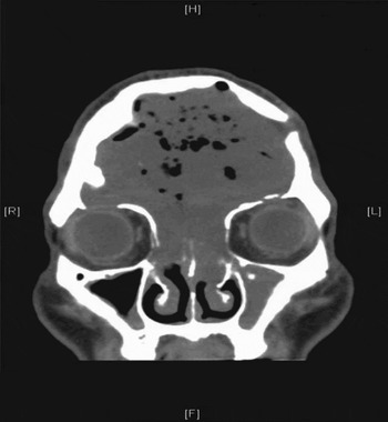
A sinus CT scan demonstrated a large soft tissue lesion approximately 8 cm in diameter within the frontal sinus, with extensive bony expansion, gas retention, compression of the adjacent frontal lobes and dura exposure, inferior erosion or thinning of the ethmoid plates, and cortical interruption at the anterior frontal sinus wall (Figure 1).
Coronal computed tomography sinus scan showing a large soft tissue lesion in the frontal sinus, with bony expansion, gas retention, compression of the adjacent frontal lobes and dura exposure, inferior erosion or thinning of the ethmoid plates, and cortical interruption at the anterior frontal sinus wall. H = head; F = feet; R = right; L = left

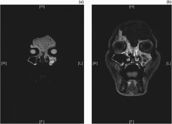
Magnetic resonance imaging was thus performed to aid the diagnosis. This revealed a large soft tissue lesion filling the frontal sinus, with scattered low signal intensity air bubbles. The lesion showed low signal intensity on T1-weighted images and high signal intensity on T2-weighted images (Figure 2). Gadolinium administration revealed polypoidal, strongly enhancing soft tissue adherent to the mucosa of the fused frontal sinuses, with downward extension via the frontal sinus outlets into both nasal cavities, together with bilateral erosion of the anterior ethmoid sinuses.
(a) Coronal, T1-weighted magnetic resonance imaging (MRI) scan showing a large soft tissue lesion filling the frontal sinus, with low signal intensity and scattered air bubbles. (b) Coronal, T2-weighted MRI scan showing a large soft tissue lesion filling the frontal sinus, with high signal intensity. H = head; F = feet; R = right; L = left

The patient agreed to undergo endoscopic sinus surgery. Transnasal, exclusively endoscopic sinus surgery was conducted under general anaesthesia. The maxillary and ethmoid sinus were found to be bilaterally associated with a polypoidal mass and mucopus. The frontal sinus was found to be filled by a shiny, soft, whitish, cholesteatoma-like mass, with extensive bony erosion and dura exposure. The whole tumour was removed endoscopically, using a modified Lothrop procedure, and sent for histopathological analysis.
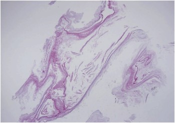
Histological examination revealed a lamellated cluster of keratinous material (Figure 3). The pathological diagnosis was sinusitis with nasal polyps and keratoma of the frontal sinus.
Photomicrograph showing aggregation of keratinous material (H&E; ×20).

The patient's post-operative course was uneventful.
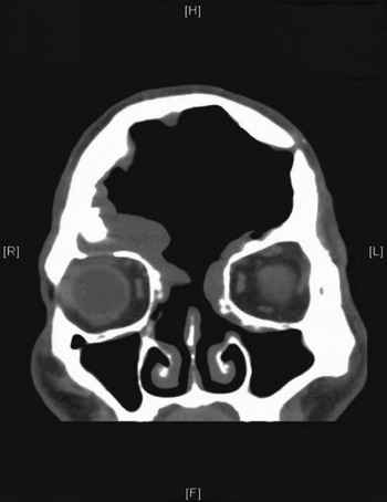
No complications were noted over a two-year follow-up period. A follow-up CT scan demonstrated clearance of the previous soft tissue lesion in the frontal sinus, with persistent frontal sinus bony expansion and frontal sinus recess mucosal thickening (Figure 4). A sinoscopic examination revealed swollen sinus mucosa with mucus but no evidence of keratin accumulation.
Follow-up coronal computed tomography scan showing persistent frontal sinus bony expansion and frontal sinus recess mucosal thickening. H = head; F = feet; R = right; L = left

Discussion
Keratomas of the paranasal sinuses occur when the normal respiratory epithelium that lines the sinus is partially or total replaced by hyperkeratotic squamous epithelium, leading to the formation of lamellar sheets of keratin.Reference Storper and Newman1 Four basic theories of the pathogenesis of keratoma have been proposed: the theory of congenital epithelial rests, the metaplasia theory, the immigration theory and the implantation theory. However, the pathogenesis of keratomas is still unclear.
Keratomas of the paranasal sinuses are rare, and keratoma of the frontal sinus is thus seldom diagnosed pre-operatively. Although keratomas are not biological neoplasms, they do have the capacity to erode bone and to expand into adjacent areas, due to the release of a number of proteolytic enzymes.Reference Hansen, Sørensen, Stage, Mouritzen and Cayé-Thomasen3–Reference Huisman, de Heer, Ten Dijke and Grote5 The symptoms and complications of keratomas are caused by lesion expansion, bony erosion and infection. In our patient's case, the frontal sinus keratoma extended into the ostiomeatal complex, causing rhinorrhoea and sinusitis.
The differential diagnosis of a frontal sinus keratoma includes both benign lesions (both neoplastic and non-neoplastic) and malignant lesions. Relevant benign non-neoplastic lesions include mucocele, pyocoele, mucus retention cyst, haematoma, sebaceous cyst and empyema. Benign neoplastic lesions include papilloma, meningioma, schwannoma, haemangioma, juvenile nasal angiofibroma and fibrous dysplasia. Malignant lesions include squamous cell carcinoma, neuroblastoma, ameloblastoma and sarcoma.
Computed tomography and MRI may be useful to establish the definitive diagnosis. On CT, keratoma appears as a non-enhancing, homogeneous and expansile lesion. On MRI, it shows low signal intensity on T1-weighted images and high signal intensity on T2-weighted images. Our patient's CT and MRI images clearly demonstrate these features.
• Keratoma of the paranasal sinuses occurs when the normal respiratory epithelium lining the sinus is partially or totally replaced by hyperkeratotic squamous epithelium
• Keratoma of the frontal sinus is extremely rare, and should be differentiated from other slowly expanding lesions of the frontal sinus
• This case report describes the first successful application of a modified endoscopic Lothrop procedure for removal of an extensive frontal sinus keratoma
A review of the English language literature indicated that surgical resection is currently the treatment of choice for keratoma. For frontal sinus keratoma, an external surgical approach via a brow or coronal incision is recommended, depending on the location and extension of the keratoma.Reference Hansen, Sørensen, Stage, Mouritzen and Cayé-Thomasen3 Obliteration of the frontal sinus is necessary in cases with associated sinusitis blocking the nasofrontal duct, or those with a disproportionately large frontal cavity.Reference Storper and Newman1 Cranioplasty and prosthesis insertion can be considered in special cases.Reference Hansen, Sørensen, Stage, Mouritzen and Cayé-Thomasen3 If a frontal dura defect exists, primary closure or repair with a fascia graft may be performed. Post-operatively, the patient should be monitored using sinoscopy and CT to exclude recurrence.
Due to advances in endoscopic techniques and instruments, we have now refined our existing, published endoscopic technique and successfully used it to resect different types of tumour.Reference Wu, Tsao, Yang and Chen6–Reference Chen, Tsai, Chen and Chen13 In the reported patient, a modified endoscopic Lothrop procedure was used to remove a frontal sinus keratoma and to enlarge the frontal sinus outlet. There was no recurrence of keratoma over a two-year follow-up period.
Conclusion
Keratoma of the frontal sinus is an extremely rare condition. This lesion is biologically benign but continuously desquamates and expands; this leads to erosion of surrounding bony structures and may result in severe, life-threatening medical complications. Keratoma should be differentiated from other slowly expanding lesions of the frontal sinus. The present report includes characteristic CT and MRI images. In addition, it represents, to the best of our knowledge, the first description of the removal of such an extensive frontal sinus keratoma via a modified endoscopic Lothrop procedure. Therefore, we believe that the indications for endoscopic sinus surgery may be expanded to include frontal sinus keratoma removal. In the presented patient, we found the procedure to be curative, with reduced operating time, reduced risk of wound-related complications, shorter hospital stay and a better cosmetic outcome.