Introduction
Teleostei is the most numerous and diverse vertebrate group. Egg structure and developmental type may be among the prerequisites of their evolutionary success. They have polylecithal telolecithal eggs with yolk separated from an ooplasm. This egg type is characterized by structural plasticity: the amount of yolk and the yolk–ooplasm ratio vary significantly in different species. Such egg organization determines meroblastic cleavage and influences further developmental processes (Soin, Reference Soin1981; Ivanova-Kazas, Reference Ivanova-Kazas1995). Teleostei diversity is displayed already during the early developmental stages (Kunz, Reference Kunz2004; Alix et al., Reference Alix, Chardard, Ledoré, Fontaine and Schaerlinger2015; Desnitskiy, Reference Desnitskiy2015). The uncleaved yolk mass is utilized with a specialized transient structure, the YSL. Structures, analogous to the teleost YSL, have been found in other animal groups with meroblastic cleavage (Nagai et al., Reference Nagai, Sezaki, Kakiguchi, Nakaya, Lee, Ladher, Sasanami, Han, Yonemura and Sheng2015; Bruce, Reference Bruce2016; Kondakova et al., Reference Kondakova, Efremov and Nazarov2016).
The yolk syncytial layer (YSL) is a dynamic polyfunctional system. It is a symplast with numerous polymorphic yolk syncytial nuclei (YSN). The YSL forms during the blastula period. In the majority of species, it forms from the marginal blastomeres confluent with the yolk sphere (Carvalho & Heisenberg, Reference Carvalho and Heisenberg2010). As shown in D. rerio, during marginal blastomere division, cytokinesis between daughter nuclei is absent and pre-existing membranes regress (Chu et al., Reference Chu, Fong, Kondrychyn, Loh, Ye and Korzh2012). YSL formation depends on actin filaments and microtubules. Cytoskeleton activity is regulated by Rock1 and Slc3a2; however data on the molecular mechanism of YSL formation are still incomplete (Chu et al., Reference Chu, Fong, Kondrychyn, Loh, Ye and Korzh2012; Takesono et al., Reference Takesono, Moger, Farooq, Cartwright, Dawid, Wilson and Kudoh2012). The YSL becomes an active component of the yolk sphere that also consists of a yolk cytoplasmic layer (YCL), yolk mass and oil globule (if present) (Fuentes & Fernández, Reference Fuentes and Fernández2010). Initially the YSL is a ring around the blastoderm margin, and the internal YSL (I-YSL) forms later. The YSN divide and form several rows after which time mitotic divisions cease. The YSN become polyploid (Kimmel et al., Reference Kimmel, Ballard, Kimmel, Ullmann and Schilling1995; Kageyama, Reference Kageyama1996; Williams et al., Reference Williams, Müller, Lavender, Orbán and Maclean1996). During early developmental stages the YSN display highly organized movements (D'Amico & Cooper, Reference D'Amico and Cooper2001, Carvalho et al., Reference Carvalho, Stühmer, Bois, Kalaidzidis, Lecaudey and Heisenberg2009); The YSL performs morphogenetic, nutritional and immune functions. Its region at the prospective dorsal side functions as a Niewkoop centre equivalent. During the gastrula period, the YSL is involved in specification of endoderm and ventrolateral mesoderm. The YSL participates in epiboly, serves as a scaffold for cell migration and provides the compounds involved in it. The YSL is necessary for liver and heart morphogenesis and regulation of development of the blood vessels. The nutritional YSL function includes yolk metabolism and transport of ions from the yolk (Carvalho & Heisenberg, Reference Carvalho and Heisenberg2010; Lepage & Bruce, Reference Lepage and Bruce2010; Avraham-Davidi, et al., Reference Avraham-Davidi, Ely, Pham, Castranova, Grunspan and Malkinson2012; Bruce, Reference Bruce2016). The YSL also synthesizes several factors of innate immunity, such as C3 (Huttenhuis et al., Reference Huttenhuis, Grou, Taverne-Thiele, Taverne and Rombout2006). Despite the crucial importance of YSL, its structure has been described only in several model and commercially valuable species (Kondakova & Efremov, Reference Kondakova and Efremov2014).
This study describes the YSL of Misgurnus fossilis (Cobitidae). Like Danio rerio, M. fossilis belongs to Cypriniformes, the most diverse group of freshwater Teleostei (Mayden et al., Reference Mayden, Chen, Bart, Doosey, Simons, Tang and Clements2009). The development of M. fossilis is generally similar to the development of zebrafish (Kostomarova, Reference Kostomarova, Dettlaff and Vassetzky1991; Kimmel et al., Reference Kimmel, Ballard, Kimmel, Ullmann and Schilling1995). Based on our data and data in the literature we propose that YSL organization is fundamentally conservative, however its particular features can vary in phylogenetically related species (Mani-Ponset et al., Reference Mani-Ponset, Guyot, Diaz and Connes1996; Kondakova et al., Reference Kondakova, Efremov and Nazarov2016). The aim of this study was to find out the similarities and important differences between loach and zebrafish.
Misgurnus fossilis has been a model organism for a long time. Many important early research studies, including studies on maternal–zygotic transition (formerly known as morphogenetic function of nuclei), beginning of rRNA synthesis and other aspects of early development, were carried out with the use of M. fossilis (Neyfakh, Reference Neyfakh1959, Reference Neyfakh1964; Kafiani et al., Reference Kafiani, Timofeeva, Neyfakh, Melnikova and Rachkus1969, Reference Kafiani, Akhalkatsi and Gasaryan1973; Korzh, Reference Korzh2009; Korzh & Minin, Reference Korzh and Minin2010). Nuclear transplantation experiments have been also performed in this species (Gasaryan et al., Reference Gasaryan, Hung, Neyfakh and Ivanenkov1979). At present M. fossilis is still actively used for both scientific and educational purposes (Sleptzova et al., Reference Sleptzova, Neklyudova, Korvin-Pavlovskaya and Burlakova2000).
YSL formation in loach and the spatio-temporal characteristics of intercellular junctions between the YSL and blastoderm cells have been described previously (Rozanova & Bozhkova, Reference Rozanova and Bozhkova1995; Bozhkova & Voronov, Reference Bozhkova and Voronov1997). YSN ploidy and shape have been studied in living embryos (Korzh et al., Reference Korzh, Bajkova and Dmitrevskaya1989, Reference Korzh, Dmitrevskaya and Kononenko1990). The morphogenetic function of the yolk sphere has been studied in loach using explantation experiments (Kostomarova, Reference Kostomarova1969). An electron-microscopic study of early embryos has been also carried out, however the authors focused their attention on the distribution of ribosomes and did not describe the localization and peculiarities of organelles, yolk inclusions and other YSL characteristics (Aitkhozhin et al., Reference Aitkhozhin, Belitsina and Spirin1964).
In this paper we describe for the first time the YSL structure in M. fossilis at sequential early embryonic stages.
Materials and Methods
Breeding of Misgurnus fossilis
The adult M. fossilis L. were taken from the wild and kept in the laboratory. The eggs and sperm were obtained by means of hCG injection (Kostomarova, Reference Kostomarova, Dettlaff and Vassetzky1991). The embryos developed at 19–20°C.
Histology
The embryos were fixed in Bouin's liquid and stored in 70% ethanol. Ten embryonic stages of blastula, gastrula and segmentation periods were examined (according to Kostomarova, Reference Kostomarova, Dettlaff and Vassetzky1991): 8 (n = 8), 9 (n = 6), 9+ (n = 7), 10 (n = 6), 11 (n = 11), 14 (n = 8), 16 (n = 10), 18 (n =13), 20 (n = 10), 21 (n = 5). Total number of specimen examined was 84. Two egg batches were used. The samples were embedded into a paraplast. Serial sections were cut at 5–7 mm using a sleigh microtome Leitz 1208 and Leica SM2010R at the Centre for Molecular and Cell Technologies, Saint Petersburg State University. Sections were stained with Carazzi's hematoxylin and eosin (erythrosin) or Heidenhain's iron hematoxylin. Preparations were viewed and analysed with a Carl Zeiss Primo Star microscope. Digital photographs of sections were taken using a Leica DMPXA microscope equipped with a Leica DC 500 digital camera in the resource research centre ‘Chromas’, Saint Petersburg State University. Images were processed using Adobe Photoshop 7.0.
Morphometry
For morphometry, the sections were photographed using Leica DMI6000 and Leica DM4000 microscopes at the ‘Centre for Molecular and Cell Technologies’, Saint Petersburg State University. Measurements were made using a Leica LAS Core and Fiji instrument (Schindelin, et al., Reference Schindelin, Arganda-Carreras, Frise, Kaynig, Longair, Pietzsch, Preibisch, Rueden, Saalfeld, Schmid, Tinevez, White, Hartenstein, Eliceiri, Tomancak and Cardona2012). The YSL thickness was measured at stage 11 (n = 4), 16 (n = 4) in the dorsal, animal and ventral regions; 20 parasagittal sections per embryo were analysed. The differences between dorsal and ventral YSL regions were evaluated using the Mann–Whitney U-test. The level of significance was fixed at P < 0.05. The thickness of the YSL in dorsoanterior and caudal regions was measured at stage 21 (n = 3); 10–18 parasagittal sections per embryo were used. The diameter or length of the YSN and nuclei of embryonic cells were measured at stages 10, 14 and 21 (four embryos per stage, 50 deep cell nuclei and 50 YSN per gastrula, 50 embryonic cell nuclei and 21–30 YSN per embryo at stage 21). To minimize the risk of one YSN being measured twice, we left not less than three sections between the measured sections at gastrulation stages and not less than five sections at stage 21. All data are presented as the mean ± standard error of the mean.
Results
Yolk inclusions and yolk platelets are generally eosinophilic or stained black with iron haematoxylin, but some of these acquire a greyish or brownish colour respectively. The term ‘yolk inclusion’ refers to the yolk particles within the YSL cytoplasm, and the ‘yolk platelet’ is a component of yolk mass, that is not internalized by the YSL. The yolk platelets are small in proximity of the YSL.
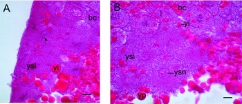
At stage 8 (late high blastula) the I-YSL formation is still not complete. At stage 9 (late epithelial blastula) there are syncytial regions and numerous energids within the yolk under the blastoderm, and during this stage the formation of the I-YSL finishes. It becomes a ‘layer’ in a strict sense. The YSL contains both interphase YSN and mitotic figures (Fig. 1 A, B). The contour of the basal YSL surface is very uneven. The epiboly begins between stages 9 and 10 (early epiboly, initiation of gastrulation). We did not observe mitotic figures at this intermediate stage. During blastula and gastrula periods the YSL thickness is variable (Fig. 2).
YSL of M. fossilis at stage 9, stained with Carazzi's haematoxylin–eosin. (A) E-YSL. One of the mitotic figures is indicated with an arrowhead. (B) The forming I-YSL. blastoderm cell (bc), yolk inclusion (yi), yolk platelet (yp), yolk syncytial layer (ysl), yolk syncytial nuclei (ysn). Scale bars = 10 μm.

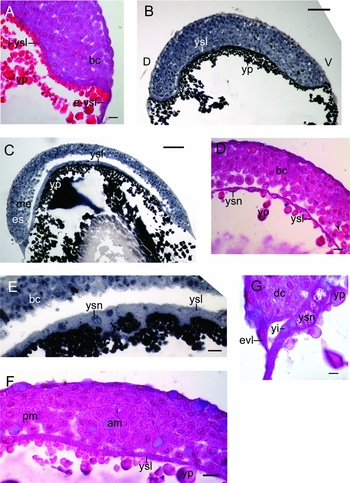
YSL of M. fossilis during epiboly and gastrulation. (A, D, F, G) Stained with Carazzi's haematoxylin and eosin (erythrosin). (B, C, E) Stained with Heidenhain's iron haematoxylin). (A) Stage 10. Longitudinal section. E-YSL contains less yolk inclusions (yi) than I-YSL. (B) Stage 11. Sagittal section. Dorsal (D) and ventral (V) sides of an embryo are indicated. (C) Stage 16, sagittal section. (D) Stage 16. Longitudinal section. The thin YSL region. (E) Stage 16. Parasagittal section. The thickened animal-most YSL region with protuberances. (F) Stage 18. Transverse section. The YSL under the axial structures. (G) Stage 18. Longitudinal section. The E-YSL with giant YSN (ysn) and yolk inclusions. axial mesoderm (am), embryonic shield (es), enveloping layer (evl), mesendoderm (me), presomitic mesoderm (pm). Scale bars = 20 μm (A, D, E–G), 100 μm (B, C).

During gastrulation the YSL is thickened at the dorsal side and in the animal-most region (Fig. 2 B, C). The mean thickness of the dorsal, animal and ventral I-YSL regions at stage 11 is 13.04 ± 0.4 µm, 11.36 ± 0.58 µm and 5.79 ± 0.16 µm respectively. At stage 16 the mean thickness of the dorsal, animal and ventral I-YSL regions is 14.65 ± 0.28 µm, 16.75 ± 0.44 µm and 7.25 ± 0.14 µm, respectively. Mann–Whitney U-test indicates that the thicknesses of the dorsal and ventral I-YSL regions differ significantly. The basal YSL surface forms the protuberances with 1–2 YSN into the yolk mass (Fig. 2 E). The syncytial ‘islands’ with 1–2 YSN in the yolk mass are also observed. Both E-YSL and I-YSL contain yolk inclusions, but they were more numerous in the I-YSL (Fig. 2 A, E, G). In the course of epiboly and gastrulation, the amount of yolk inclusions in the YSL decreases, and its contour becomes smoother. At stage 18 (nearly 88% epiboly) the YSL remains thickened in the animal region. The YSL region under the axial structures became thinner compared with previous stages (Fig. 2F).
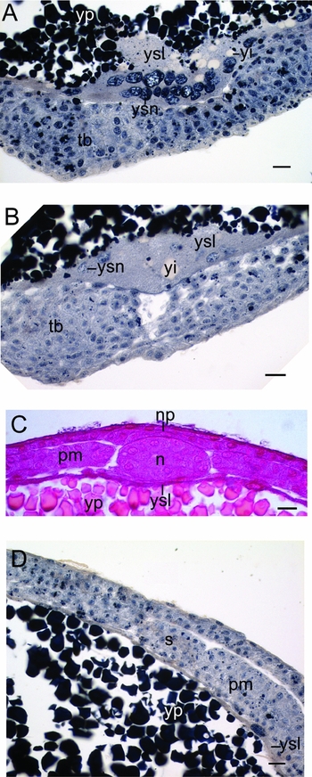
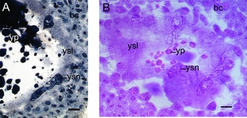
At stage 20 (tail bud stage) the epiboly finishes. A large portion of the YSL cytoplasm accumulated at the region of yolk plug closure and contained numerous YSN, including giant ones (3A, B). The YSL remains significantly thickened in the posterior region of an embryo at stage 21 (1-somite stage). During these stages the YSL is also thickened in the ventral region, and is relatively thinner in other regions including the region under the axial structures (Fig. 3 C, D). At stage 21 the mean thickness of the YSL is 2.44 ± 0.08 µm in the antero-dorsal region and 44.92 ± 3.71 µm in the region of yolk plug closure and tail bud.
YSL of M. fossilis during stages 20 and 21 (A, B, D) Stained with Heidenhain's iron haematoxylin. (C) Stained with Carazzi's haematoxylin and erythrosin. (A) Stage 20. Transverse section. Region of yolk plug closure. (B) Stage 21. Parasagittal section. Region of yolk plug closure. (C) Stage 20. Transverse section. The YSL under the axial structures. (D) Stage 21. Parasagittal section. The YSL under paraxial mesoderm. Anterior is to the left. notochord (n), neural plate (np), presomitic mesoderm (pm), somite (s), tail bud (tb), yolk inclusion (yi), yolk platelet (yp), yolk syncytial layer (ysl), yolk syncytial nuclei (ysn). Scale bar = 20 μm.

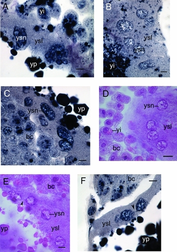
The YSN, which are nearly two times larger than the blastoderm nuclei are seen at stage 9, but they were few. The giant YSN appear sporadically from the intermediate stage between stages 9 and 10. The number of large (more than 18 µm) YSN increases by stage 14. The YSN of blastulae and gastrulae generally have regular, round and elliptical shapes (Fig. 4 B–D). The YSN with constrictions are numerous (Fig. 4 D). There are also YSN with complex lobed shapes. The ones connected with bridges are very rare (Fig. 4 E, F). The YSN have a reticulate chromatin structure. The sizes of the YSN and complexity of their shapes increase during development (Fig. 5 A, B). The mean lengths of the YSN at stages 11, 14 and 21 are 12.05 ± 0.15 µm, 14.05 ± 0.21 µm and 16.87 ± 0.35 µm respectively. In comparison, the mean lengths of the nuclei of embryonic cells is 9.51 ± 0.07 µm, 9.24 ± 0.07 µm and 6.726 ± 0.07 µm. There are lucent YSN and more heterochromatic ones, which are stained darker (Figs 4 and 5).
The YSN. (A, B, C, F) Stained with Heidenhain's iron haematoxylin, (D, E) stained with Carazzi's haematoxylin and erythrosin. (A) Stage 11. The large irregular-shaped YSN. (B) Stage 14. Elliptical lucent YSN with reticulate chromatin structure. (C) Stage 14. The heterochromatic YSN. (D) Stage 14. The round YSN and the YSN with constriction. (E) Stage 14. The YSN, connected with bridges, are indicated with an arrowhead. (F) Stage 16. The YSN, connected with bridges, are indicated with an arrowhead. blastoderm cell (bc), yolk inclusion (yi), yolk platelet (yp), yolk syncytial layer (ysl), yolk syncytial nuclei (ysn). Scale bars = 10 μm.

YSN at the stage 20. (A) The giant YSN compared with the nuclei of blastoderm cells (Heidenhain's iron haematoxylin). (B) The giant lucent YSN with reticulate chromatin (Carazzi's haematoxylin–eosin). blastoderm cell (bc), yolk inclusion (yi), yolk platelet (yp), yolk syncytial layer (ysl), yolk syncytial nuclei (ysn). Scale bars = 20 μm.

The blastoderm cells contain yolk inclusions during all stages studied. There are individual cells heavily loaded with yolk inclusions. Interestingly, the individual embryos differ from each other in amount and characteristics of the yolk inclusions in the YSL and diploid cells and amount of protuberances and syncytial ‘islands’.
Discussion
M. fossilis is an important model organism, however, little information is known about its morphology during development. Data on its YSL formation, structure and functioning are also incomplete. Formation of the YSL in M. fossilis occurs from stage 6 (early blastula) to the intermediate stage between stages 8 and 9 (Rozanova & Bozhkova, Reference Rozanova and Bozhkova1995). The syncytial regions under the blastoderm have been described for M. fossilis (Rozanova & Bozhkova, Reference Rozanova and Bozhkova1995). It is proposed that, in M. fossilis, it is formed as a result of fusion of the central basal blastoderm cells with the yolk sphere (Rozanova & Bozhkova, Reference Rozanova and Bozhkova1995). Our data are in agreement with observations made by Rozanova & Bozhkova (Reference Rozanova and Bozhkova1995). Studies using living transgenic embryos and immunohistochemical studies are necessary to test this proposal.
The YSL thickness is variable in different regions of an embryo. In loach embryos the dorsal and ventral sides of an embryo are distinguishable before embryonic shield formation (Neklyudova et al., Reference Neklyudova, Korvin-Pavlovskaya and Cherdantsev2007). Thickening of the YSL at the prospective dorsal region is seen from stage 11. Differences in the I-YSL thickness in dorsal and ventral regions at stages 11 and 16 were shown to be statistically significant.
We observe protuberances of the basal YSL surface and cytoplasmic islands with 1–2 YSN within the yolk mass. The syncytial cytoplasm within the yolk has been described previously in Solea senegalensis larvae (Padrós et al., Reference Padrós, Villalta, Gisbert and Estévez2011). The YSN within the yolk mass have been also described in Elasmobranchii (Jollie & Jollie, Reference Jollie and Jollie1967). The protuberances and ‘syncytial islands’ may serve to increase the area of the syncytium–yolk interaction. These islands probably arise from energids present in the yolk mass at the blastula period. Another possibility is that the ‘islands’ detach from the YSL. They are reminiscent of vitellophages – the energids with polyploid nuclei that metabolize yolk in particular arthropods (Kimble et al., Reference Kimble, Coursey, Ahmad and Hinsch2002).
Although the E-YSL contains yolk inclusions, they are more numerous in the I-YSL. A similar distribution of yolk inclusions is observed in zebrafish and indicates the functional regionalization of the YSL during blastula and gastrula periods (Kimmel et al., Reference Kimmel, Ballard, Kimmel, Ullmann and Schilling1995; Kondakova & Efremov, Reference Kondakova and Efremov2014). During gastrulation, the yolk inclusions in the YSL become less abundant. A decrease in the number of yolk inclusions has been described previously in F. heteroclitus (Lentz & Trinkaus, Reference Lentz and Trinkaus1967). In Teleostei, the yolk utilization rate is lower during early developmental stages compared with late embryonic and larval stages, when yolk circulation is established (Finn & Fyhn, Reference Finn and Fyhn2010).
Cytoplasm accumulation at the region of yolk plug closure has been shown for D. rerio (Kondakova & Efremov, Reference Kondakova and Efremov2014). This region contains numerous YSN. The YSL remains thickened in the caudal region during early segmentation in both species (Kondakova & Efremov, Reference Kondakova and Efremov2014).
During the blastula period, mitotic figures and interphase YSN are present. Similar observations have been made in Oryzias latipes and zebrafish (Kageyama, Reference Kageyama1996; Kondakova & Efremov, Reference Kondakova and Efremov2014). As shown in living loach embryos, the YSN can either be round, teardrop-shaped or irregular-shaped. The latter can have one or more constrictions and branch. YSN, connected with nuclear bridges, were also shown. It was proposed that this could be a pathological state caused by the vital dye, but embryos used in our study developed under normal conditions. YSN with nuclear bridges have been found in morphologically normal Cyprinus carpio koi larvae studied by histological methods (Kondakova et al., Reference Kondakova, Efremov and Nazarov2016) and living zebrafish gastrulae (D'Amico & Cooper, Reference D'Amico and Cooper2001). The reticulate chromatin structure of the YSN has been shown previously using Hoechst 33258 staining in living loach embryos, and was confirmed by our data. Our measurements of YSN on histological sections agree with the data on linear dimensions of YSN in living embryos (Korzh et al., Reference Korzh, Bajkova and Dmitrevskaya1989). At the beginning of epiboly there were nearly 150–200 YSN (Korzh et al., Reference Korzh, Dmitrevskaya and Kononenko1990).
Yolk inclusions in blastoderm cells were present during all stages studied. In comparison, in D. rerio, they are present until the 50% epiboly stage (Thomas, Reference Thomas1968), and in Salmo gairdneri embryonic tissues contain yolk until the beginning of blood circulation in yolk sac vessels (Sire et al., Reference Sire, Babin and Vernier1994).
There is individual variability in early loach embryos (Cherdantsev & Tsvetkova, Reference Cherdantsev and Tsvetkova2005; Cherdantsev & Korvin-Pavlovskaya Reference Cherdantsev and Korvin-Pavlovskaya2016). We have observed slight individual variation in YSL morphological characteristics, such as the number of protuberances or yolk inclusions and in yolk inclusion staining.
Misgurnus fossilis and D. rerio are phylogenetically related, but there are interesting differences in YSL formation and morphofunctional organization between these model species. YSL formation is prolonged in M. fossilis compared with zebrafish, and the mode of I-YSL formation is probably different, because central basal blastomeres may contribute to its formation (Rozanova & Bozhkova, Reference Rozanova and Bozhkova1995). In zebrafish, the I-YSL is presumably formed as a result of YSL spreading under the blastodisc (Kimmel et al., Reference Kimmel, Ballard, Kimmel, Ullmann and Schilling1995). Bozhkova & Voronov (Reference Bozhkova and Voronov1997) have shown similar, but not identical, dynamics of gap junctions between YSL and blastoderm cells in loach and zebrafish. YSL thickness in the late blastulae and gastrulae of the loach is variable. It is of note that YSL thickness is different in prospective dorsal and ventral sides of a loach embryo already during early gastrulation. In contrast, in zebrafish, dorsal and ventral YSL regions do not differ from each other (Kondakova & Efremov, Reference Kondakova and Efremov2014). It is important to establish whether the dorsal thickening of the loach YSL is linked to the oriented movements of the YSN (D'Amico & Cooper, Reference D'Amico and Cooper2001, Carvalho et al., Reference Carvalho, Stühmer, Bois, Kalaidzidis, Lecaudey and Heisenberg2009). The protuberances of the basal YSL surface and syncytial islands within the yolk mass are also characteristic of the loach. M. fossilis has larger eggs than those of zebrafish and these eggs develop at a lower temperature. The optimal temperature for M. fossilis development is 21.7° С, and for D. rerio it is 28.5° С. The diameters of yolk sphere are 1.17–1.3 mm and 0.5–0.6 mm respectively. These peculiarities may be among the factors that determine the differences in YSL structure (Kostomarova, Reference Kostomarova, Dettlaff and Vassetzky1991; Kimmel et al., Reference Kimmel, Ballard, Kimmel, Ullmann and Schilling1995; Bruce, Reference Bruce2016).
Data on early development, including timing of YSL formation and epiboly in Misgurnus anguillicaudatus, another representative of genus Misgurnus, have been published previously (Fujimoto et al., Reference Fujimoto, Kataoka, Otani, Saito, Aita, Yamaha and Arai2004, Reference Fujimoto, Kataoka, Sakao, Saito, Yamaha and Arai2006). The M. fossilis species probably arose by a tetraploidization event (Raicu & Taisescu, Reference Raicu and Taisescu1972). It would be interesting to compare YSL morphology in these two species.
Organization of the temporary extraembryonic structure as a syncytial layer with numerous polymorphic polyploid nuclei is conserved among Teleostei (reviewed in Kunz, Reference Kunz2004; Jaroszewska & Dabrowski, Reference Jaroszewska and Dabrowski2011; Kondakova & Efremov, Reference Kondakova and Efremov2014), which points at its efficiency. However, variations in its structure may be linked to the evolutionary plasticity of this group.
Study of the YSL structure in M. fossilis contributes to the data on teleost developmental diversity, mechanisms of early development and to the biology of temporary structures.
Acknowledgements
The authors thank Dr S.M. Efremova, Dr E.L. Gonobobleva, Dr L.A. Sleptzova, Dr E.G. Korvin-Pavlovskaya, Dr O.A. Freilikhman and A.V. Varlamova for their invaluable help. The authors acknowledge the support of the resource research centres at St Petersburg State University, the ‘Centre for Molecular and Cell Technologies’ and ‘Chromas’.
Financial support
This work was supported by the Russian Foundation for Basic Research (grant number 16–34–00391).
Statement of interest
We declare no conflict of interest.