Introduction
Materials that can replace part of a living structure or biofunction are called “biomaterials” and have been widely used for dental implants, hip implants, plastic surgery, heart valves, and cardiac pacemakers.Reference Park and Lakes1, Reference Townes2 The typical size of a cell is 10–20 μm, whereas the characteristic dimensions of cell surface structures (receptors, filopodia, and lamelopodia) that interact with biomaterials or the extracellular matrix (ECM) are in the order of a few nanometers.Reference Nakashima, Zhou, Kunkel, Zhang, Deng, Behringer and de Crombrugghe3 Thus, by introducing microscale or nanoscale topography on biomaterials without compromising the material properties, researchers want to achieve better cellular attachment. In this way, the integration of an implant into bone or other tissues can be improved. Indeed, it is very important that the implanted material integrates in the surrounding tissue and is functional in the living tissue; in other words, the implant should be “biocompatible.”
Introducing surfaces containing micro- and nanoscale features in an engineered controlled manner has already been shown to significantly affect medical implant integration into the human body.Reference Chehroudi, Gould and Brunette4 A key factor in this process is the initial cellular response toward the device; and to improve this interaction, different techniques such as chemical treatment, plasma spraying, sputtering, lithography, and organic or inorganic coatings have been applied alone or in different combinations.Reference Shin, Jo and Mikos5 One of the methods that has started to be exploited recently is laser surface engineering because of its precision, speed, and an optimal price-performance ratio.
This review describes how lasers are used to modify and enhance surface properties of biomaterials. To get a better understanding of these processes, we first discuss the relevant processes that occur at the implant interface, and then provide a short overview of different techniques used to modify the surface topography. Subsequently, we will specifically focus on the laser techniques that have been developed for surface texturing by describing in vitro, pre-clinical, and clinical trials for bone tissue replacement. Finally, we will discuss the advantages and disadvantages of laser surface engineering.
Interface and initial events
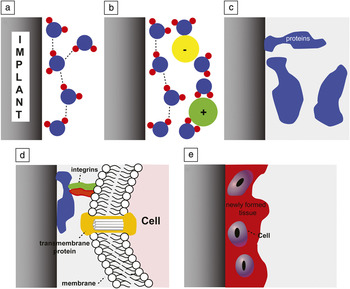
The success of bone as well as other implants depends on better cell-material interactions as it improves the fixation of an implant into the tissue. The process of implant integration into a tissue is complex in nature, and depending on the biocompatibility of the implant, it can lead to different final outcomes, which result in either full integration or implant loss.Reference Anderson, Rodriguez and Chang6 When looking at initial stages immediately after the placement of an implant into a freshly prepared surgical site, water, ions, and serum proteins arrive at the biomaterial surface (Figure 1). An important parameter for the adhesion of these molecules is the surface free energy (or wettability) of an implant surface, which is reflected by its hydrophilicity.Reference Roach, Eglin, Rohde and Perry7 Differences in surface wettability (e.g., very hydrophilic or hydrophobic) provide different opportunities for protein binding. Generally, the more hydrophilic a surface, the more proteins will be adsorbed.
Initial processes on the interface between a biomaterial and biofluid immediately after implantation. (a) The surface is first exposed to water, and the interactions are strongly affected by the surface free energy of the implant surface. (b) The surface is covered with a layer of hydrated ions followed by proteins (c). (d) Interaction of blood cells with the implant surface via cell receptor integrins. (e) Regenerated tissue at the implant-tissue interface.

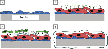
Directly after the adherence of proteins on the implant site, monocytes arrive (blood cells involved in immune response). If these cells adhere to the implant, they can mature into macrophages (Figure 2). Macrophages are the key orchestrator in the inflammation process and produce a large number of biologically active products such as proteases, coagulation factors, growth factors, and cytokines (proteins that regulate immune response). These processes trigger different events around the implant, greatly determining whether an implantation will clinically succeed.Reference Anderson, Ratner, Hoffman, Schoen and Lemons8 Degradable implants usually are resorbed in time by macrophages. After resorption, a fibrotic tissue (scar) will remain. The most common unwanted process that occurs at the implant site is encapsulation of the implant.Reference Anderson, Rodriguez and Chang6 If this occurs, macrophages will fuse to form giant cells to enhance their effectiveness. The giant cells will start releasing signals to attract connective cells (fibroblasts) from the surrounding tissue.Reference Anderson9 Fibroblasts and vascular endothelial cells will form granulation tissue and encapsulate the implant with a thin layer of collagenous matrix to isolate the implant from the surrounding tissue.Reference Ratner10 The formation of this collagen matrix layer is often called the “foreign body reaction.”Reference Anderson, Ratner, Hoffman, Schoen and Lemons8
Foreign body reaction of macrophages to implant materials. (a) Adsorption of plasma proteins (blue). (b) Macrophages attached to the implant site fuse into giant cells (red) releasing digestive molecules (green). (c) The giant cells start releasing chemotactic factors to attract collagen-secreting (dark green) fibroblasts (green). (d) Collagenous matrix proteins are released, and the implant is encapsulated (dark green).

If cells from the tissue surrounding the replacement (bone cells) are attached to the adhered proteins on the surface of the biomaterial, the implant can be integrated directly into the surrounding tissue without the formation of a fibrous capsule. This response is frequently described for bone tissue as osseointegration. During this event, platelets and macrophages start secreting proteins for bone regeneration (transforming growth factor beta [TGF-β], bone morphogenetic protein [BMP], and platelet derived growth factor [PDGF]) that will adhere to the provisional matrix. The presence of these proteins at the implant surface induces the interaction between bone cells and the protein loaded implant, which is ultimately followed by tissue regeneration at the implant site. Bone formed at the implant surface is considered a positive result, whereas fibrous encapsulation is considered a negative result.Reference Albrektsson and Sennerby11
Biomaterial properties
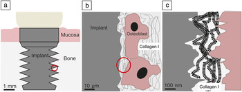
Bulk composition, mechanical characteristics, and surface properties such as surface free energy, surface charge, ion release (not discussed in this review), and perhaps even more importantly the specific surface topography of a material, determine the wound healing response and tissue repair after implantation. Micrometer and nanometer roughness first enhance the hydrophilicity of a material when compared to its smooth counterpart.Reference Rupp, Scheideler, Olshanska, de Wild, Wieland and Geis-Gerstorfer12 The importance of surface topography has already been recognized on many levels for a long time.Reference Albrektsson, Branemark, Hansson, Kasemo, Larsson, Lundstrom, McQueen and Skalak13–Reference Kasemo15 Biological systems recognize and respond to the surface through surface patterns. Implant surfaces are thus rarely smooth but generally have a certain topography, which can be either roughness (a specific topography) or texture (structured topography).Reference von Recum and van Kooten16 Surface roughness is characterized by randomly distributed surface topographies, whereas surface texture is characterized by a regular surface topography with defined dimensions and distribution. Surface topography can be divided into three levels: macro-, micro-, and nano-sized topologies. Macro-sized topography (>100 μm; Figure 3a) improves mechanical stability; however, it also introduces an increased risk of the induction of inflammatory processes.Reference Le Guehennec, Soueidan, Layrolle and Amouriq17 Microscale surface topography (1–100 μm; Figure 3b) maximizes the interlocking between ingrowth of mineralized bone and the implant surface. An advantage of micro-sized roughness is the early bone implant contact, which is especially important in patients with insufficient bone quantity or anatomical limitations. Nanoscale surface topography (<100 nm; Figure 3c) is least understood, yet it falls in the same order of size as the natural ECM, and it seems ultimately important to determine the adsorption of proteins, the adhesion of bone cells (osteoblasts), and rate of bone healing.Reference Williams18 The ECM contains mostly collagen Type I fibrils that have a diameter of 1.4 nm, a length of 300 nm, and are staggered with a periodicity of 68 nm. Additionally, compact bone has an average roughness of 32 nm, which is also in the nanometer scale.Reference Palin, Liu and Webster19 Mimicking such dimensions of the ECM on implant biomaterials by the use of nanotechnology might improve bone healing after implantation.
Overview of several topographies on a dental implant ranging from (a) macroscale (i.e., >100 μm), (b) microscale (1–100 μm), and down to (c) nanoscale (<100 nm).

Non-laser techniques used to modify the surface topography
It is important to emphasize that prior to the use of lasers for surface modification of biomaterials, a variety of techniques for random and ordered topographies were developed. The role that the micro- and nanoscale topography of a surface can play in controlling cell responses has been well documented and has led to an interest in studying cell response to submicron-scale surface features.
Random micro- and nano-roughness
Random micro- and nano-roughness can be created by several techniques, such as machining,Reference de Oliveira and Nanci20 chemical (acid) etching, anodization,Reference Fan, Cui, Hou, Xu, Chen and Lee21–Reference Pantoya and Granier23 sintering,Reference Webster and Ejiofor24 self-assembly,Reference Zhang25 and polymer demixing.Reference Park, Harrison, Chaikin, Register and Adamson26, Reference Norman and Desai27 Substrates created with such techniques display a completely random micro- and nano-roughness. The formed pattern dimensions are dependent on acid concentration and composition, current, electrolyte temperature, and time of incubation.
Ordered micro- and nano-texture
Manifold lithographic techniquesReference Senefelder28 have been used to create textured surfaces with ordered micro- and nano-roughness. Electron beam lithography (EBL),Reference Norman and Desai27, Reference Altissimo29 focused ion beam processing (FIBP),Reference Chen and Pepin30, Reference Tseng31 nanocontact printing (NP),Reference Torres, Wu, Holowka and Baird32 and nanoimprint lithography (NIL)Reference Chou, Krauss and Renstrom33, Reference Austin, Ge, Wu, Li, Yu, Wasserman, Lyon and Chou34 are widely used for this purpose, and surface features down to 5 nm can be created. The general setup for all lithographic techniques is that they require spin coaters, vacuum chambers, and optimization protocols to coat and develop specific photoresists or thin films, and they are time-consuming.
In vitro studies using nanotopography
The groups of Jansen,Reference Prodanov, te Riet, Lamers, Domanski, Luttge, van Loon, Jansen and Walboomers35 Curtis,Reference Curtis, Casey, Gallagher, Pasqui, Wood and Wilkinson36 and DalbyReference Dalby, Yarwood, Riehle, Johnstone, Affrossman and Curtis37 were the pioneers in the field of surface texturing and performed in vitro studies on substrates bearing several different microtopographical (semi-) ordered patterns (pits, pillars, and grooves). More recently, several studies demonstrated the beneficial effects of nanophase materials compared to conventional materials on osteoblast adhesion, proliferation, and differentiation.Reference Webster and Ejiofor24, Reference Webster, Siegel and Bizios38–Reference Dulgar-Tulloch, Bizios and Siegel44 Dalby et al.Reference Dalby, Childs, Riehle, Johnstone, Affrossman and Curtis45–Reference Dalby, Riehle, Johnstone, Affrossman and Curtis50 performed many studies on nano-sized islands. Such islands can be formed using spontaneous mixing of two polymers (polystyrene and poly(4-bromostyrene)), with the height of the features determined by the polymer concentration. Results demonstrated that fibroblast and endothelial cell adhesion and focal adhesion formation were mostly enhanced on islands with dimensions around 10–13 nm. In addition, studies of osteoblast behavior on polymer demixed islands demonstrated that spreading and differentiation were specifically enhanced on 10 nm high islands.Reference Dalby, McCloy, Robertson, Agheli, Sutherland, Affrossman and Oreffo51
Thus far, several types of nanotextures created with EBL have been used for cell culture assays, such as pits, pillars, and grooves. First Curtis et al.Reference Curtis, Casey, Gallagher, Pasqui, Wood and Wilkinson36 and later Dalby and Biggs et al.Reference Dalby, Riehle, Johnstone, Affrossman and Curtis49, Reference Biggs, Richards, Gadegaard, McMurray, Affrossman, Wilkinson, Oreffo and Dalby55–Reference Dalby, Gadegaard and Wilkinson59 used large arrays of pits or pillars in silicon with a diameter of 120 nm or less and a center-to-center spacing of 300 nm or less. Using an optimized method of EBL, 1 × 1 cm2 areas were created within one hour. These were used as a master for replication into either polymethylmethacrylate (PMMA), poly(caprolactone) (PCL), or (poly)carbonate (PC). Using the nanopatterned replicas, Dalby et al. demonstrated that the 13-nm islands produced fibroblasts with the biggest area, and 95 nm islands produced cells with the smallest areas.Reference Dalby, Riehle, Johnstone, Affrossman and Curtis49 Loesberg et al.Reference Loesberg, te Riet, van Delft, Schon, Figdor, Speller, van Loon, Walboomers and Jansen60 studied fibroblast alignment on a polystyrene “biochip” containing 50 different nanotextured patterns in a range of groove dimensions, nanopits, and squares. The study showed that fibroblasts sense patterns with a feature size down to 35 nm. Recently, we studied initial osteoblast behavior on the same substrates and demonstrated that osteoblasts are more sensitive to nanopatterns than fibroblasts and respond to patterns with a feature size down to 18 nm. The same study also demonstrated that groove depth and width are key factors in determining the osteoblast response and calcium formation by osteoblasts.Reference Lamers, van Horssen, te Riet, van Delft, Luttge, Walboomers and Jansen61
Finally, nanocontact printing experiments on both micro- and nanoscale (protein) patterns have been performed to study the behavior of cells (i.e., neuronal, melanoma, and capillary endothelial cells). Patterns influenced many cellular processes, such as motility, adhesion, spreading, and differentiation.Reference Lenhert, Meier, Meyer, Chi and Wiesmann62–Reference Hoover, Lee and Yousaf66
In vivo studies using techniques excluding laser treatment
Only preliminary studies have been reported assessing the biocompatibility of nanotextured biomaterials in vivo. Giavaresi et al. studied the effect of polymeric nanopits and nanocylinders on fibrous capsule formation and vascularity by implanting the biomaterials subcutaneously into rats. The study demonstrated that nanopits increased the development of fibrous capsules, while nanocylinders increased the cellularity of the fibrous capsule and the vascular density.Reference Giavaresi, Tschon, Daly, Liggat, Sutherland, Agheli, Fini, Torricelli and Giardino67
In another in vivo study, Popat et al. first studied the in vivo biocompatibility of nanotubular substrates with a pore size of approximately 80 nm by subcutaneous implantation of the biomaterial into rats. The study demonstrated that TiO2 nanotubular substrates did not induce fibrous tissue formation around the implant and concluded that the biomaterials were biocompatible.Reference Popat, Leoni, Grimes and Desai68
Laser surface engineering
LSE is a material processing method to melt, heat, or modify the surface of different types of materials. In general, the laser beam is focused to a small spot and has a power density that is sufficiently high for the material to absorb it, rather than reflect it. Briefly, lasers are devices that transform light of varying frequencies into a single intensity in the infrared, visible, ultraviolet, or x-ray regions of the spectrum and can be based on liquids, gases, solid-state materials, and excimers.Reference Semak and Dahotre69–Reference Probst72
For texturing applications, various lasers are used depending on the features to be fabricated and the type of material to be processed. Most of the in vitro studies on cell-implant interactions applied the laser surface melting (LSM) technique with neodymium doped yttrium aluminium garnet (Nd:YAG) or CO2 lasers or used excimer lasers for ablation. The actions of CO2 and Nd:YAG lasers are generally thermal processes, whereas the actions of the different excimer lasers produce almost no heat.Reference Kurella and Dahotre73 In this section, the potential of LSE using laser texturing methods will be described.
Lasers surface melting
Generally in LSM, a thin surface layer is rapidly melted by a predetermined energy density laser beam into a focused spot on the surface, followed by rapid ablation and solidification to produce a microstructure. Usually the laser is used in a continuous wave, meaning that its output is nominally constant over an interval of seconds or longer. Prior to ablation of the material, the absorption of radiation at the surface raises its temperature, and this results in melting at the surface. As the temperature continues to rise, vaporization of the material occurs, and finally it solidifies when the temperature drops, leaving a patterned surface behind.Reference Asthana, Kumar and Dahotre74 Thus, melting, evaporation, and solidification are the series of steps involved in generation of new surface topography, and the rapid cooling rate results in homogenization and the refinement of microstructures or nanostructures. The beam from a Nd:YAG laser can be focused to a smaller spot size because of shorter wavelength compared to a CO2 laser.
Laser engineered net shaping
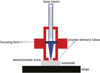
The LENS process uses metal powders that are deposited on the surface to modify metal parts. A high-powered pulsed Nd:YAG laser is mostly used, emitting near infrared laser radiation to create a melt pool on a metallic substrate of interest. Pulsed lasers concentrate their output energy into brief high-power bursts. The laser beam is focused to a small spot by one or more lenses. Metal powders are pushed around the laser with a carrier gas or by gravity, and an inert gas is often used to shield the melt pool from atmospheric oxygen. By moving the plate on which the material of interest is placed, or the head of the laser, a nanoscale pattern of the material is deposited on the surface. By repetition of this process, 3D structures can be created. LENS has been used for net shape manufacturing to fabricate porous Ti structures for hard tissue replacement, but mostly for coatings of various metallic materials (Figure 4).Reference Paital, Cao, He and Dahotre75–Reference Krishna, Bose and Bandyopadhyay78
General scheme representing the laser engineered net shaping (LENS) process. LENS uses metal powders to create nanostructures, which are deposited on the surface to modify metal parts. A high-powered pulsed laser creates a melt pool on a metallic substrate of interest. The laser beam is focused to a small spot by one or more lenses. Metal powders are pushed around the laser with a carrier gas or just by gravity. By moving the plate on which the material of interest is placed, or the head of the laser, a pattern of the material is deposited on the surface.

Selective laser melting/sintering
SLS mostly uses a CO2 high-heat laser to melt, sinter, or selectively fuse plastic, ceramic, metal, and glass powder particles. The particles can be fused layer by layer to form 3D structures. SLS is a type of additive manufacturing, meaning the item is built-up from materials into a final form. SLS also requires a computer model or description of the final piece. The heat from the laser fuses together powdered material by hitting it with very hot pulses. SLS allows the creation of parts with accurate detail with a structural resolution down to 30 μm.Reference Yadroitsev79
Laser interference lithography
A very promising technique for creating nanoscale surface patterns down to a 5 nm depth is laser interference lithography (LIL). LIL is a maskless lithographic technique using the interference pattern of two obliquely incident beams.Reference van Soest, van Wolferen, Hoekstra, de Ridder, Worhoff and Lambeck80, Reference Luttge81 One part of the laser beam reaches a photoresist layer directly, while the second part of the laser reaches the photoresist layer via a mirror and produces a regular interference pattern in the photoresist, followed by etching. A line pattern is created using two lasers, whereas a dot type pattern can be created by using three lasers (two interfering beams).Reference Lasagni, Acevedo, Barbero and Mucklich82 By the use of an extra interference beam (four-beams in total), periodic arrays of holes are generated forming pillar-like structures.Reference Tan, Peng, Pakarinen, Pessa, Petryakov, Verevkin, Zhang, Wang, Olaizola, Berthou and Tisserand83
In vitro experiments using laser surface treatments
On the basis of their chemical properties, bone implant materials can roughly be divided into metals, ceramics, and polymers. These different material classes provide very different mechanical characteristics.Reference Cooke84 The mechanical properties of an implant biomaterial determine stress-strain, elasticity, tension-compression, shear, and fracture resistance properties.Reference Binyamin, Shafi and Mery85 Lasers have been used to modify the surfaces in all three main groups of materials. Although some studies report complete experimental laser conditions and parameters, unfortunately most do not give the complete experimental conditions necessary for good reproducibility. It must be emphasized, however, that a careful selection of laser parameters for surface modification is of primary importance. Thus, in the following paragraphs, we will discuss only the final results obtained with different materials and different types of cells.
Titanium and titanium alloys
Because of their good mechanical properties such as biocompatibility and corrosion resistance, titanium and titanium alloys are the most commonly used medical implant materials for bone tissue replacement in oral and orthopedic surgeries. The first laser surface treatment procedure on titanium was actually performed to join titanium structures in order to avoid severe embrittlement. Cracks normally appear while joining titanium structures when using an oxygen flame. Using tungsten gas as a laser source was shown to be a better heating substitute for the oxygen used.Reference Wang and Welsch86 Using a combination of several excimer laser treatments, Berezani et al. demonstrated that in titanium samples, the original alpha-titanium crystal structure was preserved,Reference Bereznai, Pelsoczi, Toth, Turzo, Radnai, Bor and Fazekas87 whereas Sisti et al. demonstrated that such samples had better isomorphic characteristic, increased surface area, and micro-roughness.Reference Sisti, Garcia, Guastaldi, Antoniolli, Rossi, Brochado and Ade88 In another study, using laser-based optical interference and direct laser melting techniques, microtexture features were produced on Ti-6Al-4V alloys. Subsequently, improved wettability and better cellular attachment were demonstrated when MC3T3-E1 osteoblast cells were used.Reference Paital, He, Daniel and Dahotre89 Similar results with improved proliferation rate were obtained with human osteosarcoma U2-OS cells seeded on Er,Cr:YSGG laser-treated titanium.Reference Huang, Chuang, Chen, Lee and Chen90
Porosity and pore size of biomaterials play an important role in bone formation in vivo. Higher porosity and pore size result in greater bone ingrowth, implant stability, and better integration of the implant into the bone tissue. Using a direct Nd:G laser forming method on titanium, pores with a diameter of 300 μm were created, and it was shown that cells grew along the rims of the pores better than in the control samples.Reference Mullen, Stamp, Brooks, Jones and Sutcliffe91 Furthermore, using SLM, microporous titanium alloy implants created by the use of a Nd:YAG laser demonstrated very good mechanical stability. Thus lasers can be used for the purpose of bone ingrowth production or orthopedic devices.Reference Lin, Wirtz, LaMarca and Hollister92 Xue produced titanium porous implants using the LENS method and tested the implant with human osteoblast cells.Reference Xue, Krishna, Bandyopadhyay and Bose93 The results clearly demonstrated strong local adhesion, increased bone cell proliferation and differentiation, and an enhanced ECM formation compared to the non-treated controls. The results obtained also indicated that a critical pore size of 200 μm or higher was needed for cell in-growth into the pores. Below such dimensions, cells bridged the pore surface without any in-growth into the pores.
Cell interactions with laser-modified polymers and other materials
Different types of cells can be immobilized on a variety of biopolymers via covalent conjugation, hydrophobic interaction, or electrostatic interaction. Laser-modified polymer materials with nanoscale and micron-scale features produced on different types of polymers demonstrated improved grow rate, attachment, and differentiation of various type of cells. Surface KrF excimer laser irradiation on polystyrene significantly enhanced adhesion, alignment, and proliferation of human kidney cells (HEK-293) compared to unmodified controls.Reference Rebollar, Frischauf, Olbrich, Peterbauer, Hering, Preiner, Hinterdorfer, Romanin and Heitz94 Chinese hamster ovary (CHO-K1) cells and skeletal muscle cells aligned along the direction of the nanostructures, which had a periodicity between 200–430 nm and a depth of 30–100 nm. The alignment was only observed when the periodicity was above a critical value that was cell-type specific. Furthermore, using excimer laser irradiation on polyethylene terephthalate films clearly revealed that laser treatment also improved the adhesion and proliferation of human embryonic epithelial cells.Reference Mayer, Blanchemain, Dupas-Bruzek, Miri, Traisnel, Gengembre, Derozier and Hildebrand95 The maximal ablation depth on the surface of films was 1 μm, and the cell number sharply increased reciprocal to the laser fluency.
The endothelium is the thin layer of cells that lines the interior surface of blood vessels. Using KrF excimer laser radiation, polyurethane films with micropores down to a size of 1 μm were produced to study endothelialization rates. Results showed that pore size and pore density were both important; the endothelization rate increased with an increasing pore size. The highest endothelialization was found on films with micropores between 18 and 50 μm in diameter.Reference Matsuda and Nakayama96
A typical neuron possesses a cell body, dendrites, and an axon. Dendrites are branching filaments that arise from the cell body, often extending for hundreds of micrometers, and an axon is a filament that arises from the cell body and can travel for distances as far as 1 m in humans.Reference Nowakowski97 Finding new strategies to improve neuronal cell growth is important to study neurogenesis more precisely. Adult rat neural progenitor cells were seeded on polyethersulfone fibers that were treated with a KrF excimer laser to create microchannels. The results showed spontaneous cell process growth into the channels of the scaffold wall, while preventing the entrance of cell bodies.Reference Mapili, Lu, Chen and Roy98
Cones and ripples with spacings ranging from 2 to 25 μm were produced on poly(ethylene terephthalate) films by UV laser ablation to study the response of the shape and spacing of surface features on fibroblasts. Here, the highest cell attachment was observed in the regions with cones at ∼4 μm spacing. As feature spacing increased, cell spreading decreased, and the fibroblasts became more circular, indicating stress-mediated cell shrinkage.Reference Chandra, Lai, Sung, Murthy and Kohn99 In another study, fibroblasts and endothelial cells initially proliferated better on polymer nylon 12 films treated with an excimer laser.Reference Ball, Sherlock and Glynn100 Similar results were obtained on micron-scale structures generated on polyamide Kapton using an ArF excimer laser. Moreover, the same technique could be employed with a phase mask to generate nanoscale features.Reference Ball, Prendergast, O’Connell and Sherlock101
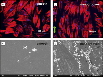
Using LIL, recently we studied the response of osteoblast-like cells to nanogrooved polystyrene substrates and demonstrated that cells aligned down to groove widths of 75 nm and depths of 35 nm. In addition, the study demonstrated that ECM produced by osteoblasts was sensitive to even smaller nanopatterns, and specific gene expression was induced by the nanopatterns (Figure 5).Reference Lamers, Frank Walboomers, Domanski, te Riet, van Delft, Luttge, Winnubst, Gardeniers and Jansen102
Fluorescent micrographs of cytoskeleton actin filaments staining (red) and nucleus staining (blue) of osteoblast-like cells grown for 48 h on a smooth and nanogrooved polystyrene substrate. Nanotextured groove patterns were made using laser interference lithography (LIL) on silica. Polystyrene replicas were made by solvent casting. In this study, patterned wafers with 130 nm wide grooves (200 nm pitch) and a depth of ∼50 nm were used. (a) Cells grown on the smooth substrate have random cell and actin orientation. (b) Note that cells and actin filaments oriented in parallel direction toward the nanogrooves. The green arrow indicates the direction of the nanogrooves. (c–d) Scanning electron microscopy micrographs of osteoblast-like cells after eight days of cell culture. Note that the calcium particles on top of the cells are found more on the nanogrooved substrate when compared to the smooth substrate and orient parallel to the groove direction.

In vivo studies using laser surface treatments
Pre-clinical development is a stage of research that begins before clinical trials (testing in humans) can begin, and during which important feasibility, testing, and safety are investigated in animal models. The success of bone but also other implants depends on better tissue-material interactions, as it improves the fixation of the implant to the bone. Thus, in vivo pre-clinical studies are very important to investigate the behavior of the implants and its fixation before real clinical studies are done. Until now, very few pre-clinical and clinical studies have been performed with laser-treated implants. In this section, we will discuss some of the results obtained so far.
Surface modification of titanium machined implants, which were additionally etched with a laser and micro-arc oxidation (plasma-electrochemical process), showed enhanced bone formation versus that seen around machined titanium implants in rabbits.Reference Guo, Zhou, Rong, Zhu and Geng103 In another study, low-level laser radiation improved the bone healing response around a titanium dental implant in the tibia of rabbits, but only after 45 days of implantation.Reference Lopes, Pinheiro, Sathaiah, Duarte and Cristinamartins104 In another similar study with a longer implantation period (six months), titanium machined implants, partly modified with nanofeatures by Nd:YAG laser irradiation, were implanted in the femurs and tibias of rabbit models and investigated. Good osseointegration of the implant in bone was observed for both implant types. The partly laser-modified titanium implants were associated with an increased removal torque after six months compared to the machined titanium implants. A different failure pattern with fractures in the bone tissue rather than at the bone implant interface also suggested better bone bonding than laser-modified surfaces.Reference Palmquist, Emanuelsson, Branemark and Thomsen105
Ethylene-propylene rubber (EPR)-based materials that were chemically etched, followed by CO2-pulsed laser treatment, implanted in the deep intramuscular and peritoneal layers (the lining of the abdominal cavity) in rabbits, showed that after eight weeks of implantation, the adhesion of the macrophages to the chemically modified laser-treated EPR samples was significantly lower than their adhesion on the unmodified surfaces. Also, no acute or chronic inflammatory reaction was observed at the site of implantation, and a thinner fibrous tissue capsule formed around the modified samples, whereas foreign body giant cells adhered to unmodified EPR.Reference Mirzadeh, Katbab, Khorasani, Burford, Gorgin and Golestani106
Porous Ti6A14V alloy implants fabricated by LENS and implanted into the femurs of rabbits demonstrated that new bone penetrated into the pores and directly bonded to the walls. This was seen together with a significant increase in calcium within the implants, indicating excellent biological tissue in-growth through the interconnected porosity when compared with the controls.Reference Bandyopadhyay, Espana, Balla, Bose, Ohgami and Davies107
The inflammatory process induced by implant surfaces is an important component of the tissue response, where knowledge is limited regarding the role of surface topography. Palmquist et al. showed that the combination of surface micro- and nano-features on laser-treated titanium implants contributes to the downregulation of early inflammatory events. The machined and laser-treated implants were implanted subcutaneously in rats for 72 hours. The results revealed that significantly fewer inflammatory cells were attracted to the sites with the laser-modified implants compared with machined titanium implants.Reference Palmquist, Johansson, Suska, Brånemark and Thomsen108
Clinical trials
After a tooth extraction, resorption of the crestal bone (i.e, beneath the tooth) can occur. By placing an implant, this process can be reduced. Tapered 2 mm dental implants treated with Laser-Lok (LL) microchannels were compared to machined (control) implants of the same design after implantation in the crestal bone of 15 patients. LL limited the bone loss to 0.59 mm as opposed to the 1.94 mm crestal bone loss reported for control implants.Reference Pecora, Ceccarelli, Bonelli, Alexander and Ricci109
Recent studies using direct laser metal sintering (DLMS) were used for mandibular reconstruction. Implantation of titanium meshes treated with DLMS in four patients showed that it can produce structures with complex geometry and consequently allow better osteoconductive properties (new bone formation). After eight weeks, the results showed a newly formed tissue composed of calcium and phosphorus. Good bone-to-implant contact was observed, as the bone-to-implant contact presented a mean of 60.5±11.6%.Reference Mangano, Piattelli, Raspanti, Mangano, Cassoni, Iezzi and Shibli110
Taken together, the pre-clinical and clinical studies using LSE for micro- or nanoscale surface topographical features indicate that structured surfaces promote long-term bone bonding and interfacial strength. However, more in vivo studies are needed to investigate other mentioned biomaterials. In addition, studies comparing implants treated with different techniques need to be taken into account to find the best candidates.
Summary
The advantages of using laser techniques for texturing surfaces over other conventional methods (chemical etching, anodization) are that they are rapid and extremely clean, focus great amounts of energy in one spot for a short time (pulse), and generate complex nano-features with high resolution. Unlike the different lithographic techniques, which require time-consuming steps such as class 100 clean rooms, spin coaters, vacuum chambers, and optimization protocols to coat and develop specific photoresists or thin films, laser-based techniques can avoid them and shorten these steps. The laser techniques can be used to selectively modify surface composition and microstructures without affecting the surrounding material properties or causing serious thermal distortion. The efficiency of absorption is linked to both the physical state of the material and the power density at the surface. As expected, higher power density is required to vaporize rather than to melt the material. Because of their ability to show no, or very little, thermal effects, excimer laser beams with higher density are particularly suited to photoablation. However, a laser beam with sufficient energy density has a limited beam size. Therefore, laser surface melting is less efficient than other conventional methods such as thermal spraying, in terms of coverage rate. Most laser surface engineering (LSE) processes appear expensive compared to other technologies; still, in many cases, they can be adapted to be more cost-effective because of their precision and speed.
Studies done on the effects of micro- and nano-textures on cell biocompatibility using conventional methods have shown good results in the last 30 years, particularly with regard to cellular attachment, proliferation, and differentiation of cells. LSE has just recently shown that the same effect can be achieved and gave the same biocompatibility effects. Pre-clinical and clinical studies with micro- and nanoscale laser engineered surface structures still need to be done. By understanding how physical surface parameters influence cellular attachment or how to develop a direct single-step method for creating micron or nanoscale surface texture, we can more effectively design biomaterial surfaces for a variety of tissue engineering applications.