Anomalous origin of the left coronary artery from the pulmonary artery is often fatal in infancy, but extensive collaterals permit rare adult survival despite coronary steal. Reference Yau, Singh, Halpern and Fischman1 While coronary translocation is the routine care in children, surgical ligation of the left main coronary artery arrests steal in adults, often combined with bypass grafts. Size mismatch favours saphenous veins or subclavian artery as conduits rather than mammary arteries. Reference Alexi-Meskishvili, Berger, Weng, Lange and Hetzer2
Evidence lacks in adults with extensive collaterals to show the superiority of establishing dual coronary supply over left coronary ligation. Reference Wilson, Dlabal and McGuire3 We document normalisation of perfusion abnormality after transcatheter left coronary occlusion in a symptomatic adult with inducible ischaemia.
Case report
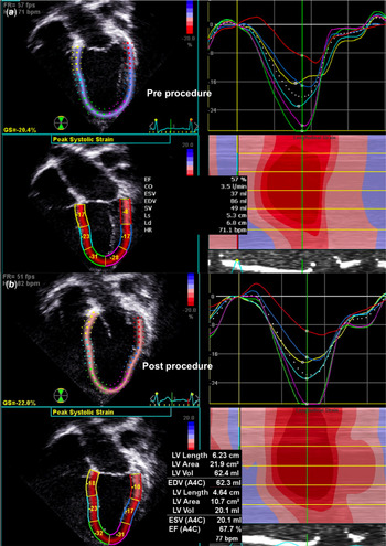
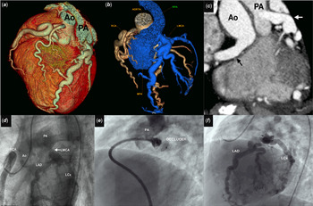
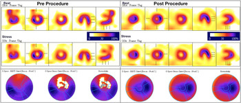
A 38-year-old female with dyspnoea after the second childbirth showed anterolateral ischaemia on the electrocardiogram. Echocardiogram diagnosed anomalous left coronary artery arising from the pulmonary artery with normal ventricular function (Fig 1). Diagnosis was confirmed on computed tomography (Fig 2). Exercise stress showed large perfusion defects (Fig 3). The patient declined surgical left coronary ligation with a venous coronary bypass, but agreed for a transcatheter left coronary occlusion with informed consent.
Before the intervention ( a ), the left ventricle was mildly dilated on a four-chamber echocardiographic view with basal lateral hypokinesia and the ejection fraction was 57% and longitudinal strain was −20%. After the intervention ( b ), there was a reduction in the left ventricular volumes along with improvement of ejection fraction to 68% and longitudinal strain to −22%.

The large dilated epicardial coronaries in volume-rendered computed tomographic image ( a ) were color coded ( b ) to separate the left (blue) and right (yellow) coronary arteries. The left main coronary artery (LMCA) measured 11mm in the multiplanar reformatted image ( c ). A still frame ( d ) in LAO 40° projection from a right coronary (RCA) rotational angiogram profiled the LMCA between the pulmonary artery (PA) and left anterior descending (LAD) and circumflex (LCx) branches. Mother and child strategy assisted cannulation and occlusion of LMCA with a long sheath ( e ). The final right coronary angiogram ( f ) showed adequate filling of the left coronary system without any steal. Ao=aorta.

Technetium sestamibi myocardial perfusion during rest (first row) and exercise stress (second row) showed significant resting and reversible ischaemia in three short axes, one horizontal and one vertical long-axis views. Polar map (third row) demonstrated apical and anterolateral fixed and reversible ischaemia. Post-intervention images showed a complete resolution of ischaemia at a higher exercise workload.

A rotational right coronary angiography identified 40° left anterior oblique projection to profile the coronary away from the pulmonary artery. Cannulating the coronary artery from the pulmonary sinus was challenging due to a sharp inferior angulation.
A mother and child strategy using an outer long 6F Flexor sheath (Cook Medical, Bloomington, IN), 6F Judkins left coronary guide catheter and inner 4F Judkins left coronary diagnostic catheter facilitated deep cannulation. An 8 mm Amplatzer muscular VSD occluder (Abbott medical, Plymouth, MN) with 16 mm retention skirts occluded the vessel (Fig 2, Supplementary video). The myocardial perfusion after 9 minutes and 40 seconds of exercise in Bruce protocol was normal at a 3-month follow-up (Fig 3).
Discussion
Adults with anomalous left coronary artery origin from the pulmonary artery present with angina, dyspnoea or sudden death due to coronary steal and myocardial scar despite extensive collaterals. Reference Yau, Singh, Halpern and Fischman1 Reestablishing dual coronary supply is superior to left coronary ligation in children, but is performed only in two-thirds of the adults. Reference Yau, Singh, Halpern and Fischman1–Reference Wilson, Dlabal and McGuire3 Isolated left coronary ligation without bypass grafts is practiced in one-fourth of adults, especially in emergent surgeries. Reference Yau, Singh, Halpern and Fischman1–Reference Kreutzer, Schlichter, Roman and Kreutzer4
Addition of a venous bypass to left coronary ligation in adults as well as graft occlusions after bypass do not alter surgical outcomes; but occasional late mortality after isolated ligation may favour dual coronary flows. Reference Wilson, Dlabal and McGuire3 Histological ischaemic changes indicating coronary steal persist despite coronary bypass due to slow-flow in the venous grafts. Reference Kubota, Endo and Ishii5 Saphenous venous grafts are unlikely to retain adequate patency beyond few years. Reference Wilson, Dlabal and McGuire3
Even though patient refusal for surgery forced this intervention, the resolution of ischaemia after left coronary occlusion showed the utility of this strategy as a surgical alternative. Acute angulation of the left coronary artery offered technical challenges, unlike a previous report of a similar device occlusion of the left coronary artery arising from the right pulmonary artery. Reference Collins, Colman, Benson, Hansen, Merchant and Horlick6 Despite the advantages of dual coronary perfusion with an additional bypass graft, clinicians may opt for left coronary occlusion due to high surgical risks, graft occlusions on follow-up, slow-flow in venous grafts and non-suitability of mammary grafts. Long-term follow-up of these adults and more experience with catheter closure are needed to test if this transcatheter strategy can prove to be a surgical alternative.
Acknowledgements
None.
Financial support
This research received no specific grant from any funding agency, commercial or not-for-profit sectors.
Conflicts of interest
None.
Ethical standards
The authors assert that all procedures contributing to this work comply with the Ethical Standards of the Indian Council of Medical Research and with the Helsinki Declaration of 1975, as revised in 2008, and has been approved by the Institutional Committee of Madras Medical Mission, Chennai, India.
Supplementary material
To view supplementary material for this article, please visit https://doi.org/10.1017/S1047951120003418