Introduction
Widespread availability of ultrasound imaging has led to more frequent recognition of cystic liver disease (CLD). There is a wide range of possible causes. Cystic disease of infective origin is usually caused by an echinococcal species (Forbes & Murray-Lyon, Reference Forbes and Murray-Lyon1991). Echinococcosis is a zoonosis caused by the larval stage of tapeworms belonging to the genus Echinococcus (family Taenidae), which live in the intestine of domestic dogs and wild carnivores. Echinococcus granulosus and Echinococcus multilocularis, which cause cystic echinococcosis (CE) and alveolar echinococcosis (AE), respectively, are species of major medical and public health importance. Echinococcus granulosus infects both humans and domestic animals worldwide. Social behaviour and poor hygienic conditions favour the transmission of infection (Craig et al., Reference Craig, Rogan and Allan1997). CE, also called hydatidosis, has been reported in age groups from 1 to 75 years (Eckert & Deplazes, Reference Eckert and Deplazes2004).
During the past three decades a number of CE control programmes have led to a significant decrease in the incidence of human hydatidosis in some endemic areas (Larrieu et al., Reference Larrieu, Costa, Cantoni, Alvarez, Aquino, Giménez and Pérez1994; Palmer et al., Reference Palmer, Biffin, Craig and Walters1996). A study in Dalmatia, a well-known endemic area of CE, has shown a decrease in the incidence from the mid-1950s until 1990 in most Mediterranean parts of Croatia (Morović, Reference Morović1997). In Croatia, reporting of echinococcosis to the Epidemiology Reference Centre of the Croatian National Institute of Public Health (CNIPH) has been mandatory since 1965 (Morović, Reference Morović1997).
The diagnosis of CE is mainly suggested by imaging techniques, predominantly ultrasonography, computed tomography and X-ray examinations. For confirmation of CE it is essential to detect specific E. granulosus antibodies by immunodiagnostic tests (Grimm et al., Reference Grimm, Maly, Lü and Llano1998; Liance et al., Reference Liance, Janin, Bresson-Hadni, Vuitton, Houin and Piarroux2000; Eckert et al., Reference Eckert, Gemmell, Meslin and Pawlowski2001; Eckert & Deplazes, Reference Eckert and Deplazes2004). Enzyme-linked immunosorbent assay (ELISA), indirect haemagglutination antibody assay, latex agglutination test and immunoblot test are the most commonly used immunological methods (McManus et al., Reference McManus, Zhang, Li and Bartley2003). In routine laboratory practice, usually at least two different tests are used to obtain the most reliable results (Grimm et al., Reference Grimm, Maly, Lü and Llano1998; Eckert et al., Reference Eckert, Gemmell, Meslin and Pawlowski2001).
The aim of this study was to determine the seroprevalence of E. granulosus infection in Croatian patients presenting with CLD.
Materials and methods
Between January 2008 and July 2009, a total of 540 serum samples were collected from patients with hepatic cysts detected by imaging methods.
Collection and analysis of serum samples
Serum samples were obtained from patients residing in rural and urban areas of the Croatian mainland. Serological analysis was performed at the Parasitology Department, CNIPH, using a commercial semiquantitative ELISA (Echinococcus IgG-ELISA, Bioactiva Diagnostica GmbH, Germany), according to manufacturer's recommendations. ELISA reactive samples were confirmed using Echinococcus Western blot (WB) IgG test (LDBIO diagnostics, Lyon, France). According to WB manufacturer's instructions, the presence of the 7 and/or the 26–28 kDa bands is indicative for the presence of Echinococcus-specific IgG in the serum sample. Differential diagnosis between E. granulosus and E. multilocularis was made by detecting specific bands between 7 and 28 kDa. There are five typical immunoblot patterns (P1 to P5). Immunoblot patterns P1 (only the 7 kDa band) and P2 (the 7 kDa band and a large, fuzzy band at 16–18 kDa) are species specific for E. granulosus, while pattern P3 (26–28 kDa band plus two narrow bands at 16 and/or 18 kDa) is specific for E. multilocularis. Patterns P4 (only the 26–28 kDa band) and P5 (bands at 7 kDa and 26–28 kDa) can not distinguish these two species.
Statistical analysis
Data analysis was performed using the chi-square test. A P value less than 0.05 was considered to be significant.
Results
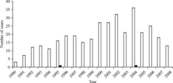
The annual number of reported cases of echinococcosis in Croatia in the past two decades is shown in fig. 1 (unpublished data from the Reference Epidemiology Centre, CNIPH).
Annual number of reported human cases of echinococcosis in Croatia, 1990–2008. White bars, number of reported human cases; black bars, number of deaths. (Unpublished data from the Reference Epidemiology Centre, CNIPH.)

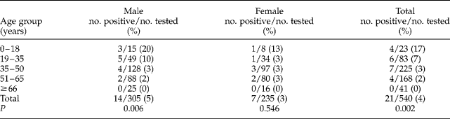
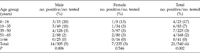
Of 540 analysed serum samples, 305 (56%) were from male and 235 (44%) from female patients. In 51 (9%) serum samples from patients with CLD, IgG antibodies to Echinococcus were detected by ELISA. Of 51 positive serum samples only 21 (3.9%) were confirmed by WB as E. granulosus infection (immunoblot patterns P1 or P2). The E. granulosus seroprevalence rate was 4.6% (14/305) among male and 2.9% (7/235) among female patients with no significant difference (P = 0.541).
The seroprevalence rate in participants residing in rural regions was 5% (8/151), compared to 3% (13/389) of participants residing in urban regions. This difference was not statistically significant (P = 0.291).
According to age, there was a significant difference in overall CE seropositivity between age groups (P = 0.002). According to gender, the CE seropositivity rate differs among age groups only in male patients (P = 0.006). The highest CE seroprevalence rate was reported in the youngest age group (up to 18 years), both in male and female patients (20% and 13%, respectively) (table 1).
Seroprevalence of Echinococcus granulosus in Croatian patients relative to age and gender.
Discussion
This is the first report of the seroprevalence of E. granulosus in patients with CLD in Croatia, with an overall seroprevalence rate of 3.9%. In a similar study conducted in Slovenia (Logar et al., Reference Logar, Soba and Kotar2008), the seropositivity in patients with suspected CE was 2.6%. This study showed that male and female patients up to 18 years of age, as well as younger males from 19 to 35 years of age, were at highest risk, with seroprevalences of 20%, 13% and 10%, respectively. These results are similar to those in some other endemic areas, in which most cases are recorded in the age groups between 21 and 40 years, with the highest morbidity found in younger individuals aged between 6 and 20 years (Ammann & Eckert, Reference Ammann and Eckert1996; Eckert & Deplazes, Reference Eckert and Deplazes2004). In two large published studies, most patients presenting with symptomatic disease were between the ages of 4 and 15 (Menghebat et al., Reference Menghebat, Jiang, Chai, Andersen, Chai and Liu1993; McManus et al., Reference McManus, Zhang, Li and Bartley2003), while in North Africa and Dalmatia, most cases are related to adults aged 21–40 and 20–60, respectively (Craig, Reference Craig, Rogan and Allan1997; Morović, Reference Morović1997). In the rural region of central Uruguay, seroprevalence was detected to be higher in the >60-year-old group (>11%) than in 4- to 6-year-old group (1.1%). Additionally, the 20- to 29-year-old group had a significantly higher CE rate, compared with younger and older age groups (Cohen et al., Reference Cohen, Paolillo, Bonifacino, Botta, Parada, Cabrera, Snowden, Gasser, Tessier, Dibarboure, Wen, Allan, Soto de Alfaro, Rogan and Craig1998).
In studies conducted in the Mediterranean part of Croatia, a known endemic area, most patients with human hydatidosis lived in rural areas (Barić, Reference Barić1994; Morović, Reference Morović1997). In this study no significant difference in CE seroprevalence was found between patients from rural areas compared to patients from urban areas, although seroprevalence in rural areas was slightly higher than in urban areas (5% and 3%, respectively).
In this study male patients were affected more frequently (67%) than females, but with no significant difference (P = 0.541), while the previous study from Dalmatia reported different results, with 60% of affected patients being females (Barić, Reference Barić1994; Morović, Reference Morović1997). Logar et al. (Reference Logar, Soba and Kotar2008) found no significant difference between the CE-positive patients in regard to their gender, although there were slightly more positive sera from female patients (24/34). In most of the published studies, there were no significant differences in the gender ratios of individuals with CE (Perdomo et al., Reference Perdomo, Alvarez, Monti, Ferreira, Chiesa, Carbó, Alvez, Grauert, Stern, Carmona and Yarzabal1997; Cohen et al., Reference Cohen, Paolillo, Bonifacino, Botta, Parada, Cabrera, Snowden, Gasser, Tessier, Dibarboure, Wen, Allan, Soto de Alfaro, Rogan and Craig1998; Eckert et al., Reference Eckert, Gemmell, Meslin and Pawlowski2001), which is in agreement with the results of this study.
During the past decades (especially during the 1970s and 1980s), a significant decrease in the incidence of human hydatidosis has been observed in some endemic areas (Larrieu et al., Reference Larrieu, Costa, Cantoni, Alvarez, Aquino, Giménez and Pérez1994; Palmer et al., Reference Palmer, Biffin, Craig and Walters1996; Morović, Reference Morović1997). However, the observed decline in the number of new cases was partially attributed to under-notification (Serra et al., Reference Serra, Araya, Araneda and Serra1995; Morović, Reference Morović1997).
The initial phase of primary Echinococcus infection is always asymptomatic. Small cysts usually remain asymptomatic for many years, if not permanently (McManus et al., Reference McManus, Zhang, Li and Bartley2003). The presence of a cyst-like mass in the liver of a person with a history of exposure to sheepdogs in areas where E. granulosus is endemic supports the diagnosis of cystic echinococcosis. However, echinococcal cysts must be differentiated from benign cysts, cavitary tuberculosis, mycoses, abscesses, and benign or malignant neoplasms. The diagnosis is mainly suggested by imaging techniques, but has to be confirmed using serological assays (Liance et al., Reference Liance, Janin, Bresson-Hadni, Vuitton, Houin and Piarroux2000). Despite the development of molecular methods to prepare antigens, nowadays there is no standard, highly sensitive and specific test available for antibody detection in cases of CE (Siracusano & Bruschi, Reference Siracusano and Bruschi2006). Cross-reactivity with antigens from other parasites, notably other taeniid cestodes, is a major problem (McManus et al., Reference McManus, Zhang, Li and Bartley2003). To exclude false-positive results we used a commercially available assay, Echinococcus WB IgG, which is proven to be reliable for serological confirmation of human echinococcosis (Liance et al., Reference Liance, Janin, Bresson-Hadni, Vuitton, Houin and Piarroux2000), as the confirmatory assay. Echinococcus multilocularis infection was not found in this study.
Human echinococcosis is an endemic parasitic disease in many areas of the world, including Mediterranean countries. Humans are usually aberrant hosts for E. granulosus and CE may develop after accidental ingestion of tapeworm eggs excreted in the faeces of infected animals (Logar et al., Reference Logar, Soba and Kotar2008), usually by direct hand-to-mouth transfer. Children are most commonly infected, probably due to unintentional soil consumption and contact with infected dogs/puppies. Accumulated experience shows that sustained CE control can only be achieved by well-planned and realized programmes (Larrieu et al., Reference Larrieu, Costa, Cantoni, Alvarez, Aquino, Giménez and Pérez1994; Palmer et al., Reference Palmer, Biffin, Craig and Walters1996). According to the results of this study, especially findings that the highest CE seroprevalence rate occurred in children and younger adults, it can be concluded that CE still represents a public health problem in Croatia. Preventive measures should be used to control Echinococcus infections, including avoidance of contact with infected dogs, egg-contaminated soil or plants; control and treatment of dogs with antihelmintics; hand washing, improved sanitation and health education.
Acknowledgements
No financial support was received for this study. The authors declare no possible conflict of interest. This study was approved by the Ethical Committee of the Croatian National Institute of Public Health.