Introduction
The genus Brachyspira (formerly Treponema and later Serpulina) is a member of the family Brachyspiraceae (Ochiai et al., Reference OCHIAI, ADACHI and MORI1997) within the class Spirochaetes (Paster and Dewhirst, Reference PASTER and DEWHIRST2000). Spirochaetes are gram-negative, long, thin, helical, and motile bacteria. Brachyspira spp. are obligate anaerobes that attach to the mucin layer of the colon and can cause inflammation in some animal species and humans (Lux et al., Reference LUX, MOTER and SHI2000; Hirsh, Reference HIRSH2004).
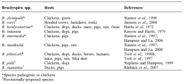
There are relatively few studies on Brachyspira and avian intestinal spirochaetosis (AIS). At present, there are nine species of Brachyspira isolated from birds (Table 1). Among them, B. hyodysenteriae, B. intermedia, B. pilosicoli and B. alvinipulli are considered pathogenic. The pathogenic potential of the rest of the Brachyspira spp. isolated from chickens is difficult to establish because this bacterium requires specialised techniques and methods to isolate. In addition, both pathogenic and ‘non-pathogenic’ species of Brachyspira can be detected in infected animals making it difficult to establish the clinical significance of ‘non-pathogenic’ Brachyspira spp. (Duhamel et al., Reference DUHAMEL, TROTT, MUNIAPPA, MATHIESEN, TARASIUK, LEE and HAMPSON1998). For instance, B. innocence, B. murdochii, and B. pulli, the presumed non-pathogenic species in chickens, have been isolated from chickens showing signs of AIS in conjunction with pathogenic species (Feberwee et al., Reference FEBERWEE, HAMPSON, PHILLIPS, LA, VAN DER HEIJDEN, WELLENBERG, DWARS and LANDMAN2008). In addition to B. pulli, there are two other provisionally proposed species (i.e. a species in which the name has been suggested but is not official) that have been isolated from birds (B. corvi and B. suanatina); however, their clinical significance is questionable (Stephens and Hampson, Reference STEPHENS and HAMPSON1999; Råsbäck et al., Reference RÅSBÄCK, JANSSON, JOHANSSON and FELLSTROM2007; Jansson et al., Reference JANSSON, FELLSTROM and JOHANSSON2008). To date, B. corvi, which has been isolated from corvid birds, has not been associated with AIS and B. pulli, which has been isolated from chickens and dogs, is presumed to be non-pathogenic (Feberwee et al., Reference FEBERWEE, HAMPSON, PHILLIPS, LA, VAN DER HEIJDEN, WELLENBERG, DWARS and LANDMAN2008). Brachyspira suanatina has been recovered from mallard ducks, and caused diarrhoea in experimentally infected pigs (Råsbäck et al., Reference RÅSBÄCK, JANSSON, JOHANSSON and FELLSTROM2007).
Brachyspira spp. reported in avian species and mammals.
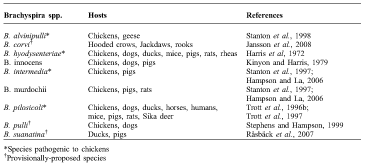
*Species pathogenic to chickens
†Provisionally-proposed species
All of the pathogenic species, except B. alvinipulli, have been shown experimentally to cause AIS (Trott et al., Reference TROTT, MCLAREN and HAMPSON1995; Hampson and McLaren, Reference HAMPSON and MCLAREN1999; Jamshidi and Hampson, Reference JAMSHIDI and HAMPSON2003). Avian intestinal spirochaetosis is a disease complex of mainly layer chickens (Adachi et al., Reference ADACHI, SUEYOSHI, MIYAGAWA, MINATO and SHOYA1985; Swayne et al., Reference SWAYNE, BERMUDEZ, SAGARTZ, EATON, MONFORT, STOUTENBURG and HAYES1992). In chickens, AIS can be manifested by reduced growth rate, delayed onset of egg laying, chronic diarrhoea, faecal staining of egg shells, weight loss, reduced egg production, and increased mortality (Davelaar and Smit, Reference DAVELAAR and SMIT1986; Griffiths et al., Reference GRIFFITHS, HUNT, LISTER and LAMONT1987; Swayne et al., Reference SWAYNE, BERMUDEZ, SAGARTZ, EATON, MONFORT, STOUTENBURG and HAYES1992; Stephens and Hampson, Reference STEPHENS and HAMPSON1999; Burch et al., Reference BURCH, HARDING, ALVAREZ and VALKS2006; Bano et al., Reference BANO, MERIALDI, BONILAURI, DALL'ANESE, CAPELLO, COMIN, CATTOLI, SANGUINETTI, HAMPSON and AGNOLETTI2008). Of the pathogenic species, only B. alvinipulli is exclusively pathogenic to poultry (Swayne et al., Reference SWAYNE, EATON, STOUTENBURG, TROTT, HAMPSON and JENSEN1995). The other pathogenic species infect other animals as well. For instance, B. hyodysenteriae causes swine dysentery in pigs, whereas B. pilosicoli causes porcine intestinal spirochaetosis (Trott et al., Reference TROTT, STANTON, JENSEN, DUHAMEL, JOHNSON and HAMPSON1996b). Brachyspira pilosicoli has a wider host range, and has been isolated from a number of other species of animals, including humans with symptoms of intestinal spirochaetosis (Hampson et al., Reference HAMPSON, OXBERRY and LA2006b).
Pathogenic Brachyspira spp.
As indicated, pathogenic species of Brachyspira cause gastrointestinal symptoms in a range of animals. A brief review of each pathogenic species follows.
brachyspira hyodysenteriae
Adachi et al. (Reference ADACHI, SUEYOSHI, MIYAGAWA, MINATO and SHOYA1985) initially observed lesions in the caecal mucosa of chickens exposed to B. hyodysenteriae. However, the first natural infection of chickens with this species was not confirmed until 2007, where it was identified from three commercial layer flocks in the Netherlands that exhibited signs of AIS (Feberwee et al., Reference FEBERWEE, HAMPSON, PHILLIPS, LA, VAN DER HEIJDEN, WELLENBERG, DWARS and LANDMAN2008). Subsequently it has been recovered from layer hens with a history of AIS in Hungary (Ivanics et al., Reference IVANICS, GLAVITS, THUMA, SIMON, BERTA, KASZANYITZKY, SAMU, DENCSO, URSU and DAN2009). Brachyspira hyodysenteriae has also been isolated from rheas and mallards with typhlocolitis (Jensen et al., Reference JENSEN, STANTON and SWAYNE1996; Jansson et al., Reference JANSSON, BROJER, GAVIER-WIDEN, GUNNARSSON and FELLSTROM2001).
Brachyspira hyodysenteriae is highly virulent in pigs (Jensen et al., Reference JENSEN, STANTON and SWAYNE1996), in which it causes swine dysentery in growers and finishers. Signs include chronic diarrhoea, weight loss, and high mortality (Taylor, Reference TAYLOR1997). Mortality can reach up to 30%, incurring a significant economic loss (Harris et al., Reference HARRIS, GLOCK, CHRISTEN and KINYON1972). Recovered pigs continue to shed the bacteria, serving as a source of infection for susceptible pigs (Songer and Harris, Reference SONGER and HARRIS1978). Pigs recovered from swine dysentery are resistant to re-infection (Hirsh, Reference HIRSH2004). Brachyspira hyodysenteriae has been detected from the faeces of dogs, rats, and mice living on swine farms, indicating possible cross-species transmission (Hirsh, Reference HIRSH2004), which may pose a risk to poultry farms as well.
brachyspira pilosicoli
Brachyspira pilosicoli causes severe reduction of egg production (Stephens and Hampson, Reference STEPHENS and HAMPSON2002b) and increased mortality in layers (Burch et al., Reference BURCH, HARDING, ALVAREZ and VALKS2006). Brachyspira pilosicoli can be isolated from various animal species and humans (Munshi et al., Reference MUNSHI, TAYLOR, MIKOSZA, SPENCER and HAMPSON2003; Smith, Reference SMITH2005). Brachyspira pilosicoli isolates from infected humans resulted in wet faeces when inoculated into chickens (Jamshidi and Hampson, Reference JAMSHIDI and HAMPSON2003). Typhlocolitis has been observed in turkeys colonised by this species (Shivaprasad and Duhamel, Reference SHIVAPRASAD and DUHAMEL2005). Although the association between clinical signs and colonisation was not established, this bacterium has been isolated from faecal samples of water fowl, mainly ducks (Oxberry et al., Reference OXBERRY, TROTT and HAMPSON1998). Brachyspira pilosicoli has been associated with intestinal spirochaetosis in pigs, dogs, cattle, horses, sika deer, wild mice, rats, and humans (Joens and Kinyon, Reference JOENS and KINYON1982; Trott et al., Reference TROTT, STANTON, JENSEN, DUHAMEL, JOHNSON and HAMPSON1996b; Shibahara et al., Reference SHIBAHARA, HIKITA, WADA, SUEYOSHI, OHYA, ISHIKAWA and KADOTA2000a; Reference SHIBAHARA, WADA, SUEYOSHI, OHYA, ISHIKAWA and KADOTA2000b; Reference SHIBAHARA, OHYA, ISHII, OGIHARA, MAEDA, ISHIKAWA and KADOTA2002b; Jamshidi and Hampson Reference JAMSHIDI and HAMPSON2003; Hirsh, Reference HIRSH2004; Hampson et al., Reference HAMPSON, LESTER, PHILLIPS and LA2006a). It has also been isolated from animals with diarrhoea, including colts (Shibahara et al., Reference SHIBAHARA, KUWANO, UENO, ANZAI, KUWAMOTO, SATO, MAEDA, ISHIKAWA and KADOTA2002a), puppies (Oxberry and Hampson, Reference OXBERRY and HAMPSON2003), and pigs (Trott et al., Reference TROTT, HUXTABLE and HAMPSON1996a). In pigs, it causes porcine intestinal spirochaetosis, which is manifested primarily by typhlocolitis and watery diarrhoea (Trott et al., Reference TROTT, HUXTABLE and HAMPSON1996a). Natural spread among humans and animals was established after ducks, pigs, and dogs living around infected humans were found positive for this species (Trott et al., Reference TROTT, HUXTABLE and HAMPSON1996a; Reference TROTT, COMBS, MIKOSZA, OXBERRY, ROBERTSON, PASSEY, TAIME, SEHUKO, ALPERS and HAMPSON1997). The zoonotic importance of B. pilosicoli will be discussed later in this review.
brachyspira intermedia
Brachyspira intermedia was originally recognized as a new species of Serpulina in 1997 (Stanton et al., Reference STANTON, FOURNIEAMAZOUZ, POSTIC, TROTT, GRIMONT, BARANTON, HAMPSON and SAINTGIRONS1997) and was later classified under the genus Brachyspira in 2006 (Hampson and La, Reference HAMPSON and LA2006). Brachyspira intermedia affects mainly poultry and pigs (Jordan and Hampson, Reference JORDAN and HAMPSON2008). It is commonly isolated from older layer flocks with chronic diarrhoea (Myers et al., Reference MYERS, DUNN, PHILLIPS, LA and HAMPSON2009). The within-flock prevalence in layers older than 46 weeks ranged from 10 to 100%, and it was detected in 80% of the flocks tested (Myers et al., Reference MYERS, DUNN, PHILLIPS, LA and HAMPSON2009). This species is responsible for significant economic losses due to reduced egg production and faecal staining of eggs (Hampson and McLaren, Reference HAMPSON and MCLAREN1999). An average weekly drop of 1.4% in egg production and 1.16 grams reduction of egg weight has been reported in experimentally infected flocks (Hampson and McLaren, Reference HAMPSON and MCLAREN1999).
brachyspira alvinipulli
A fourth distinct virulent species of Brachyspira in chickens was described in 1995 (Swayne et al., Reference SWAYNE, EATON, STOUTENBURG, TROTT, HAMPSON and JENSEN1995). Thereafter, the name B. alvinipulli was proposed (Stanton et al., Reference STANTON, POSTIC and JENSEN1998). Following this first report from the United States (U.S.) (Stanton et al., Reference STANTON, POSTIC and JENSEN1998), B. alvinipulli was reported in layer flocks in Hungary (Nemes et al., Reference NEMES, GLAVITS, DOBOS-KOVACS, LVANICS, KASZANYITZKY, BEREGSZASZI, SZEREDI and DENCSO2006) and the Netherlands (Feberwee et al., Reference FEBERWEE, HAMPSON, PHILLIPS, LA, VAN DER HEIJDEN, WELLENBERG, DWARS and LANDMAN2008). Brachyspira alvinipulli was the most commonly detected species in geese with signs of typhlocolitis (Nemes et al., Reference NEMES, GLAVITS, DOBOS-KOVACS, LVANICS, KASZANYITZKY, BEREGSZASZI, SZEREDI and DENCSO2006; Glavits et al., Reference GLAVITS, IVANICS, NEMES, DAN, KASZANYITZKY, SAMU, THUMA, BEREGSZASZI, SIMON, ALADICS, BERTA and DOBOS-KOVACS2008).
Non-pathogenic species of Brachyspira
The presumably non-pathogenic species of Brachyspira can be found in the faeces of animals with or without signs of spirochaetosis (Hirsh, Reference HIRSH2004). The three known species that colonise chickens are B. innocens, B. murdochii, and B. pulli (Kinyon and Harris, Reference KINYON and HARRIS1979). One-day-old white leghorn chicks experimentally inoculated with B. innocens and followed for 21 days did not show any clinical signs, whereas those inoculated with B. pilosicoli and B. hyodysenteriae exhibited stunted growth and diarrhoea (Trott et al., Reference TROTT, MCLAREN and HAMPSON1995). Of broiler breeder hens inoculated with B. innocens and B. pilosicoli at 17 weeks of age, only those inoculated with B. pilosicoli showed decreased egg production and diarrhoea (Stephens and Hampson, Reference STEPHENS and HAMPSON2002b). In contrast, decreased egg production due to B. innocens in free-range layer flocks has been reported (Burch, Reference BURCH2010). Brachyspira murdochii, which is an apparently non-pathogenic species, was found to be slightly pathogenic to pigs when present in high numbers in the intestine (Weissenböck et al., Reference WEISSENBÖCK, MADERNER, HERZOG, LUSSY and NOWOTNY2005; Jensen et al., Reference JENSEN, CHRISTENSEN and BOYE2010). This observation led to the conclusion that under certain unidentified conditions, all Brachyspira spp. could be potential pathogens in animals (Weissenböck et al., Reference WEISSENBÖCK, MADERNER, HERZOG, LUSSY and NOWOTNY2005).
Zoonotic importance of Brachyspira spp.
Brachyspira pilosicoli is a potentially zoonotic species that colonises the large intestine of humans and is considered to be the agent of human intestinal spirochaetosis (HIS) (Trivett-Moore et al., Reference TRIVETT-MOORE, GILBERT, LAW, TROTT and HAMPSON1998; Mikosza and Hampson, Reference MIKOSZA and HAMPSON2001). Common symptoms of HIS are rectal bleeding, abdominal cramps, and chronic diarrhoea (Trott et al., Reference TROTT, STANTON, JENSEN, DUHAMEL, JOHNSON and HAMPSON1996b; Smith, Reference SMITH2005). A person who voluntarily ingested B. pilosicoli developed nausea, abdominal pain, and headache in addition to shedding the bacteria until therapy was initiated on day 52 (Oxberry et al., Reference OXBERRY, TROTT and HAMPSON1998). To date, there is no evidence that Brachyspira is transmitted through the consumption of eggs from affected hens. It has been suggested that this is because of the anaerobic nature of the bacteria and its inability to survive the dry environment on the egg surface (Jamshidi and Hampson, Reference JAMSHIDI and HAMPSON2005). Smith (Reference SMITH2005) hypothesised that ingesting meat from B. pilosicoli infected animals might be a source of infection to humans. A recent study of end-of-lay hens found contamination of carcasses with pathogenic and non-pathogenic Brachyspira species, including B. Pilosicoli, and live Brachyspira cells were isolated from 59% of the carcasses (Verlinden et al., Reference VERLINDEN, PASMANS, GARMYN, DE ZUTTER, HAESEBROUCK and MARTEL2012). Transmission to humans likely occurs by the faecal–oral route from the faeces of animals, such as dogs and birds (Hampson et al., Reference HAMPSON, OXBERRY and LA2006b).Wild birds and rodents are recognised as potential sources of transmission by contaminating water sources with faecal material (Oxberry et al., Reference OXBERRY, TROTT and HAMPSON1998; Jansson et al., Reference JANSSON, BROJER, GAVIER-WIDEN, GUNNARSSON and FELLSTROM2001).
Reported prevalence estimates of B. pilosicoli in humans vary depending on geographic location; estimates range from 2 to 7% in western countries to as high as 30% in developing countries (Trott et al., Reference TROTT, COMBS, MIKOSZA, OXBERRY, ROBERTSON, PASSEY, TAIME, SEHUKO, ALPERS and HAMPSON1997). A lower prevalence was reported in children younger than two years of age compared to those who were older than two years of age (Trott et al., Reference TROTT, COMBS, MIKOSZA, OXBERRY, ROBERTSON, PASSEY, TAIME, SEHUKO, ALPERS and HAMPSON1997).
Prevalence estimates vary depending on the immune status of individuals. Generally, a higher prevalence was observed among immune-compromised individuals (Fournieamazouz et al., Reference FOURNIEAMAZOUZ, BARANTON, CARLIER, CHAMBREUIL, COHADON, COLLIN, JOLIVET, HERMES, LEMARIE and SAINTGIRONS1995). Prevalence estimates as high as 54% have been reported among HIV-infected patients (Trivett-Moore et al., Reference TRIVETT-MOORE, GILBERT, LAW, TROTT and HAMPSON1998), and spirochaetemia has been observed in terminally ill subjects (Fournieamazouz et al., Reference FOURNIEAMAZOUZ, BARANTON, CARLIER, CHAMBREUIL, COHADON, COLLIN, JOLIVET, HERMES, LEMARIE and SAINTGIRONS1995; Smith, Reference SMITH2005). Spirochaetemia occurs due to injury to the gut mucosa that facilitates mucosal invasion (Trott et al., Reference TROTT, COMBS, MIKOSZA, OXBERRY, ROBERTSON, PASSEY, TAIME, SEHUKO, ALPERS and HAMPSON1997; Bait-Merabet et al., Reference BAIT-MERABET, THILLE, LEGRAND, BRUN-BUISSON and CATTOIR2008).
Antimicrobials, such as metronidazole, mebendazole, and erythromycin, were found to be successful in eradicating B. pilosicoli from the faeces of infected individuals (Ruane et al., Reference RUANE, NAKATA, REINHARDT and GEORGE1989; Oxberry et al., Reference OXBERRY, TROTT and HAMPSON1998; Calderaro et al., Reference CALDERARO, BOMMEZZADRI, GORRINI, PICCOLO, PERUZZI, VILLANACCI, ZAMBELLI, DETTORI and CHEZZI2007).
Epidemiology of Brachyspira in chickens and other avian hosts
prevalence
Brachyspira spp. have been identified in chicken flocks in Australia, Sweden, Italy, Poland, the United Kingdom, the Netherlands, Hungary, Iran, the U.S, and Canada (Davelaar and Smit, Reference DAVELAAR and SMIT1986; Dwars et al., Reference DWARS, SMIT, DAVELAAR and VANTVEER1989; Swayne et al., Reference SWAYNE, BERMUDEZ, SAGARTZ, EATON, MONFORT, STOUTENBURG and HAYES1992; Jannson et al., 2008; Kizerwetter-Świda et al., Reference KIZERWETTER-ŚWIDA, RZEWUSKA and BINEK2005; Burch et al., Reference BURCH, HARDING, ALVAREZ and VALKS2006; Bano et al., Reference BANO, MERIALDI, BONILAURI, DALL'ANESE, CAPELLO, COMIN, CATTOLI, SANGUINETTI, HAMPSON and AGNOLETTI2008; Ivanics et al., Reference IVANICS, GLAVITS, THUMA, SIMON, BERTA, KASZANYITZKY, SAMU, DENCSO, URSU and DAN2009; Jamshidi et al., Reference JAMSHIDI, BASSAMI, KASAEI and MOHAMADI2009, Medhanie et al., Reference MEDHANIE, MCEWEN, WEBER, SANEI, COOLEY, HOUGHTON, SLAVIC and GUERINunpublished). Although AIS due to Brachyspira spp. is particularly a problem of layer chickens, it can also affect broiler breeders (Stephens and Hampson, Reference STEPHENS and HAMPSON1999). The prevalence of Brachyspira spp. among broiler breeder and layer flocks from eastern Australia was 43% and 68%, respectively (Stephens and Hampson, Reference STEPHENS and HAMPSON1999). Globally, the prevalence of Brachyspira spp. among layer flocks has been reported as 32% in the Netherlands (Dwars et al., Reference DWARS, SMIT, DAVELAAR and VANTVEER1989), 35% in Western Australia (McLaren et al., Reference MCLAREN, HAMPSON and WYLIE1996), 50% in Poland (Kizerwetter-Świda et al., 2005), 71% in Italy (Bano et al., Reference BANO, MERIALDI, BONILAURI, DALL'ANESE, CAPELLO, COMIN, CATTOLI, SANGUINETTI, HAMPSON and AGNOLETTI2008), and 17% in Iran (Jamshidi et al., Reference JAMSHIDI, BASSAMI, KASAEI and MOHAMADI2009). Higher prevalence has been observed among chicken flocks with AIS compared to those without. For example, 28% of the positive layer flocks in the Netherlands and 70% of the positive layer and broiler breeder flocks in eastern Australia had symptoms of AIS, whereas only 4% of the positive layer flocks in the Netherlands and 15% of the positive layer and broiler breeder flocks in eastern Australia did not show signs of AIS (Dwars et al., Reference DWARS, SMIT, DAVELAAR and VANTVEER1989; Stephens and Hampson, Reference STEPHENS and HAMPSON1999; Kizerwetter-Świda et al., 2005). A strong positive association was observed between the presence of Brachyspira spp. and dirty egg shells among Ontario layer chicken flocks; of the brachyspira-positive flocks, 79% and 21% were from the dirty and clean egg cohorts, respectively (Medhanie et al., unpublished) Reported within-flock prevalence estimates of Brachyspira in eastern Australia, Italy, and the U.S. range from 10 to 100% (Stephens and Hampson, Reference STEPHENS and HAMPSON1999; Bano et al., Reference BANO, MERIALDI, BONILAURI, DALL'ANESE, CAPELLO, COMIN, CATTOLI, SANGUINETTI, HAMPSON and AGNOLETTI2008; Myers et al., Reference MYERS, DUNN, PHILLIPS, LA and HAMPSON2009).
Of the colonised flocks in the eastern Australian study (Stephens and Hampson, Reference STEPHENS and HAMPSON1999), 56% were colonised with pathogenic species; 12.5% were colonised with B. intermedia, 31% with B. pilosicoli, and 12.5% with both species. Bano et al. (Reference BANO, MERIALDI, BONILAURI, DALL'ANESE, CAPELLO, COMIN, CATTOLI, SANGUINETTI, HAMPSON and AGNOLETTI2008) reported that, of the brachyspira-positive farms in northern Italy, 41.3% were colonised with pathogenic species; B. intermedia was the most frequent species (27.5%) followed by B. pilosicoli (13.8%). Similarly, a study from the U.S. reported that the two most prevalent pathogenic species were B. intermedia and B. pilosicoli, affecting 81% and 24% of the flocks, respectively; for several flocks, both species were isolated concurrently (Myers et al., Reference MYERS, DUNN, PHILLIPS, LA and HAMPSON2009). In Ontario, Canada, among brachyspira-positive flocks, 26% were positive for B. pilosicoli; all were negative for B. hyodysenteriae and B. intermedia (Medhanie et al., unpublished). Brachyspira intermedia was reported as the most prevalent species in Western Australia and Poland (Kizerwetter-Świda et al., 2005; Phillips et al., Reference PHILLIPS, LA and HAMPSON2005). Even in eastern Australia, where B. pilosicoli was observed to have higher occurrence, B. intermedia was found to be more prevalent when gel electrophoresis detection techniques were employed (Stephens and Hampson, Reference STEPHENS and HAMPSON1999).
Survival in the environment
Brachyspira survives for prolonged periods in aquatic environments. Brachyspira pilosicoli was recovered for up to 66 days in lake water at 4°C and for 4 days at 25°C (Oxberry et al., Reference OXBERRY, TROTT and HAMPSON1998). The documented experimental survival time for B. pilosicoli and B. intermedia in porcine faeces mixed with soil at 10°C was up to 210 and 119 days, respectively (Boye et al., Reference BOYE, BALODA, LESER and MOLLER2001). However, a shorter survival time was recorded under natural conditions above 10°C, especially when the bacteria were directly exposed to sunlight (Boye et al., Reference BOYE, BALODA, LESER and MOLLER2001). In chicken faeces, Brachyspira remained viable for up to 3.5 days at 4°C and 2 to 17 hours at 37°C, depending on the concentration of the bacteria (Phillips et al., Reference PHILLIPS, LA and HAMPSON2003). The authors indicated that the shorter survival time could be due to the acidic nature of chicken faeces and the small bulk that did not provide a protective habitat. A recent study documented a five day survival time of B. intermedia on packed chicken meat (Verlinden et al., Reference VERLINDEN, PASMANS, GARMYN, DE ZUTTER, HAESEBROUCK and MARTEL2012).
Risk factors associated with Brachyspira spp. in chickens and other hosts
age
Several studies have shown a significant positive association between age and Brachyspira colonisation (Phillips et al., Reference PHILLIPS, LA and HAMPSON2005; Myers et al., Reference MYERS, DUNN, PHILLIPS, LA and HAMPSON2009). In particular, pathogenic Brachyspira spp. have been isolated in a higher proportion among layer flocks older than 40 weeks compared to those younger than 40 weeks (Bano et al., Reference BANO, MERIALDI, BONILAURI, DALL'ANESE, CAPELLO, COMIN, CATTOLI, SANGUINETTI, HAMPSON and AGNOLETTI2008; Myers et al., Reference MYERS, DUNN, PHILLIPS, LA and HAMPSON2009). The odds of Brachyspira spp. among flocks aged 60 weeks or older were nine times higher than flocks aged 34 weeks or younger (Medhanie et al., Reference MEDHANIE, MCEWEN, WEBER, SANEI, COOLEY, HOUGHTON, SLAVIC and GUERIN2013). Within-flock prevalence increased to 100% as flocks became older (Jordan and Hampson, Reference JORDAN and HAMPSON2008).
Others have identified a positive association between age and Brachyspira prevalence, although, in these cases, the association failed to reach statistical significance. For example, the among-flock prevalence in chickens 10 to 39 weeks of age was reported to be 41%, increasing to 81% in those 40 to 100 weeks of age, whereas chickens less than 10 weeks of age tested negative for Brachyspira spp. (Stephens and Hampson, Reference STEPHENS and HAMPSON1999). Jordan and Hampson (Reference JORDAN and HAMPSON2008) documented that pullets up to 15 weeks of age were rarely colonised. Experimentally infected, one-day-old white leghorn chicks did not exhibit diarrhoea until they were 21 days of age, although they started shedding at 7 days old (Trott et al., Reference TROTT, MCLAREN and HAMPSON1995).
This age-associated increase in Brachyspira colonisation has also been noted in other avian species. For instance, mortality due to Brachyspira in ducks and geese increased with age (Nemes et al., Reference NEMES, GLAVITS, DOBOS-KOVACS, LVANICS, KASZANYITZKY, BEREGSZASZI, SZEREDI and DENCSO2006; Glavits et al., Reference GLAVITS, IVANICS, NEMES, DAN, KASZANYITZKY, SAMU, THUMA, BEREGSZASZI, SIMON, ALADICS, BERTA and DOBOS-KOVACS2008). There is no clear explanation why younger birds are not easily infected; however, it is possible that colonisation could be a result of long exposure time to the bacteria (Stephens and Hampson, Reference STEPHENS and HAMPSON1999). Once the bacterium is introduced into a farm there can be cross-contamination within and between flocks by means of equipment and personnel (Phillips et al., Reference PHILLIPS, LA and HAMPSON2005). Signs, such as chronic diarrhoea, have been observed approximately four weeks after exposure (Dwars et al., Reference DWARS, SMIT and DAVELAAR1991).
housing and manure disposal
Several studies have shown that flocks with outdoor access were at a higher risk of contracting Brachyspira spp. than caged flocks (Griffiths et al., Reference GRIFFITHS, HUNT, LISTER and LAMONT1987; Bano et al., Reference BANO, MERIALDI, BONILAURI, DALL'ANESE, CAPELLO, COMIN, CATTOLI, SANGUINETTI, HAMPSON and AGNOLETTI2008; Jansson et al., Reference JANSSON, FELLSTROM and JOHANSSON2008). However, a Dutch study found that the risk of colonisation with Brachyspira did not differ between flocks housed in floor barns with access to litter and flocks housed in cages (Dwars et al., Reference DWARS, SMIT, DAVELAAR and VANTVEER1989). The chance of Brachyspira spp. Infection among Ontario layer flocks housed in A-frame cages fitted with manure curtains were 20 times higher than flocks housed in conventional stacked cages (Medhanie et al., Reference MEDHANIE, MCEWEN, WEBER, SANEI, COOLEY, HOUGHTON, SLAVIC and GUERIN2013). Flocks from multi-age farms were positively associated with the presence of Brachyspira spp. (Burch et al., Reference BURCH, HARDING, ALVAREZ and VALKS2006; Medhanie et al., Reference MEDHANIE, MCEWEN, WEBER, SANEI, COOLEY, HOUGHTON, SLAVIC and GUERIN2013). No significant association has been reported between flock size and the presence of Brachyspira (Bano et al., Reference BANO, MERIALDI, BONILAURI, DALL'ANESE, CAPELLO, COMIN, CATTOLI, SANGUINETTI, HAMPSON and AGNOLETTI2008; Myers et al., Reference MYERS, DUNN, PHILLIPS, LA and HAMPSON2009).
Layer flocks housed in systems that used belts for manure disposal had a significantly lower risk of Brachyspira spp. infection than flocks housed in systems using deep pits. However, there was no association between the presence of pathogenic Brachyspira spp. and manure disposal system (Bano et al., Reference BANO, MERIALDI, BONILAURI, DALL'ANESE, CAPELLO, COMIN, CATTOLI, SANGUINETTI, HAMPSON and AGNOLETTI2008). Interestingly, the odds of Brachyspira spp. among flocks housed in newer barns were 10 times higher than flocks housed in older barns; although the reason was unclear, the authors suggested it could be related to differences in building materials and ease of cleaning, or differences in barn cleaning and disinfecting procedures or products (Medhanie et al., Reference MEDHANIE, MCEWEN, WEBER, SANEI, COOLEY, HOUGHTON, SLAVIC and GUERIN2013).
feed and medication
Wheat-based feed has been reported to enhance colonisation by Brachyspira in layer flocks (Phillips et al., Reference PHILLIPS, LA, PLUSKE and HAMPSON2004). Layers experimentally infected with B. intermedia that were fed a wheat diet had a significantly higher risk of colonisation than layers that were fed barley, sorghum, or a combination of barley and sorghum based diets (Phillips et al., Reference PHILLIPS, LA, PLUSKE and HAMPSON2004). Higher numbers of dirty eggs were produced from hens that were fed wheat and rye compared to those fed a corn-based diet (Lazaro et al., Reference LAZARO, GARCIA, ARANIBAR and MATEOS2003). Adding an enzyme supplement, which hydrolyses the non-starch polysaccharide in wheat, was shown to significantly decrease the number of faecal-stained eggs (Lazaro et al., Reference LAZARO, GARCIA, ARANIBAR and MATEOS2003). Colonisation of laboratory mice by B. pilosicoli was observed only when the mice were fed a special diet that increased fermentation in the large intestine (Jamshidian et al., Reference JAMSHIDIAN, LA, PHILLIPS and HAMPSON2004). Furthermore, although the feed ingredients did not differ, pelleted feed was associated with a higher occurrence of B. pilosicoli and B. innocens in pigs compared to non-pelleted feed (Stege et al., Reference STEGE, JENSEN, MOLLER, BAEKBO and JORSAL2001).
Several studies have examined the effect of antimicrobials on the occurrence of Brachyspira. Zinc bacitracin (ZnB) mixed with feed at a dose of 100 ppm decreased colonisation in layer hens experimentally infected with B. Intermedia. Of the hens on this diet, 16.7% were positive for B. intermedia, whereas the number of colonised hens increased to 60% after removal of ZnB from the feed (Hampson et al., Reference HAMPSON, OXBERRY and STEPHENS2002a). Zinc bacitracin at a dose 50 ppm also inhibited colonisation with B. intermedia (Hampson et al., Reference HAMPSON, PHILLIPS and PLUSKE2002b). In contrast, 50 ppm ZnB augmented colonisation with B. pilosicoli (Jamshidi and Hampson, Reference JAMSHIDI and HAMPSON2002; Stephens and Hampson, Reference STEPHENS and HAMPSON2002a). Among layers experimentally infected with B. pilosicoli, 70% of those that were treated with ZnB remained positive for B. pilosicoli compared to 10% of those that were not treated with ZnB (Jamshidi and Hampson, Reference JAMSHIDI and HAMPSON2002). Myers et al. (Reference MYERS, DUNN, PHILLIPS, LA and HAMPSON2009), reported that flocks that were treated by producers with tetracycline and bacitracin within 3 months before the study were all (100%) positive for B. intermedia and 14% of the flocks were positive for B. pilosicoli.
Flocks were found negative for Brachyspira after being treated with 60 mg/kg oxytetracycline for four consecutive days (Stephens and Hampson, Reference STEPHENS and HAMPSON1999). Treatment with 25 mg/kg tiamulin for five consecutive days also eliminated B. pilosicoli from an experimentally infected flock, however, 90% of the chickens tested positive after five weeks (Hampson et al., Reference HAMPSON, OXBERRY and STEPHENS2002a). In another study involving experimentally infected broiler breeder chickens, B. pilosicoli appeared to have been permanently eradicated by treating all of the chickens with 25 mg/kg tiamulin and 20 mg/kg lincomycin (Stephens and Hampson, Reference STEPHENS and HAMPSON2002a). The authors concluded that re-infection might be the result of contamination from untreated birds rather than from internal residual infection.
reservoir animals
Various birds and mammals are considered reservoirs of Brachyspira (Buckles et al., Reference BUCKLES, EATON and SWAYNE1997; Hirsh, Reference HIRSH2004). Pigs and rodents are well established reservoirs of B. hyodysenteriae (Joens and Kinyon, Reference JOENS and KINYON1982). Non-caged pullets that were living near pigs exhibited some symptoms of AIS (Griffiths et al., Reference GRIFFITHS, HUNT, LISTER and LAMONT1987). It has been reported that experimentally infected mice were able to shed B. hyodysenteriae for up to 180 days and were able to transmit the pathogen to swine five to seven days after being infected (Joens, Reference JOENS1980). In addition, B. hyodysenteriae have been found in the caeca of wild rodents, suggesting that rodents can be potential carriers of spirochaetes (Joens and Kinyon, Reference JOENS and KINYON1982).
other management factors
It has been suggested that transmission of Brachyspira spp. in barns can be controlled with proper hygiene and management, such as cleaning and disinfecting the premises, and by effective control of rodents and wild birds (Stephens and Hampson, Reference STEPHENS and HAMPSON2001). Allowing downtime of a few days between flocks in combination with disinfection has been shown to eradicate Brachyspira (Phillips et al., Reference PHILLIPS, LA and HAMPSON2003). The maximum survival time for B. intermedia and B. pilosicoli when exposed to common disinfectants under in vitro conditions was only a few minutes (Phillips et al., Reference PHILLIPS, LA and HAMPSON2003). Quaternary ammonium, iodine (active), chlorine, glutaraldehyde, and hydrogen peroxide at the manufacturer's recommended concentration have been shown to destroy the bacteria in less than one minute (Phillips et al., Reference PHILLIPS, LA and HAMPSON2003).
Conclusions
Avian intestinal spirochaetosis is an economically important disease in chickens, especially layers and breeders, caused by pathogenic Brachyspira spp. Understanding the risk factors associated with the colonisation of chickens with these bacteria is important to establish control and preventive measures. In this regard, age, housing, and manure disposal are the most important risk factors thus far identified. The wide range of animal species including humans that can be colonised by these bacteria makes control quite challenging. Therefore, studies aimed at addressing factors influencing inter-species transmission, as well as understanding the effect of age, are required to reduce transmission and minimise the economic impact of Brachyspira spp. associated with AIS.