Introduction
Symbiotic interactions between bacterial symbionts and insects are ubiquitous. Many plant-sucking insects (Hemiptera) are associated with a diversity of microbial symbionts (Douglas, Reference Douglas2013; Bistolas et al., Reference Bistolas, Sakamoto, Fernandes and Goffredi2014). These mutualistic interactions, ranging from obligate intracellular to extracellular symbioses, are present in four suborders, Sternorrhyncha, Auchenorrhyncha, Coleorrhyncha, and Heteroptera (Sudakaran et al., Reference Sudakaran, Kost and Kaltenpoth2017). The symbionts of Sternorrhyncha and Auchenorrhyncha are generally bacteriocyte-associated and passed to the next generation in a transovarial manner (Prado & Zucchi, Reference Prado and Zucchi2012). The best-known association is observed between Buchnera and aphids (Shigenobu et al., Reference Shigenobu, Watanabe, Hattori, Ishikawa and sakaki2000) which the symbiont provides its host with necessary nutrients, such as amino acids and vitamins (Engel & Moran, Reference Engel and Moran2013). Many of hemipteran symbionts, on the other hand, are orally and vertically transmitted to newborn nymphs by post-hatching transmission mechanisms such as egg smearing, symbiont-containing capsules, coprophagy, or acquisition from the environment ‘de novo’ every generation (Buchner, Reference Buchner1965; Fukatsu & Hosokawa, Reference Fukatsu and Hosokawa2002; Prado & Zucchi, Reference Prado and Zucchi2012). Naturally, first-instar nymphs probe on the egg surface and orally ingest the bacteria (Buchner, Reference Buchner1965; Prado et al., Reference Prado, Hung, Daugherty and Almeida2010). The symbionts inoculate and colonize the gut lumen or specialized posterior midgut structures, called crypts or caeca, (Fukatsu & Hosokawa, Reference Fukatsu and Hosokawa2002; Karamipour et al., Reference Karamipour, Fathipour and Mehrabadi2016) which appear to be the ancestral symbiont-bearing structures (Sudakaran et al., Reference Sudakaran, Kost and Kaltenpoth2017). This transmission route has evolved in many stinkbug symbionts, therefore, it seems that to be a conserved (Hosokawa et al., Reference Hosokawa, Hironaka, Mukai, Inadomi, Suzuki and Fukatsu2012; Hayashi et al., Reference Hayashi, Hosokawa, Meng, Koga and Fukatsu2015) and an intermediate mode between environmental acquisition of Burkholderia in alydids (Kikuchi et al., Reference Kikuchi, Hosokawa and Fukatsu2007) and deposition of bacteria-containing capsules with eggs in plataspid bugs (Fukatsu & Hosokawa, Reference Fukatsu and Hosokawa2002).
Hemipteran species are associated with several bacterial taxa, including Actinobacteria, Alphaproteobacteria, Bacteroidetes, Betaproteobacteria, Gammaproteobacteria, and Firmicutes (Sudakaran et al., Reference Sudakaran, Salem, Kost and Kaltenpoth2012, Reference Sudakaran, Kost and Kaltenpoth2017). Recent molecular and phylogenetic studies certainly indicated that one of several distinct lineages of vertically transmitted Gammaproteobacteria have predominantly related with many species in the suborder Heteroptera (Moran et al., Reference Moran, Dale, Dunbar, Smith and Ochman2003; Hosokawa et al., Reference Hosokawa, Hironaka, Mukai, Inadomi, Suzuki and Fukatsu2012; Kikuchi et al., Reference Kikuchi, Hosokawa, Nikoh and Fukatsu2012; Bansal et al., Reference Bansal, Michel and Sabree2014; Bistolas et al., Reference Bistolas, Sakamoto, Fernandes and Goffredi2014; Hayashi et al., Reference Hayashi, Hosokawa, Meng, Koga and Fukatsu2015; Karamipour et al., Reference Karamipour, Fathipour and Mehrabadi2016; Sudakaran et al., Reference Sudakaran, Kost and Kaltenpoth2017). In addition, association between Gammaproteobacteial symbionts and other hemipteran suborders were commonly reported in different species of aphids, whiteflies, and psyllids (Moran et al., Reference Moran, Russell, Koga and Fukatsu2005, Reference Moran, McCutcheon and Nakabachi2008).
Many pentatomid bugs are important crop pests cause quantitative and qualitative losses to agricultural products and vectoring serious disease agents to plants. One species of this family, the pistachio green stink bug, Acrosternum heegeri Fieber (Hemiptera: Pentatomidae) is a highly polyphagous, causes severe damages to pistachio production due to its abundance, quantitative and qualitative damages (Mehrnejad, Reference Mehrnejad2001; Bigham & Hosseininaveh, Reference Bigham and Hosseininaveh2010). This pest has 3–5 generations per year (Ramzi & Hosseininaveh, Reference Ramzi and Hosseininaveh2010) and overwinter as an adult. Adult or nymphs (five instars) of the bug attack host plants by inserting their piercing-sucking mouthparts into soft-shelled pistachios cause epicarp lesion on the hull in spring (Mehrnejad, Reference Mehrnejad2001; Bagheri et al., Reference Bagheri, Talebi and Hosseininaveh2010), and kernel necrosis in the midsummer as well as transmission of the fungal pathogen Nematospora coryli in pistachio nuts (Bigham & Hosseininaveh, Reference Bigham and Hosseininaveh2010). Although the importance and some biological and physiological characteristics of pistachio stinkbugs were investigated (Mehrnejad, Reference Mehrnejad2001; Bagheri et al., Reference Bagheri, Talebi and Hosseininaveh2010; Bigham & Hosseininaveh, Reference Bigham and Hosseininaveh2010; Ramzi & Hosseininaveh, Reference Ramzi and Hosseininaveh2010; Mohammadpour et al., Reference Mohammadpour, Ziaaddini, Jalali, Hashemirad and Mohammadi-Khoramabadi2016; Pourkhatoon et al., Reference Pourkhatoon, Ziaaddini, Alizadeh, Jalali and Ebrahimi2016), the symbiotic interactions with bacteria are poorly understood.
Experimental deprivation of the symbiotic bacteria in temperate pentatomids illustrate their necessity for successful growth and reproduction of the host. The symbiont-free insects exhibited a decrease in fitness, retarded growth and/or higher nymph mortality, sterility and reduced fecundity (Prado et al., Reference Prado, Hung, Daugherty and Almeida2010; Kikuchi et al., Reference Kikuchi, Hosokawa, Nikoh and Fukatsu2012; Bistolas et al., Reference Bistolas, Sakamoto, Fernandes and Goffredi2014; Taylor et al., Reference Taylor, Coffey, DeLay and Dively2014; Karamipour et al., Reference Karamipour, Fathipour and Mehrabadi2016). These observations highlight a probable, though controversial, beneficial of host-bacteria relationships in pentatomids (Su et al., Reference Su, Zhou and Zhang2013) which was suggested to upgrade nutrient-poor diets of their insect hosts by providing essential nutrients (Akman et al., Reference Akman, Yamashita, Watanabe, Oshima, Shiba, Hattori and Aksoy2002; Hosokawa et al., Reference Hosokawa, Kikuchi, Nikoh, Shimada and Fukatsu2006; Engel & Moran, Reference Engel and Moran2013; Su et al., Reference Su, Zhou and Zhang2013; Kenyon et al., Reference Kenyon, Meulia and Sabree2015).
In the present study, we explored A. heegeri gut regions histologically and characterized the predominant bacterial symbiont by fluorescence in situ hybridization (FISH) and molecular phylogenetic analyses. Then, the importance of the predominant bacterium in the host biology was investigated using egg surface sterilization. These experiments suggested that the symbiont is vertically transmitted to the next generation and could be the primary gut symbiont in this insect.
Materials and methods
Insect materials
Naturally, A. heegeri are found in pistachio orchards of Kerman province, Iran. Populations of this pest were received from Pistachio Research Center (Rafsanjan, Iran), which insect adults were collected from pistachio orchards around Rafsanjan, Kerman province, Iran (30° 21′ N, 56° 0′ W). The insects were kept alive for natural history observations, molecular experiments and symbiont elimination procedures. Rearing was performed on pistachio nuts and Salsola kali L. (Amaranthaceae, insect second host plant) replaced every two days and in plastic cages (50 × 25 × 35 cm for rearing) coved by tissue papers. The insects were reared for three generations within incubator (Binder KBF 240, Germany) which was set at 25°C, RH of 65 ± 5%, and a 16:8 (L:D) photoperiod. Egg masses laid by insects within 24 h were collected and subjected to biological characteristics analysis.
For the insect dissection, the fifth instar nymphs and adult insects were cold immobilized and their alimentary canals were dissected. For removing surface contaminations, the preserved insects were immersed in 70% ethanol (2 minutes) and then rinsed with sterile de-ionized water. The bug abdomen was incised on both sides; the alimentary canal was pulled carefully under sterile de-ionized water. The alimentary canal and different regions of midgut were placed into separated 1.5 ml tubes for histological and microbiological assays, respectively.
Light and electron microscopy
After dissection, the insect alimentary canals and the fourth midgut sections containing the midgut crypts were isolated for the light and transmission electron microscopy analyses, respectively. For the light microscopy analyses the gut tissues were cut at 5 μm thickness in a Minot microtome, then stained with 0.5% toluidine blue and observed in a light microscope Olympus BH2.
For the transmission electron microscopy, the midgut crypts were fixed in 2.5% glutaraldehyde (1.5 h, 4°C) and washed in phosphate buffer two times (first: 10 min, second: 3 h). Following this, the materials were post-fixed in 2% osmium tetroxide for 60 min at room temperature and washed in phosphate buffer for 5 min. Following fixation, samples were dehydrated in a graded ethanol series and infiltrated with propylenoxid and resin. Then, the samples were embedded in pure resin. Ultra-thin sections (80–100 nm) were made on an ultramicrotome (OmU3, Reichert, Austria) and mounted on copper grids. The samples were stained in 2% uranyl acetate and 2% lead citrate and observed under a transmission electron microscope (Philips CM30 300 kv).
For the scanning electron microscopy, the extrachorion matrix surrounding the eggs were crushed using a combination of vigorous vortexing (1X PBS solution in a 1.5-ml tube, vortexing for 10–15 s) and manual crushing by pipetting (Kenyon et al., Reference Kenyon, Meulia and Sabree2015). The eggs were then rinsed thoroughly with 1X PBS to remove any residual extrachorion matrix. The fixation was performed in 2.5% glutaraldehyde, 2% paraformaldehyde in 0.1 M sodium cacodylate buffer, pH 7.2 (PB) over night, and subsequently rinsed three times for 10 min with PB and postfixed in 1% osmium tetroxide in PB for 1 h. After two PB washes, samples were dehydrated in a graded ethanol series and critical point dried. Samples were then gold coated and examined using a AIS-2100 scanning electron microscope.
DNA extraction
The dissected tissues of the insects (i.e. the gut different regions) were individually subjected to DNA extraction using the CinnaPure DNA kit for tissues and bacteria (Sinaclon, Tehran, Iran). Concentrations of the extracted DNA samples were quantified by Epoch Microplate Spectrophotometer (BioTek, USA). Subsequently, the extracted DNA from each sample was used for the real-time PCR and/or sequencing analysis.
Quantitative PCR (qPCR)
We used qPCR to analyze bacterial community structure in different gut regions of A. heegeri. To do this, the DNA samples from different gut regions were subjected to qPCR by using a pair of universal primers as well as taxon-specific primers targeting the major phylogenetic groups of bacteria including α-Proteobacteria, β-Proteobacteria, γ-Proteobacteria, Actinobacteria, Firmicutes, Bacteroidetes, and Acidobacteria, (table 1). Moreover, 18S rRNA gene was used as the reference gene (table 1). PCR conditions for each primer pair were optimized using gradient PCRs with a pooled A. heegeri gut sample as the template.
PCR and qPCR primers used in the present study.

QPCRs were performed on a Mic real time PCR (BMS, Australia) using SYBR Green Mix without ROX (Ampliqon, Denmark) and the extracted DNA samples from the insect or sterile water as a negative control. The midgut regions of 10 insects were used for this analysis and each sample and primer replicated three times. For qPCR, the following cycling parameters were used: an initial denaturation step at 95°C for 15 min, followed by 35 cycles of amplification consisting of 15 s at 95°C, 15 s at the annealing temperature, followed by 30 s at 72°C for extension. An additional melting curve analysis was also performed for primer specificity monitoring, where fluorescence was measured as the temperature increased from 50 to 100°C. The control reactions lacking template DNA were run to ensure that primer dimers were not contributing to the overall signal. The qPCR threshold values (Ct) were calculated for each primer and replication and normalized using the reference gene, 18S rRNA and the ΔΔCt method (Livak & Schmittgen, Reference Livak and Schmittgen2001).
PCR amplification and sequencing
The DNA samples were subjected to PCR amplification of bacterial symbiont 16S rRNA and groEL genes using general primers listed in table 1. PCR was carried out using an Eppendorf 5331 MasterCycler Gradient Thermal Cycler (VWR, Radnor, USA) by using AmpliTaqGold DNA polymerase (Applied Biosystems) and its supplemented buffer system. PCR reactions were conducted under cycle parameters of 10 min at 95°C, followed by 30 cycles of 95°C for 30 s, 1 min at the annealing temperature (table 1), and 72°C for 1 min, and a final extension time of 10 min at 72°C. The PCR products were subjected to 1% agarose gel electrophoresis (130 V, 30 min) and fragments with the expected size excised from gel, purified and sequenced bidirectional using Sanger method with four replications.
Molecular phylogenetic and evolutionary analysis
Using nucleotide BLAST (Basic Local Alignment Search Tool) analyses of 16S rRNA gene sequence, and Blastx analysis of GroEL protein sequence, the closest homologous relatives were acquired and identified using phylogenetic analysis. Multiple alignments of the nucleotide sequences were generated using ClustalW algorithm implemented in MEGA6. The ambiguously aligned sites were removed by correcting the alignments manually. Phylogenetic analyses were conducted by three methods: maximum parsimony (MP), maximum likelihood (ML), and Bayesian. MP and ML trees were constructed using the program MEGA6. Maximum parsimony (MP) analysis was conducted for near full-length representatives and closest relatives. All sites and character changes were weighted equally in the MP analysis. The Akaike criterion selected GTR + G + I and LG + G models as the best-fit substitution model for the maximum likelihood trees of 16S rRNA and GroEL trees, respectively. The bootstrap values were calculated with 100 replications for ML and MP methods to assign confidence levels to nodes. For the Bayesian analysis, the programs MrBayes (v3.2b4) and MrModeltest (v2.3) were used. Totally, 6000 and 7500 trees were obtained for the 16S rRNA (ngen = 120 000, samplefreq = 10) and GroEL (ngen = 130 000, samplefreq = 10), respectively. Based on the data, a 50% majority-rule consensus tree was produced.
FISH analyses
Insect samples (V4 section of the midgut) were treated based on the method previously described (Koga et al., Reference Koga, Tsuchida and Fukatsu2009; Kashkouli et al., Reference Kashkouli, Fathipour and Mehrabadi2018) and were hybridized using hybridization buffer containing the specific probe, Cy5-labeled Pantoea specific probe (‘Pantoea-specific’: 5′-Cy5-GACCCGGTTATTAACCGC-3′). Then 4′,6-diamidino-2-phenylindole (DAPI) were added for nuclei detection and the treated samples were mounted on slides (Kashkouli et al., Reference Kashkouli, Fathipour and Mehrabadi2018). These slides were observed using a fluorescent microscopy (Cytation 5 cell imaging multi-mode reader, Biotek, USA).
Egg surface sterilization
The egg masses estimated to hatch within 24 h were chosen for this experiment. A total of 51 egg masses were randomly assigned into two treatment groups: the control group, in which the egg masses were untreated and not manipulated in any way, or the sterilized group, in which the egg masses were sterilized by in 96% ethanol followed by bleach (12% NaOCl) to remove the symbionts. For the egg surface sterilization, the egg masses were first treated with 96% ethanol for 5 min followed by treatment with bleach (12% NaOCl) for 7 min; the eggs were then rinsed thoroughly in a separate 96% ethanol bath and then in a sterile water bath, approximately two days before egg hatching (Kashkouli et al., Reference Kashkouli, Fathipour and Mehrabadi2018). After air-drying, these egg masses and the control eggs were returned to the original rearing containers and allowed to develop to adulthood or died.
To validate that the surface sterilization procedure was successful, we randomly selected the control and surface sterilized eggs for DNA extraction (as described above). Subsequently, qPCRs were performed by using specific primers targeting the groEL gene of the bacterium (table 1); the 18S rRNA gene was also used as the reference as before.
Fitness analysis
The aposymbioitic and symbiotic isolines were obtained from the surface-sterilized and the control egg clutches of A. heegeri, respectively. The insects from each line were subjected to fitness analysis. Observations of newborn nymph behaviors were performed because it was suggested that symbiont presence or absence may influence the behavior of newly-hatched nymphs (Hosokawa et al., Reference Hosokawa, Kikuchi, Shimada and Fukatsu2007; Taylor et al., Reference Taylor, Coffey, DeLay and Dively2014). We recorded the number of wandering nymphs per egg mass within 3, 12, 24 and 48 h post hatching. The number of nymphs resting nearby the eggshells and physically touching and probing the egg mass were regarded as ‘resting’ and compared with wandering. The data were statistically analyzed by using independent sample T test (SPSS v. 23).
After this assessment, all the hatchlings were separated and examined every day until all of them died. Then, the percentages of survived nymphs on each stage and stage compositions (during 10 day intervals) were measured for symbiont and aposymbiont A. heegeri sibling populations. The data were statistically analyzed by using Fisher's exact probability test (SPSS v. 23). All graphs were plotted using Graph Pad Prism (v.7.01).
Nucleotide sequence accession numbers
The 16S rRNA and groEL bacterial sequences of A. heegeri have been submitted to the GenBank database under accession numbers KX247623 and MH559130, respectively.
Results
The A. heegeri alimentary canal and histological observations related to acquisition and transmission of bacterial symbionts
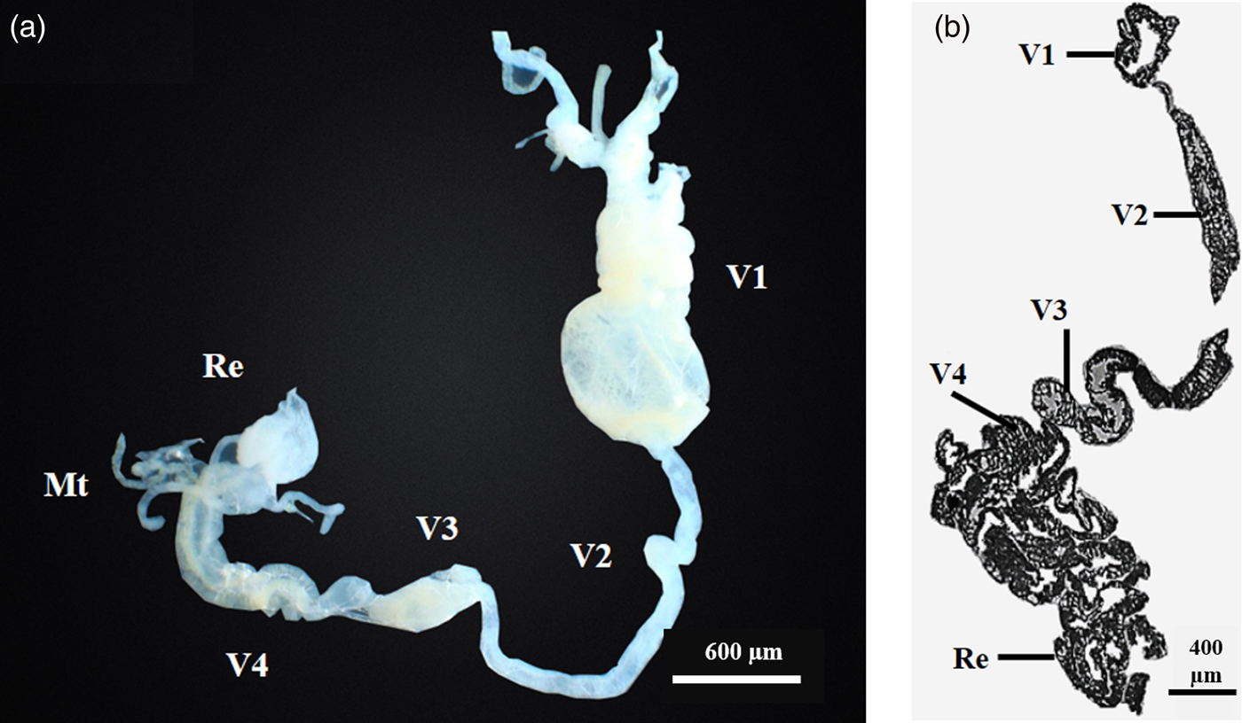
A. heegeri digestive tract is divided into three morphologically distinct regions including a narrow foregut, an enlarged midgut and a short hindgut (fig. 1a, b). The midgut is the large and distinct part of the insect alimentary canal which was composed of four different sections (V1–V4) being V4 connected to hindgut (fig. 1a, b). The first section (V1) was dilated with a stomach-like shape, filled with liquid material and providing space for ingested food (fig. 1a, b). The second region (V2) was a long and narrow tube, while the third section (V3) was slightly expanded and shorter than V2 (fig. 1a, b).
Micrographs from Acrosternum heegeri digestive tract. (a) Alimentary canal of A. heegeri (b) Longitudinal section from whole digestive tract with different midgut regions and rectum. V1-V4: midgut regions; Mt: Malpighian tubules; Re: Rectum.

The fourth part (V4) was equipped by a number of sac-like tissues, called crypts, which were arranged in rows, and fused to each other into a helical assemblage (figs 1a, b and 6a). At the peripheral zone of these crypts, the symbiont cells were closely integrated with the host epithelial cells in a mixed manner, meanwhile, at the central region of the crypts, the symbionts were in dispersed locations (fig. 2a).
Micrographs from the fourth midgut region of A. heegeri. Light micrographs: (a) Longitudinal section of the fourth ventricle showing a crypt with symbiont bacteria into the lumen and epithelial cells. (b) More detailed portion of part A (c) Transmission electron micrographs of A. heegeri showing longitudinal and transversal sections of the bacterial symbiont found in the lumen of the midgut fourth ventricle. EP: columnar epithelium; SB: symbiont bacteria; IM: inner and OM: outer symbiont cell membranes.

Light microscopy and transmission electron microscopy (TEM) observations revealed that the lumen of the swollen crypts was filled with rod-shaped bacterial cells, which were morphologically uniform (fig. 2b, c) and with a length of 4–5 µm (fig. 2c). Double cell wall organization, typical of gram-negative bacteria, was found in the symbiotic bacterial cells (fig. 2c). Close observations of the microscopic images revealed that the bacterial cells were actually confined not in the cytoplasm but in the extracellular spaces (fig. 2b). On the other hand, scanning electron microscopy confirmed the presence of rod shape bacterial cells on the egg surface (fig. 3).
Egg mass and scanning electron micrographs of the egg surface of A. heegeri. (a) An egg clutch. (b) An enlarged image of an egg crushed using a combination of vigorous vortexing and manual crushing. (c) A number of symbiont cells (arrows) on the egg surface.

Bacterial communities in different midgut regions, egg and ovary of the insect host
The microbiotas of the mid-gut regions, egg and ovary of A. heegeri were characterized using qPCR. Results showed that the greatest titer of Eubacteria and Gammaproteobacteria were present in the V4 region which contained crypts (fig. 4a). Comparing other parts, the V1 and egg represent higher Eubacterial and Gammaproteobacterial titers (fig. 4a). The occurrences of the other bacterial taxa, Alphaproteobacteria, Bacteroidetes, Betaproteobacteria, Actinobacteria and Firmicutes were measured relatively less than one (fig. 4b). The egg exhibited a diverse bacterial community (fig. 4b), while the V2, V3 and ovary showed low titers of different bacterial taxa (fig. 4a, b).
Relative frequency of different bacterial groups along the midgut regions, egg, and ovary of A. heegeri. (a) The extracted DNAs were subjected to qPCR by using a pair of universal primers as well as taxon-specific primers, γ-Proteobacteria. The V4 region showed the most quantity of Eubacteria and Gammaproteobacteria. (b) The occurrence of the major phylogenetic groups of bacteria including α-Proteobacteria, β-Proteobacteria, γ-Proteobacteria, Actinobacteria, Firmicutes, Bacteroidetes, and Acidobacteria were examined in the insect body parts (V1, V4, ovary) and egg by using taxon-specific primers. For each bacterial taxon, means followed by different letters are significantly different according to one-way ANOVA and Tukey's test (P < 0.05).

Phylogenetic placement of the symbiont
The symbiont 16S rRNA and groEL genes were amplified and sequenced from the fourth region of the insect midgut. The top BLAST hit for 16S rRNA gene sequence and top Blastx hit for groEL sequence were the γ- Proteobacterial representatives and specifically the genus Pantoea. The highest identities of the symbiont genes were with 16S rRNA gene sequence of Acrosternum arabicum endosymbiont clone Rafsanjan1 [MG725752.1] (99% identity) and groEL sequence of P. stali Gammaproteobacterium symbiont strain B/Ps-OERBm1 [BAQ20500] (90% identity).
Molecular phylogenetic trees including representative sequences of in- and out-group taxa were reconstructed on the basis of 16S rRNA gene and GroEl protein sequences (fig. 5a, b). The trees revealed that the bacterial symbiont of A. heegeri formed a monophyletic group with some other Pantoea sp. symbionts of pentatomids (fig. 5a, b). This clade constructed a sister group with the bacterial symbiont of other hemipteran families (fig. 5a, b).
Molecular phylogenetic analysis of the A. heegeri bacterial symbiont and representatives in the γ-Proteobacteria. The trees were reconstructed based on (a) 16S rRNA gene sequences, and (b) and GroEl protein sequences. Maximum likelihood trees inferred from the results for a total of 1151 and 286 aligned nucleotide sites are shown for 16S rRNA and groEL sequences, respectively, while other trees exhibited substantially the same topologies. On each of the nodes, bootstrap probabilities determined in the maximum likelihood and maximum parsimony analyses, and posterior probabilities determined in the Bayesian analysis are shown in that order. Asterisks indicate bootstrap values lower than 50%. Sequence accession numbers were shown in brackets and the taxonomic affiliations were mentioned in parentheses. The AT contents of the nucleotide sequences were also indicated.

FISH analyses
To confirm the presence and density of Pantoea genus symbiont, we performed 16S rRNA-targeted in situ hybridization using a Pantoea -specific fluorescent probe. The FISH Analyses detected dense signals of bacteria, belonged to Pantoea genus in the content of the midgut crypts (fig. 6).
Fluorescent in situ hybridization (FISH) targeting 16S rRNA of the symbiont in V4. (a) Light micrograph, (b) Bright field observation, (c) Red Pantoea stained with CY5-conjugated Pantoea-specific, Texas red filter. (d) Violet indicates the combination of signals of the CY5-conjugated Pantoea-specific probe and blue signals show host insect nuclei stained with DAPI.

Symbiont elimination by egg surface sterilization in A. heegeri
In an attempt to experimentally confirm the possibility of symbiont transmission via egg surface contamination and to explore the potential of inhabiting gut microbes to change the behavior and fitness of A. heegeri, we divided the egg masses estimated to hatch within 24 h into the two groups: one of the halves was left untreated while the other part was surface sterilized. Then we evaluated the relative densities of the bacterial symbiont in the control and surface sterilized eggs by performing qPCR analyses. The qPCR results indicated that the density of bacterial symbiont in the sterilized eggs was 20.35 fold lower than that of the control (fig. 7).
qPCR analyses for bacterial symbiont intensities in the control and surface sterilized eggs. To eliminate the bacterial symbiont, the egg masses were treated with bleach, 12% NaOCl and 96% ethanol. The egg surface sterilization reduced the density of bacterial symbiont in the sterilized eggs 20.35 folds lower than that of the control.***: significant difference between the control and the aposymbiont insects (independent-samples t-test).

Symbiont elimination effects on fitness and behavior of the insect host
Using the mentioned surface sterilization procedure, we successfully obtained sibling populations of symbiotic and aposymbiotic A. heegeri. We observed that the untreated A heegeri new born nymphs (control) remain aggregated on the eggshell after hatching for about two days, likely due to acquisition of the symbiont that already transmitted during the oviposition (fig. 8a). We found no significant difference in wandering behavior between symbiotic and aposymbiotic nymphs (fig. 8b).
Wandering behavior of newborn nymphs in the symbiotic and aposymbiotic isolines of A. heegeri. (a) newly hatched nymphs aggregate around the eggshell, (b) Mean number (±SE) of wandering nymphs per egg mass within 3, 12, 24 and 48 h post-hatching. The n.s. indicates no significant differences between the control and the aposymbiont insects (independent-samples t-test).

The nymphs from the sterilized group exhibited significantly lower survivability than those from the control group (fig. 9a). In addition, significantly slower growth was observed in aposymbiont A. heegeri than the control insects (fig. 9b, c).
Impact of bacterial symbiont elimination on the insect host fitness. (a) Survival rates of nymphal stages in symbiont and aposymbiont A. heegeri sibling populations. The number of survived nymphs is presented on each column. (b–c) Growth profiles of symbiotic (b) and aposymbiotic insects (c). Fisher's exact probability test: n. s., not significant,*: P < 0.05 and***: P ≤ 0.001.

Discussion
Here we investigated the gut microbiota of A. heegeri, a member of pentatomid stinkbugs. The result indicated that the A. heegeri, like many other hemipteran species (Moran et al., Reference Moran, McCutcheon and Nakabachi2008), was associated with Gammaproteobacteial symbiont. QPCR microbiota analyses along the midgut of A. heegeri and insect eggs showed the largest populations of Eubacteria and highly numbers of a specific Gammaproteobacteial symbiont in V4 and egg. Our previous work also demonstrated high density of Eubacteria in the fourth midgut region of A. heegeri (Kashkouli et al., Reference Kashkouli, Fathipour and Mehrabadi2018). The fourth midgut region shared bacterial taxa with all preceding regions, probably because of the passing of bacteria from the former parts with the food material to the rectum (Sudakaran et al., Reference Sudakaran, Salem, Kost and Kaltenpoth2012). We also characterized the microbiota inhabiting the first midgut region which exhibited a diverse bacterial community, suggesting that some detected bacterial taxa in this region were taken up with the food as reported for the pyrrhocorid bug, Pyrrhocoris apterus (Sudakaran et al., Reference Sudakaran, Salem, Kost and Kaltenpoth2012). The V2 and V3 showed low titers of Eubacteria probably because of their structure which lack a bacterial collective space.
BLAST analyses of the V4 region bacterial 16S rRNA gene and GroEL protein sequences showed the highest identity of the symbiont with plant-dwelling bacteria in the genus Pantoea confirming also by phylogenetic and FISH analyses. The phylogenetic study of A. heegeri symbiont based on the 16S rRNA gene and GroEl protein sequences revealed that this symbiont is related to bacterial symbionts of Pentatomomorpha bugs while exhibited polyphyletic evolutionary origins with them. In many insect-microbe obligate symbiotic interactions, the symbiont tends to form a monophyletic group and symbiont-insect co-speciation (Moran et al., Reference Moran, McCutcheon and Nakabachi2008), suggesting a single symbiont acquisition by the common ancestor of the insects (Hosokawa et al., Reference Hosokawa, Kikuchi, Nikoh, Shimada and Fukatsu2006; Kikuchi et al., Reference Kikuchi, Hosokawa, Nikoh, Meng, Kamagata and Fukatsu2009). In crypt dwelling pentatomid stink bugs, by contrast, the symbionts exhibited polyphyletic evolutionary origins and generally do not exhibit accurate host-symbiont co-speciation (Prado & Almeida, Reference Prado and Almeida2009b; Prado et al., Reference Prado, Hung, Daugherty and Almeida2010; Hosokawa et al., Reference Hosokawa, Ishii, Nikoh, Fujie, Satoh and Fukatsu2016).
Previously, two hypotheses including (i) symbiont replacement and (ii) symbiont multiple-origins were presented to describe the complication of symbiont phylogeny in pentatomid bugs (Prado & Almeida, Reference Prado and Almeida2009a). They suggested that the symbiont replacement hypothesis is more plausible (Prado & Almeida, Reference Prado and Almeida2009a) because other heteropteran bugs also have a crypt-bearing posterior midgut inhabiting with symbionts in a monoculture (Prado & Almeida, Reference Prado and Almeida2009a; Wernegreen, Reference Wernegreen2012). Symbiont replacement hypothesis is based on the unique symbiont transmission route in pentatomidae (including A. heegeri in the present study). Although vertical transmission occurs via egg surface contamination, bacteria are outside their host within a short phase that may be prone to contamination by or competition with other environmental microbes (Prado & Almeida, Reference Prado and Almeida2009a, Reference Prado and Almeidab). As many pentatomid symbionts are closely related to plant-associated bacteria in the genera Erwinia and Pantoea (Prado & Almeida, Reference Prado and Almeida2009b; Prado et al., Reference Prado, Hung, Daugherty and Almeida2010), it was suggested that only closely related bacterial taxa are acceptable to hosts or capable of maintaining infections over host generations (Prado & Almeida, Reference Prado and Almeida2009a).
Although the exact mechanism of symbiont transmission in the superfamily Pentatomoidea is poorly understood, it was demonstrated that the females excrete symbiont-containing materials from the anus, which are orally acquired by their offspring for vertical symbiont transmission (Kaltenpoth et al., Reference Kaltenpoth, Winter and Kleinhammer2009; Hosokawa et al., Reference Hosokawa, Hironaka, Inadomi, Mukai, Nikoh and Fukatsu2013; Hayashi et al., Reference Hayashi, Hosokawa, Meng, Koga and Fukatsu2015). The lack of transovarial transmission in these bugs may explain the lack of Eubacteria in A. heegeri ovaries. This type of ‘post hatch’ transmission behavior is considered to be due to the extracellular nature of the symbiont which adds an extra challenge to both the host and symbiont (Prado & Almeida, Reference Prado and Almeida2009b; Taylor et al., Reference Taylor, Coffey, DeLay and Dively2014). In particular, the symbiont experience environmental stresses such as desiccation, high temperature and ultraviolet irradiation (Kikuchi et al., Reference Kikuchi, Hosokawa and Fukatsu2007) outside the host, so they may require high stability genome to keep the genes required for living in a variable environment (Prado & Zucchi, Reference Prado and Zucchi2012). The host challenge is to re inoculate itself each generation. These challenges may be responsible for the low host-symbiont co-speciation or symbiont multiple acquisition observed in heteropteran insects (Prado & Zucchi, Reference Prado and Zucchi2012).
Results of this study provide direct evidence that surface sterilization of the eggs results in disruption of symbiont transmission to newborn nymphs. Symbiotic and aposymbiotic newborn nymphs of A. heegeri showed no significant differences in wandering behaviors. In the stinkbugs Megacopta punctatissima and H. halys; however, negative and positive correlations between the supply of symbiont available and the number of wandering nymphs were observed, respectively (Hosokawa et al., Reference Hosokawa, Kikuchi, Shimada and Fukatsu2008; Taylor et al., Reference Taylor, Coffey, DeLay and Dively2014). It was suggested that wandering behavior in M. punctatissima is evolutionarily favored behavior by selection pressures acting on both host and symbiont (Hosokawa et al., Reference Hosokawa, Kikuchi, Shimada and Fukatsu2008). More research needs to be done on a species to species basis to fully understand the possible host behavior modification by a symbiont in heteropteran mutualistic relationships.
Insects from the sterilized treatment group exhibited retarded growth and lower nymphal survivability. In the previous study, we examined the effects of two symbiont elimination strategies, high temperature and sterilization agents, on the demographic parameters of A. heegeri (along with two other pistachio bugs, Brachynema germari and A. arabicum) (Kashkouli et al., Reference Kashkouli, Fathipour and Mehrabadi2018). We found that the insects exhibited severe fitness defects, including significantly lower fecundity, r, R 0 and GRR values in aposymbionts. Together, these results suggested that the insect symbiont can be regarded as obligate mutualism. Actually, many species of stinkbugs in the infraorder pentatomomorpha are highly dependent on their microbial partners, and experimental removal of obligate symbiont has been shown to result in strong negative effect on the insect fitness (Prado et al., Reference Prado, Hung, Daugherty and Almeida2010; Kikuchi et al., Reference Kikuchi, Hosokawa, Nikoh and Fukatsu2012; Bistolas et al., Reference Bistolas, Sakamoto, Fernandes and Goffredi2014; Taylor et al., Reference Taylor, Coffey, DeLay and Dively2014; Kashkouli et al., Reference Kashkouli, Fathipour and Mehrabadi2018). On the other hand, we have to indicate that using sterilization agents may have damaged the symbiont as well as the host to some extent. This aspect can consider as a limitation of the present study, shared with many insect symbiont researches.
In conclusion, here we reported that A. heegeri harbored a primary symbiotic bacterium in the crypts that belongs to gram-negative gammaproteobacterium, Pantoea genus, and found that the symbionts are vertically transmitted via egg surface smearing upon oviposition, and, furthermore, fitness consequences of the gut symbiont elimination. On the whole, the results indicated the symbiont features and demonstrated symbiont-dependent A. heegeri growth and development. It can be suggested that A. heegeri symbiont may have a role in provision of essential nutrients necessary to support the host. However, more studies are needed to know the biological roles played by the symbiont. As the pistachio stink bug, A. heegeri is abundant and serious pest of pistachio nuts in Iran, further investigations may lead to symbiosis-based management tactics for this important pest.
Acknowledgments
This study is part of the Ph. D. research of the first author that was financially supported by Tarbiat Modares University, grant number 9230462005, which is greatly appreciated.