Introduction
Cystic echinococcosis (CE) is a zoonotic disease caused by Echinococcus granulosus sensu lato (s.l.), characterized by the development of the metacestode stage which is known as a hydatid cyst, in many mammalian species including humans (Thompson, Reference Thompson2017). Humans become infected by oral intake of eggs excreted from infected canids through direct contact with animals or consumption of contaminated water, vegetables, uncooked food or direct hand–mouth fecal transmission, whereas cases of acquiring parasite eggs by direct contact with dogs are especially common in children (Thompson et al., Reference Thompson, McManus, Eckert, Gemmell, Meslin and Pawlowski2001; Craig and Larrieu, Reference Craig and Larrieu2006). After eggs are ingested by an intermediate host, oncospheres are released from the eggs into the upper gastrointestinal tract. They then penetrate the intestinal wall, enter the portal vein and travel to the liver. In some cases, a few oncospheres may pass the liver and reach the lungs, or more rarely, hydatid cysts can develop in other organs such as the kidneys, spleen, heart, bone and brain (Moro and Schantz, Reference Moro and Schantz2009).
Important inter-species variations have been identified, and five cryptic species have been identified in E. granulosus s.l. These species include E. granulosus s.s. (G1 and G3), E. equinus (G4), E. ortleppi (G5), E. canadensis (G6/7 and 8/10) and E. felidis. Nevertheless, the classification for the G6/7/8/10 genotypes of E. canadensis is still controversial (Nakao et al., Reference Nakao, Lavikainen and Hoberg2015). Echinococcus granulosus s.s. is especially significant as it is widespread (Deplazes et al., Reference Deplazes, Rinaldi, Rojas, Torgerson, Harandi, Romig, Antolova, Schurer, Lahmar and Cringoli2017) and because it is responsible for most human cases in the world (Rojas et al., Reference Rojas, Romig and Lightowlers2014). However, even within E. granulosus s.s., haplotype differences have emerged (Khan et al., Reference Khan, Ahmed, Simsek, Liu, Yin, Wang, Shen and Cao2020; Mehmood et al., Reference Mehmood, Simsek, Celik, Kesik, Gunyakti Kilinc and Ahmed2020).
In endemic areas, the annual prevalence of CE cases might reach up to 10% (Budke et al., Reference Budke, Carabin, Ndimubanzi, Nguyen, Rainwater, Dickey, Bhattarai, Zeziulin and Qian2013; WHO, 2017). According to reports from neighbouring countries of Turkey, the prevalence of human CE cases was stated as 5.71/100 000 in Bulgaria (Jordanova et al., Reference Jordanova, Harizanov, Kaftandjiev, Rainova and Kantardjiev2015), 4.5/100 000 in Iraq (Abdulhameed et al., Reference Abdulhameed, Habib, Al-Azizz and Robertson2018) and 1.18–3/100 000 in Iran (Rokni, Reference Rokni2008; Tavakoli et al., Reference Tavakoli, Bayat and Kousha2008). CE, although a reportable disease in Turkey, information on human CE cases is collected mainly from hospital records upon surgical detection. Therefore, the prevalence of these cases is likely to be grossly underestimated (Anadol et al., Reference Anadol, Göçmen, Kiper and Özçelik1998; Altintas, Reference Altintas2003). The incidence of human CE cases from 1990 to 2005 was estimated to be 0.8–2 per 100 000 inhabitants (Yolasigmaz et al., Reference Yolasigmaz, Reiterová, Turk, Reyhan, Bozdag, Karababa, Altintas and Altintas2006; Altintas, Reference Altintas2008). The reports from the Turkish Ministry of Health show an incidence of up to 12 per 100 000 in Turkey (Gonlugur et al., Reference Gonlugur, Ozcelik, Gonlugur, Arici, Celiksoz, Elagoz and Cevit2009). From 2001 to 2011, 59 children with CE were studied in a paediatric hospital in Iran (Aslanabadi et al., Reference Aslanabadi, Zarrintan, Abdoli-Oskouei, Salehpour, Zarrintan, Beheshtirouy, Abdollahi and Badebarin2013). In a serological study, anti-hydatid antibodies were detected in 6.7% of children in Iran (Sarkari et al., Reference Sarkari, Arefkhah, Ghorbani, Meskini, Yektaeian, Shahriarirad and Mostafavi-Pour2020). In an ultrasound scanning study, abdominal CE was detected in 31 (including 6 cases under 20 years) of 8602 people screened in Bulgaria, 35 (2 under 20 years) of 7461 screened in Romania and 53 (6 under 20 years) of 8618 screened in Turkey (Tamarozzi et al., Reference Tamarozzi, Akhan, Cretu, Vutova, Akinci, Chipeva, Ciftci, Constantin, Fabiani and Golemanov2018). Using portable ultrasound scanners, the prevalence of CE was found to be 0.15% among 6093 elementary school students in Manisa (Ok et al., Reference Ok, Ozkol, Kilimcioglu, Dinc, Bayindir, Ostan, Pabuscu, Ozcan, Korkmaz and Coskun2007). In a study conducted in the Sivas province of Turkey, it was determined that a total of 316 surgically confirmed patients had CE in the period of 1997–2004, and the average annual surgical incidence was predicted to be 6.4 per 100 000. Interestingly, 17.4% of the patients with CE were found to be under 20 years of age (Gonlugur et al., Reference Gonlugur, Ozcelik, Gonlugur, Arici, Celiksoz, Elagoz and Cevit2009). However, there are limited reports about the genotyping process of paediatric CE cases (Ergin et al., Reference Ergin, Saribas, Yuksel, Zengin, Midilli, Adas, Arikan, Aslan, Uysal and Caliskan2010; Simsek et al., Reference Simsek, Kaplan and Ozercan2011b; Bakal et al., Reference Bakal, Simsek and Kazez2015).
The aim of this study was to investigate the genetic variation of hydatid cyst isolates obtained from surgically confirmed paediatric cases that originated from two different provinces in eastern Turkey.
Material and methods
Cases
All paediatric CE cases (n = 17) were operated with open surgery at the Pediatric Surgery Department in Firat University Hospital between March 2017 and December 2020. Each patient's medical history was retrospectively reviewed for the results of their physical examination, serum biochemistry, abdominal ultrasound, chest radiography, computed tomography or magnetic resonance imaging tests. All patients were treated with albendazole (10 mg kg−1) for 7 days before surgery.
Surgery
Depending on the location of the hydatid cyst, a right subcostal or midline incision was performed in laparotomy. Starting from the puncture site, cystotomy was performed, by extracting the germinal membrane and daughter vesicles. All patients were treated with albendazole (10 mg kg day−1) after surgery for 6 months to prevent secondary cysts. Postoperative follow-ups were continued for the first 3 months, and then, every 3 months until the end of the first postoperative year.
Collection of hydatid cyst samples
Germinal layers were removed from the hydatid cysts, placed into Petri dishes containing a physiological saline solution, transferred to the laboratory and then washed with PBS a few times (pH = 7.4). Then, 70% ethanol was added for the preservation of the germinal layers, and the layers were stored at −20°C until further analyses.
Genomic DNA isolation
Total genomic DNA (gDNA) was isolated from each individual cyst sample using the Hibrigen Tissue Kit (Hibrigen, Turkey) according to the manufacturer's instructions with some minimal modifications. The germinal layers were crushed by a surgical blade, and then, the tissue samples were placed in 1.5 mL Eppendorf tubes following the washing of the samples by PBS (pH = 7.4) for at least five times to eliminate traces of ethanol, and each time, the supernatant in the tubes was discarded. Then, 300 μL of the DHP solution was added and mixed with vortex until it became homogeneous. Subsequently, 120 DA solution and 20 μl Proteinase-K (20 mg mL−1) and samples were first mixed by vortex and then subjected to a water bath overnight for incubation at 65°C. The samples were mixed by vortex to get effective results of digestion for at least 2–3 times every 1 h. The next day, the gDNA was isolated using the kit procedures and then stored at −20°C until being used.
PCR amplification
A 875 bp fragment of the mt-CO1 gene of E. granulosus s.l. was amplified using the following primers: 5’-TTGAATTTGCCACGTTTGAATGC-3’ and 5’-GAACCTAACGACATAACATAATGA-3’ (Nakao et al., Reference Nakao, Sako, Yokoyama, Fukunaga and Ito2000), with the cycle parameters as follows: an initial denaturation step at 94°C for 10 min; 30 cycles at 94°C for 30 s, 52°C for 45 s and 72°C for 1 min, followed by a final extension step at 72°C for 10 min. All PCR reactions were set up with the negative control (distilled water). After agarose gel electrophoresis (1.4%), all PCR products were purified from the agarose gel, and unidirectional sequence analyses were performed using the forward primer (F/CO1) (BM Labosis, Turkey).
Sequence analysis
All nucleotide sequences handled in this study were deposited in the GenBank database under the following accession numbers MT537158–MT537165 and MW881257–MW881265. The nucleotide sequences were then subjected to BLAST searches (http://www.ncbi.nlm.nih.gov/blast/), and they were analysed with each other and E. granulosus s.l. reference sequences downloaded from GenBank using ClustalW.
Data and phylogenetic analysis
The mt-CO1 sequence data were downloaded from GenBank in the FASTA format and uploaded to the MEGA X program (Kumar et al., Reference Kumar, Stecher, Li, Knyaz and Tamura2018). ClustalW was used to analyse the sequences of different lengths and produce a number of output formats such as FASTA suitable for subsequent analyses. All aligned sequences were analysed, and sequences with 785 bp of length were selected, containing the mt-CO1 gene region without indel problems. The reference sequences (China n = 35, Mongolia n = 11, Russia n = 6, Ethiopia n = 6, Iran n = 2) which were published in PubMed were downloaded, our sequences (n = 16) were truncated at both ends, and the lengths of the DNA sequences were equalized to 785 bp. Afterwards, the sequences were analysed in the MEGA X program by the Akaike Information Criterion (AIC) and Bayesian Information Criterion analysis, and the most suitable base change models were obtained. The phylogenetic tree was obtained with the Tamura-Nei γ distribution (TN93 + G) model, which was selected by a search for the best model using the Find Best DNA Model (Kumar et al., Reference Kumar, Stecher, Li, Knyaz and Tamura2018). To assess the statistical power of the topology of the trees, the bootstrap method with 1000 replications was used. Echinococcus canadensis (G6/G7) (KX510134, KX010859, MT380299), E. canadensis (G8) (LC184604), E. canadensis (G10) (LC184606), E. ortleppi (MN058591), E. equinus (KP101615), E. felidis (EF558356) and E. multilocularis (AB688135) were used for construction of the phylogenetic tree. The Taenia saginata (AB821273) sequence was used as the outgroup, as well.
Haplotype network and population genetics analysis
All sequence data were uploaded onto DnaSP 6 (Rozas et al., Reference Rozas, Ferrer-Mata, Sánchez-DelBarrio, Guirao-Rico, Librado, Ramos-Onsins and Sánchez-Gracia2017). The population diversity indices [numbers of haplotypes (h), haplotype diversity (Hd), nucleotide diversity (π)], the neutrality indices [Tajima's D (Tajima, Reference Tajima1989), and Fu's statistics (Fu, Reference Fu1997)], Fu and Li's D and F tests (Fu and Li, Reference Fu and Li1993) were calculated using DnaSP 6 (Rozas et al., Reference Rozas, Ferrer-Mata, Sánchez-DelBarrio, Guirao-Rico, Librado, Ramos-Onsins and Sánchez-Gracia2017). The pairwise fixation index (FST) (Salzburger et al., Reference Salzburger, Ewing and Von Haeseler2011) values between the E. granulosus s.s. populations from Turkey and the reference countries (China, Iran, Mongolia, Russia and Ethiopia) were calculated using DnaSP 6 (Rozas et al., Reference Rozas, Ferrer-Mata, Sánchez-DelBarrio, Guirao-Rico, Librado, Ramos-Onsins and Sánchez-Gracia2017). FST values approaching 1 indicate extreme genetic differentiation between two populations. DnaSP 6 was used to generate a series of output formats, such as NEXUS, which allows the user to add additional information for the next analysis. Geographical, phenotypical or other traits associated with sequences were also included in the NEXUS (Maddison et al., Reference Maddison, Swofford and Maddison1997) files in a Traits block. The following networks were created using the Minimum Spanning Networks (MSN) method, which includes all edges visible in the minimum span tree of arrays by means of the PopART-1.7 software (Bandelt et al., Reference Bandelt, Forster and Röhl1999) (http://popart.otago.ac.nz).
Results
Cases
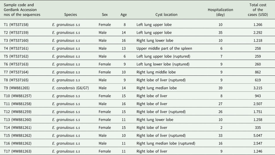
Of the 17 patients, 11 were from the Elazig province (one of them was a Syrian refugee), and 6 were from the Mus province of Turkey. The ages of the patients ranged from 6 to 16 years, while 8 of the patients were female, and 9 were male. The cysts were located in the lung (n = 9), liver (n = 7) and spleen (n = 1), and three liver and three lung cysts were ruptured (Table 1).
Age, sex, cyst location and total costs of the cases

PCR and sequence analyses
The isolates (n = 17) were successfully amplified by PCR. According to the BLAST search, 16 sequences (MT537158–MT537165 and MW881257–MW881264) were characterized as E. granulosus s.s., while only one sequence (MW881265) was identified as E. canadensis (G6/G7). A ruptured hydatid cyst was observed in the median lobe of the right lung which was identified as E. canadensis (G6/G7). This hydatid cyst belonged to a 14-year-old male patient from the Elazig province. This patient was the patient with the longest hospital stay of 39 days (Table 1).
Echinococcus granulosus s.s. Turkey isolates: nucleotide polymorphism, diversity and neutrality indices and haplotype networks
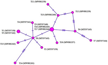
Twenty polymorphic sites were detected in the E. granulosus s.s. isolates that were sequenced in this study, and 45% (9/20) of them were parsimony informative. High haplotype diversity and low nucleotide diversity were observed in these isolates (Table 2). Tajima's D was negative in the sequences, signifying population expansion and/or purifying selection. Negative Fu's F values were determined in the sequences which indicated the presence of rare haplotypes expected from a recent population expansion or hitchhiking. The presence of unique singleton haplotypes for the mt-CO1 (11/13) sequences of E. granulosus s.s. was consistent with the structure of the haplotype networks for the isolates (Fig. 1).
Frequencies of mitochondrial cytochrome c oxidase subunit 1 (CO1) (785 base pairs) haplotypes of E. granulosus s.s. isolates from Turkey. Circle size is relative to haplotype frequency. Hatch marks indicate additional mutational steps.

Diversity and neutrality indices for E. granulosus s.s. isolates from Turkey using nucleotide data of the cytochrome c oxidase subunit 1 (CO1) (785 bp base pairs) mitochondrial genes

n, Number of isolates; hn, number of haplotypes; hd, haplotype diversity; πd, nucleotide diversity; s.d., standard deviation; FLD, Fu and Li's D test statistic; FLF, Fu and Li's F test statistic.
Echinococcus granulosus s.s. populations: Turkey vs other geographical regions
Haplotype networks
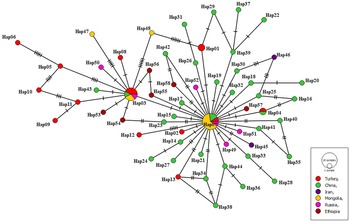
A 785 bp length fragment of the mt-CO1 gene sequence of E. granulosus s.s. isolates was analysed. Seventy-one polymorphic points were detected in the 76 analysed sequences. The overall haplotype and nucleotide diversities for the mt-CO1 sequences were 0.973 ± 0.012 and 0.00410 ± 0.00043, respectively. The overall neutrality indices were significantly negative for the mt-CO1 gene (Tajima's D −2.60975, ***P < 0.001; Fu's F −93.338, P = 0.000) (Table 3). A total of 58 haplotypes were detected in the 76 isolates within the mt-CO1 sequences of E. granulosus s.s. (Fig. 2) (Supplementary data). The network had a star-like expansion with a centrally positioned common haplotype (Hap07) that was separated from other haplotypes by one to 15 stepwise mutations and occurred in 11 (14.5%) of the 76 isolates. There was only one human isolate in Turkey (1/11) within this main haplotype. Other isolates in this haplotype were from Mongolia (n = 7), China (n = 2) and Russia (n = 1). Three of the isolates from Turkey obtained in this study settled in the second main haplotype; however, the other 9 isolates constituted a unique-single haplotype. Other unique-single haplotypes were from China (n = 31), Ethiopia (n = 6), Russia (n = 4), Mongolia (n = 2) and Iran (n = 2).
Haplotype network of E. granulosus s.s. isolates from various geographical regions using the cytochrome c oxidase subunit 1 (CO1) (785 base pairs) mitochondrial gene. Size of circles is proportional to the frequency of each haplotype. Number of mutations distinguishing the haplotypes is shown by hatch marks. Hap means haplotype.

Diversity and neutrality indices obtained by using nucleotide data of Echinococcus granulosus sensu stricto mt-CO1 gene

n, Number of isolates; hn, number of haplotypes; hd, haplotype diversity; πd, nucleotide diversity; s.d., standard deviation; FLD, Fu and Li's D test statistic; FLF, Fu and Li's F test statistic.
Phylogenetic analyses and genetic differentiations
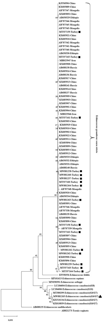
As a result of the phylogenetic tree construction, MT537164 (Hap06-Turkey) and AB787539 (Hap48-Mongolia) were designed as the two most distant isolates in two clusters owing to 15 nucleotide differences. Besides, MT537164 (Hap06) caused the second furthest branching to AB787548 (Hap47-Mongolia) and MH025946 (Hap46-Iran) due to 14 nucleotide differences (Fig. 3).
Phylogenetic tree of E. granulosus s.s. samples from Turkey (n = 16) and different (other) geographical regions (n = 60) based on a 785 bp-long fragment of the mt-CO1 gene. To assess the statistical power of the topology of the trees, the bootstrap method with 1000 replications was used. Echinococcus canadensis (G6/G7) (KX510134, KX010859, MT380299), E. canadensis (G8) (LC184604), E. canadensis (G10) (LC184606), E. ortleppi (MN058591), E. equinus (KP101615), E. felidis (EF558356) and E. multilocularis (AB688135) were used for construction of the phylogenetic tree. The Taenia saginata (AB821273) sequence was used as the outgroup, as well. MEGA X was used to construct a Maximum Likelihood tree based on the Tamura-Nei γ distribution (TN93 + G) model. To assess the statistical power of the topology of the trees, the bootstrap method with 1000 replications was used. The E. granulosus s.s. sequences (MT537158–MT537165 and MW881257–MW881264) that were analysed in this study are indicated by ‘■’, and the E. canadensis (G6/G7) sequence (MW881265) is shown by ‘▴’.

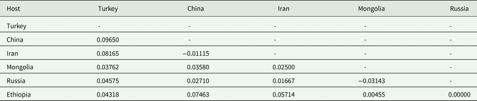
As shown in Table 4, the lowest FST value was observed between the Mongolia isolates and the Russia isolates, followed by the value between the China and Iran isolates. The highest Fixation index (Fst) (0.07463) values were observed in the China and Ethiopia populations. Using the mt-CO1 sequences, insignificant Fst values were observed when the E. granulosus s.s. sequences from this study were compared in a pairwise manner to those derived from China (0,09650), Iran (0,08165), Ethiopia (0,04318), Russia (0,04575) and Mongolia (0,03762), indication that these populations were not genetically differentiated.
Pairwise fixation index (Fst) for Echinococcus granulosus sensu stricto isolates as compared to those from various geographical regions using nucleotide data of the cytochrome c oxidase subunit 1 (CO1) mitochondrial gene

Turkey n = 16, China n = 35, Iran n = 2, Mongolia n = 11, Russia n = 6, Ethiopia n = 6.
Discussion
CE, which is listed by the WHO as a neglected tropical disease, is a zoonotic disease caused by E. granulosus s.l., which is the most common species causing CE in humans (Romig et al., Reference Romig, Deplazes, Jenkins, Giraudoux, Massolo, Craig, Wassermann, Takahashi and De La Rue2017; Thompson, Reference Thompson2017). Globally, CE affects approximately 2–3 million humans, and it is also a cause of great economic losses to livestock industries due to the condemnation of infected offal in slaughtered animals (Eckert and Deplazes, Reference Eckert and Deplazes2004; Budke et al., Reference Budke, Deplazes and Torgerson2006; Battelli, Reference Battelli2009).
Some researchers have reported that male children are more likely to be infected with hydatid cysts (Talaiezadeh and Maraghi, Reference Talaiezadeh and Maraghi2006; Montazeri et al., Reference Montazeri, Sokouti and Rashidi2007), but some others reported an equal incidence of CE in boys and girls (Cangir et al., Reference Cangir, Şahin, Enön, Kavukçu, Akay, Ökten and Yavuzer2001; Kocer et al., Reference Kocer, Gulbahar, Han, Ilhan, Dural and Sakinci2008). This may be related to behavioural differences between the sexes, with more outdoor exposure of boys in eastern countries, as suggested by Tantawy (Reference Tantawy2010). However, of our 17 cases, 9 were boys, and 8 were girls. Hence, children of both sexes are susceptible to CE.
The multi-organ presence of hydatid cysts has often occurred in humans, with liver–lung involvement at the highest frequency. The occurrence of liver hydatid cysts with one or more of other organs, for example, spleen, kidney and brain, has also been reported (Pawlowski, Reference Pawlowski2001). Hydatid cysts in paediatric cases include more frequently the lungs than the liver, but some researchers (Şehitoğulları, Reference Şehitoğulları2007) claimed that liver cysts are more widespread in children, while Talaiezadeh and Maraghi (Reference Talaiezadeh and Maraghi2006) asserted almost equal rates (41–43%) of hydatid cysts in the lung and the liver in paediatric cases. In a report from Turkey, 34.2% of lung hydatid cysts and 13.3% of liver ones were found in patients below the age of 20 (Gonlugur et al., Reference Gonlugur, Ozcelik, Gonlugur, Arici, Celiksoz, Elagoz and Cevit2009). However, among the paediatric cases in this study, 52.94% (9/17) had lung, 41.17% (7/17) had liver and 5.9% (1/17) had spleen hydatid cysts.
Surgery is the most effective treatment option for paediatric hydatid cysts of any size. Large cysts that are superficial and vulnerable to rupture, cysts in close proximity to vital organs, infected cysts and cysts exerting a substantial mass effect are treated surgically (Nouir et al., Reference Nouir, Nunez, Gianinazzi, Gorcii, Müller, Nouri, Babba and Gottstein2008). In this study, 17 hydatid cysts were treated with total cystotomy. Rupturing of hydatid cysts and spreading of protoscoleces into the peritoneum are risks of surgery. To minimize these risks, albendazole treatment was started a week before the surgery. The risk of recurrence of these cysts is as high as 11% if albendazole treatment is not performed after surgery (Gollackner et al., Reference Gollackner, Längle, Auer, Maier, Mittlböck, Agstner, Karner, Langer, Aspöck and Loidolt2000). Therefore, all patients were treated with albendazole (10 mg kg day−1) for a week before surgery and for 6 months after surgery in this study.
Many molecular techniques and targeted gene fragments have been used broadly to identify the genotypes/species within E. granulosus s.l. Genetic variation in E. granulosus s.l. has been frequently investigated using mitochondrial and/or nuclear gene sequences. Although the mt-DNA is known to be hereditary, it is considered that it contributes to genetic diversity due to its fast evolution rate (Obwaller et al., Reference Obwaller, Schneider, Walochnik, Gollackner, Deutz, Janitschke, Aspöck and Auer2004; Lymbery and Thompson, Reference Lymbery and Thompson2012). Mitochondrial DNA markers are recorded to be stronger indicators than nuclear DNA markers within E. granulosus s.l. in constructing phylogenetic relationships among closely related species because of their rapid sequence evolution (Nejad et al., Reference Nejad, Taghipour, Nochi, Mojarad, Mohebbi, Harandi and Zali2012).
Echinococcus granulosus s.s. is mostly adapted to humans, and thus, the majority of human CE cases are caused by this species (Rojas et al., Reference Rojas, Romig and Lightowlers2014). Molecular data indicate that E. granulosus s.s. is responsible for 88.44% of human CE cases worldwide (Rojas et al., Reference Rojas, Romig and Lightowlers2014). Similarly, 98.1% of human CE cases were identified as the G1 strain in China (Zhang et al., Reference Zhang, Yang, Zeng, Zhao, Liu, Piao, Jiang, Cao, Shen and Liu2014).
Echinococcus granulosus s.s. was detected after the sequence analyses of paediatric hydatid cyst isolates (n = 16) in this study. This result was similar to the results of many previous studies undertaken in Turkey and indicated a clear predominance of E. granulosus s.s. (Maillard et al., Reference Maillard, Benchikh-Elfegoun, Knapp, Bart, Koskei, Gottstein and Piarroux2007; Schneider et al., Reference Schneider, Gollackner, Edel, Schmid, Wrba, Tucek, Walochnik and Auer2008; Utuk et al., Reference Utuk, Simsek, Koroglu and McManus2008; Šnábel et al., Reference Šnábel, Altintas, D'amelio, Nakao, Romig, Yolasigmaz, Gunes, Turk, Busi and Hüttner2009; Ergin et al., Reference Ergin, Saribas, Yuksel, Zengin, Midilli, Adas, Arikan, Aslan, Uysal and Caliskan2010; Simsek et al., Reference Simsek, Balkaya, Ciftci and Utuk2011a; Eryildiz and Sakru, Reference Eryildiz and Sakru2012; Bakal et al., Reference Bakal, Simsek and Kazez2015; Eroglu et al., Reference Eroglu, Genc and Koltas2016).
Echinococcus canadensis (G6 genotype) was also recorded in two surgically confirmed human CE cases using histopathological tissue sections in the Elazig province of Turkey (Simsek et al., Reference Simsek, Kaplan and Ozercan2011b). Besides, the G7 (pig strain) and G1 genotypes were reported in a study performed on 42 human hydatid cyst samples in the Thrace region of Turkey (Eryildiz and Sakru, Reference Eryildiz and Sakru2012). Similarly, the G7 genotype was determined in a human hydatid cyst sample from the Izmir province of Turkey (Šnábel et al., Reference Šnábel, Altintas, D'amelio, Nakao, Romig, Yolasigmaz, Gunes, Turk, Busi and Hüttner2009). However, molecular characterization of human CE cases in Turkey has mostly been performed on adult hydatid cyst samples, and only a few cases about the molecular characterization of paediatric hydatid cysts were reported (Ergin et al., Reference Ergin, Saribas, Yuksel, Zengin, Midilli, Adas, Arikan, Aslan, Uysal and Caliskan2010; Simsek et al., Reference Simsek, Kaplan and Ozercan2011b; Bakal et al., Reference Bakal, Simsek and Kazez2015). Thus, this study provides data on both the molecular characterization of paediatric hydatid cyst samples and the first occurrence of E. canadensis (G6/G7) in paediatric CE cases in Turkey and haplotype analyses of the cysts.
The presence of a haplotype found in E. granulosus s.s. isolates was evident for the mt-CO1 network and suggested a common lineage for the globally common E. granulosus s.s. The little main haplotype was isolated from all countries except Iran (Turkey n = 1, China n = 2, Mongolia n = 7, Russia n = 1). There were only 2 Iranian isolates included in the study. Currently, it is well known that there is substantially greater genetic variability within E. granulosus s.s. (Vural et al., Reference Vural, Baca, Gauci, Bagci, Gicik and Lightowlers2008; Casulli et al., Reference Casulli, Interisano, Sreter, Chitimia, Kirkova, La Rosa and Pozio2012; Romig et al., Reference Romig, Ebi and Wassermann2015; Kinkar et al., Reference Kinkar, Laurimäe, Acosta-Jamett, Andresiuk, Balkaya, Casulli, Gasser, van der Giessen, González and Haag2018). In this study, determination of 58 haplotypes in the 76 isolates proved that there were significantly more genetic variabilities in E. granulosus s.s. High haplotype diversity and low nucleotide diversity were observed for the analysed mt-CO1 gene fragment. Tajima's D was negative for the mt-CO1 sequences, signifying population expansion and/or purifying selection. The significantly negative Fu's F values observed for the mt-CO1 sequences indicated the presence of rare haplotypes expected from a recent population expansion or hitchhiking.
In this study, E. granulosus s.s. was predominant in the paediatric CE cases as usual. The Hap03 and Hap07 haplotypes were highly prevalent in the analysed cases. These haplotypes showed 100% identity with the E. granulosus s.s. reference sequences and corresponded to the mt-CO1 haplotypes most frequently observed in studies on mt-CO1 diversity in E. granulosus s.s.
Conclusions
In this study, molecular characterization of hydatid cyst isolates in paediatric cases was performed, and haplotype profiles were determined. As a result, although 94.1% of the isolates were identified as E. granulosus s.s., and obtaining the different haplotypes showed that the intra-species variations in humans continued without slowing down. For this reason, haplotype differences within E. granulosus s.s. and different species such as E. canadensis (G6/G7) must be taken into account while taking control measures.
Supplementary material
The supplementary material for this article can be found at https://doi.org/10.1017/S0031182021001098.
Author contributions
Conceptualization, project administration, funding acquisition: UB, SS; investigation: F.C., H.K.K., S.G.K.; methodology: S.S., F.C.; resources: U.B., S.S., F.C.; formal analysis: F.C., H.K.K., S.G.K.; supervision: U.B., S.S.; writing – original draft, visualization: S.S., F.C.; writing – review and editing: U.B., S.S., F.C., H.K.K., S.G.K.
Financial support
This study was partially supported by a grant (119O014) from the Scientific and Technical Research Council of Turkey (TUBITAK).
Conflict of interest
All authors have read and agreed to the published version of the manuscript. The authors declare that they have no conflicts of interest.
Ethical standards
Ethics committee approval was not required in this study, since the study utilized hydatid cyst materials reserved for postoperative pathological examination (Firat University Ethical Committee report, 2018/14/1).