Pulmonary atresia with intact ventricular septum is a rare and complex congenital heart defect with great morphological variability. Right ventricular size and tricuspid valve annulus size are quite variable and are important determinants of management.Reference Choi, Seo, Choi, Yun, Kim and Lee 1
In selected cases, the percutaneous perforation of the atretic pulmonary valve and the resulting decompression of the right ventricle proved to be an effective and successful procedure.Reference Qureshi 2
In this study, we describe a new method for stable positioning of a guiding catheter in the right ventricle outflow tract to perform radiofrequency perforation and valvotomy, and we compare retrospectively the early results of two different percutaneous techniques.
Methods
From January, 2000 to December, 2010, 31 newborns with pulmonary atresia with intact ventricular septum underwent radiofrequency perforation of the pulmonary valve at our Institute. In the first 14 infants, a 5 French Judkins right coronary catheter was used to perform the procedure (Group A). Subsequently, a telescopic system consisting of Northstar® Lumax® Flex Guiding Catheters with AQ® Hydrophilic Coating (7 French) and White Lumax® coaxial Catheters (4 French) (Cook Inc., Bloomington, Indiana) was used in 17 patients to obtain the correct catheter position below the atretic pulmonary valve (Group B).
Written informed consent for radiofrequency perforation was obtained in all cases. The study was approved by the Institutional Ethics Committee at G. Gaslini Hospital.
All patients were treated under general anaesthesia; the femoral vein was percutaneously cannulated using a 5 French sheath in Group A, and a 7 French sheath in Group B; 50 units per kilogram of heparin were given intravenously once the access was obtained.
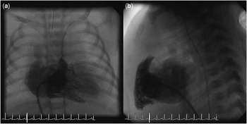
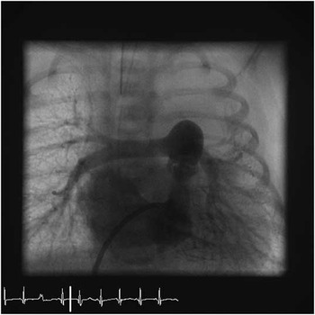
Right ventricle angiography was performed in both postero-anterior and lateral views to evaluate ventricle morphology and to exclude coronary abnormalities (Fig 1a, b). Left ventricle angiography was performed in the same views throughout the patent foramen ovalis to visualise the aorta, the arterial duct, the pulmonary arteries, and the atretic pulmonary valve. The femoral artery was percutaneously cannulated and used for retrograde aortic angiography only if right ventricle-dependent coronary circulation was suspected.
(a) Right ventricle and atretic pulmonary valve in antero-posterior view. (b) Injection into the right ventricle in lateral view.

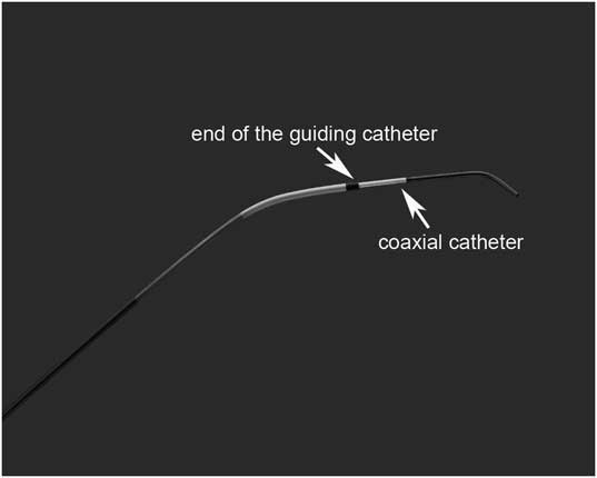
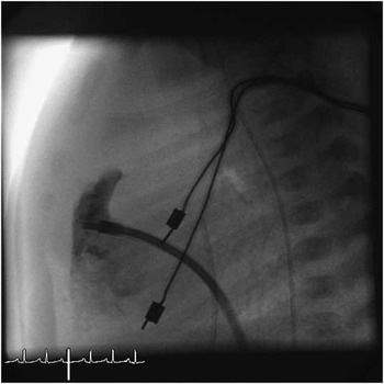
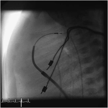
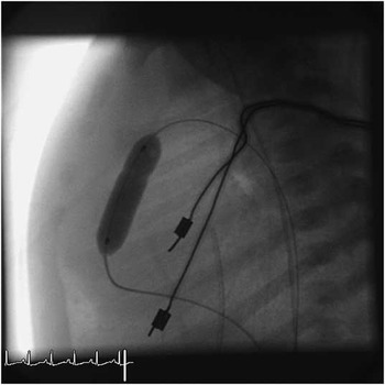
In Group A, a 5 French Judkins right coronary catheter was advanced through the femoral vein and manoeuvred directly below the atretic pulmonary valve. In Group B, we started to use a telescopic system consisting of Northstar Lumax Flex guiding catheter and Coaxial Hydrophilic Coating White Lumax Catheters (Cook; Fig 2). First, the guiding catheter was introduced into the right ventricle with the support of a 5 French multipurpose catheter and of a 0.035-inch floppy guidewire. Once the guiding catheter had reached the right ventricle, it was left in site and the multipurpose catheter was replaced with the Hydrophilic Coating White Lumax Catheter. The guiding catheter was then retracted until reaching the tricuspid valve (Fig 3), whereas the hydrophilic catheter was gently manipulated with a slight counter-clockwise rotation to reach the correct position under the atretic pulmonary valve. Radiofrequency perforation of the pulmonary valve was obtained with a Cereblate PA 120 unipolar catheter (Osypka, Rheinfelden-Herten, Germany) or with a Protrack microcatheter (0.038″) and a Nykanen radiofrequency wire connected to a radiofrequency generator (Bayliss Medical Company, Mississauga, Ontario, Canada) (Fig 4). After successful perforation of the pulmonary valve, a 0.014-inch or a 0.018-inch superfloppy high torque exchange guidewire was advanced through the arterial ductus into the descending aorta. The Hydrophilic Coating White Lumax catheter was removed to let the balloon pass through the guide and progressive dilations of the pulmonary valve were performed using a low-profile, low-pressure balloon catheter whose diameter was the same as or 1 millimetre smaller than that of the pulmonary valve (Fig 5). A 4 French end-hole catheter was then inserted over the guidewire up to the descending aorta before removing the wire to avoid damage to the ductal wall. At the end of every procedure, we measured right ventricle pressure and performed right ventricle angiography (Fig 6).
Northstar Lumax Flex guiding catheter and Coaxial Hydrophilic Coating White Lumax Catheters (Cook).

The guiding catheter advanced into the right ventricle (lateral view).

Successful radiofrequency perforation.

Balloon dilation of the pulmonary valve.

Final angiography in antero-posterior view: both pulmonary arteries are perfused.

Statistical analysis
Data are described as absolute and relative frequencies for categorical variables, whereas means with standard deviation and medians with range are used for continuous variables.
Parameters of the study groups were compared using unpaired t-test and Mann–Whitney U test for continuous variables and Fisher exact test for categorical variables. A p-value less than 0.05 was considered as statistically significant, and all p-values were based on two-tailed tests. Statistical analysis was performed using Statistical Package for the Social Sciences for Windows.
Results
There were no significant differences in patient characteristics and echocardiographic findings between the two groups.
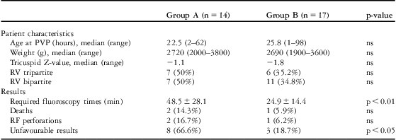
Median age at pulmonary valvotomy was 22.5 hours in Group A – with a range from 2 to 62 hours – and 25.8 in Group B – with a range from 1 to 98 hours; median weight was 2720 grams in Group A – with a range from 2000 to 3800 grams – and 2690 grams – with a range from 1900 to 3600 grams – in Group B. Median tricuspid Z-value was −1.1 in Group A and −1.8 in Group B. The right ventricle was found to be tripartite in seven patients of Group A (50%) and in six patients of Group B (35%).
The procedure was successful in all but one patient of Group A, where procedure failure was defined as the inability to perforate the atretic pulmonary valve. Procedural morbidity intended as major complications occurred in four patients. In Group A, three patients had, respectively, one immediate infundibular perforation that required surgical repair on an emergency basis by means of right ventricle outflow tract reconstruction and modified Blalock–Taussig shunt procedure, one post-procedural cardiac tamponade due to right ventricle outflow tract tear that was immediately sutured, and one iliac artery dissection that required saphenous patch angioplasty. In addition, one patient of Group B had a major intra-procedural complication: cardiac tamponade due to right ventricle outflow tract tear that was surgically repaired. In this case, the perforation of the pulmonary valve was successfully performed with a hybrid approach during the same procedure. In all the cases of infundibular perforation we reported, the Pro Track Microcatheter (0.038″) (Baylis Medical Company Inc., Montreal, QC Canada) passed across the tear and the complication was surgically managed.
Required fluoroscopy times were 48.5 plus or minus 28.1 minutes (median 45 minutes) and 24.9 plus or minus 14.4 minutes (median 21 minutes) in Group A and Group B, respectively (p < 0.01). In all, two premature infants of Group A and one patient of Group B died early from respiratory disease and lung immaturity; eight patients of Group A, including the patients with major complications, and three of Group B required a modified Blalock–Taussig shunt. Although not statistically significant, in Group A there was a higher incidence of patients in whom radiofrequency perforation was unsuccessful or needed early surgery for procedure-related complications. All data are reported in Table 1.
Comparisons between Group A and B.

PVP = pulmonary valve perforation; RF = radiofrequency; RV = right ventricle
Unfavourable results: unsuccessful procedure, major complications and/or need of early surgery
Discussion
Pulmonary atresia with intact ventricular septum is an uncommon congenital heart defect but one of the most frequent cyanotic heart defects in the neonatal period.Reference Freedom, Wilson, Trusler, Williams and Rowe 3
Owing to the anatomic heterogeneity of the disease, the therapeutic algorithm should be individualised.Reference Qureshi 2 The echocardiographic and angiographic images of the right ventricle morphology at presentation, such as tricuspid valve diameter, tricuspid valve Z-value, and coronary artery abnormalities, are critical in choosing a specific treatment for each patient.Reference Marasini, Gorrieri and Tuo 4
Biventricular repair can be achieved in the presence of adequate right heart structures, whereas univentricular repair or heart transplantation are the options for patients with hypoplastic monopartite right ventricle or right ventricle-dependent coronary circulation.Reference De Leval, Bull, Stark, Anderson, Taylor and Macartney 5 – Reference Daubeney, Wang and Delany 8
Over several decades of surgical experience, the surgical mortality has remained fairly high (15–37%).Reference Daubeney, Wang and Delany 8 , Reference Bryant, Nowicki and Mee 9
In selected cases, the initial management involves perforation of the atretic pulmonary valve with resulting decompression of the right ventricle necessary to stimulate right ventricle growth. The final objective is always to obtain a biventricular correction whenever possible. Approximately two-thirds of the affected infants may be eligible for transcatheter pulmonary valvotomy, which has been demonstrated to be an effective first-stage procedure.Reference Qureshi 2
Transcatheter perforation of an atretic pulmonary valve using the stiff end of a 0.014-inch guidewire was first described in 1991 by Latson.Reference Latson 10 Since that time, laser and radiofrequency technologies have been applied to valve perforation. The introduction of catheter treatment was associated with a relatively high mortality and possible procedural complications, which have significantly decreased in the last few years to 25–7.5%.Reference Marasini, Gorrieri and Tuo 4 , Reference Agnoletti, Piechaud and Bonhoeffer 11
Radiofrequency wire and catheter systems specifically designed for perforation of cardiac structures are now available, such as the coaxial radiofrequency system (Bayliss Medical Company), designed by Nykanen.
Proper and stable positioning of a catheter within the right ventricle outflow tract of a small, hypertrophied right ventricle is often challenging and requires complex manoeuvres and long fluoroscopy time. Small errors in catheter positioning can result in perforation of the myocardium or of the pulmonary trunk with potentially catastrophic consequences.
Some authors proposed new techniques in order to facilitate this complex manoeuvre and telescopic systems to reach difficult target lesions have been successfully used.Reference Asnes and Fahey 12 , Reference Brown, Boshoff, Eyskens, Mertens and Gewillig 13
In our experience, the complexity of catheter stabilisation under the atretic pulmonary valve was mainly due to the acute angle between the pulmonary atretic plane and the infundibular tract. In addition, the muscular hypertrophy and hypercontractility of the infundibulum are implicated in the complexity of the manoeuvre and the catheter is quite unstable and easily displaced. Furthermore, moving the catheter to reach the right position in a small and hypertrophic right ventricle can be traumatic, with the risk of lesion to the tricuspid subvalvular apparatus and to the conduction system.
We describe here a new technique that can facilitate, thanks to the two components of the telescopic system, the double movement required for correct catheter positioning under the atretic infundibulum. In this manoeuvre, regardless of the catheter employed, two opposite movements are necessary: first, the catheter must be directed from right to left and from posterior to anterior side to advance it through the tricuspid valve and to obtain the right position under the ostium infundibuli, then it must be moved from left to right and from anterior to posterior side to place its tip under the atretic pulmonary valve.
The two components of the telescopic catheter make it possible to obtain a stable position with the guide catheter and, while keeping this part of the system in the correct position, to rotate the hydrophilic catheter and position it more easily under the atretic valve with the support of the first component. Whenever the position obtained was still anterior, we retracted a little the coaxial catheter and, when necessary, made a j-shaped tipped radiofrequency guidewire to direct it more posteriorly.
We think that the advantage of telescopic systems is the availability of two differently shaped catheters allowing different orientations of the tip. Moreover, we found the hydrophilic catheter easier to be manoeuvred and less traumatic compared with a Teflon right Judkins catheter.
In reviewing our experience, we observed that in Group A, using a right Judkins catheter, correct and stable catheter position was difficult to obtain and the manoeuvre required a longer fluoroscopy time compared with Group B. In this latter group, the stabilisation of the catheter was more easily obtained and the correct catheter position was almost always maintained, which allowed a more rapid performance of the procedure.
Conclusions
In this study, we demonstrated that the telescopic catheter system can significantly decrease the fluoroscopy time of this complex procedure and consequently patients’ exposure to procedure-related risks. These encouraging results made this technique become our standard approach to radiofrequency perforation and valvotomy in patients with pulmonary atresia and intact ventricular septum.
Acknowledgments
Dr Anna Capurro and Dr Maria Calevo for their contributions to English revision and Statistical Analysis.