Case report
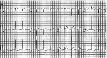
A 13-year-old boy collapsed while performing timed windsprints at a competitive sports camp. Ventricular fibrillation was documented by paramedics and defibrillation was performed successfully in the field. At hospital arrival, his blood pressure was 140/100 mmHg, and his heart rate was 93 bpm, with evidence of severe neurological devastation. Cardiovascular physical findings included regular rhythm, normal first and second heart sounds, prominent S4, grade II/VI systolic murmur at the apex with radiation to the upper sternal border, and an increased left ventricular impulse. Physical examination was notable for a muscular adolescent with Tanner IV genitalia. Electrocardiogram demonstrated sinus rhythm, left ventricular hypertrophy with T-wave inversion in the lateral praecordium, and QTc of 430 ms (Fig 1). Echocardiography demonstrated left ventricular hypertrophy, hyperdynamic left ventricular shortening fraction, and reduced left ventricular systolic dimension. Cardiac catheterization demonstrated hyperdynamic left ventricular function with left ventricular systolic and end-diastolic pressures of 130 and 14 mmHg, respectively. No left ventricular obstruction was evident at baseline or with infusion of isoproterenol. Coronary artery anatomy was normal. Standard urine toxicology tests were negative. Over the next week, serial neurological evaluations were consistent with severe diffuse hypoxic brain injury. Support was withdrawn and the patient expired.
Electrocardiogram time of admission to hospital showing left ventricular hypertrophy with inferolateral T wave inversion.

Past medical history was significant for an episode of syncope with exertion 1 week before cardiac arrest. Evaluation by his primary physician revealed a dramatic increase in the patient’s muscle mass and maturity of secondary sexual characteristics since his preceding exam, but otherwise excellent health status. He received no medications, dietary supplements, or performance-enhancing substances according to his parents’ knowledge. His coach denied knowledge of performance-enhancing supplements. His family history was negative for sudden death, hypertrophic cardiomyopathy, or heart rhythm abnormalities. All first-degree family members underwent screening with electrocardiogram and echocardiogram with normal studies.
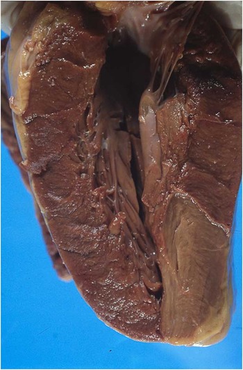
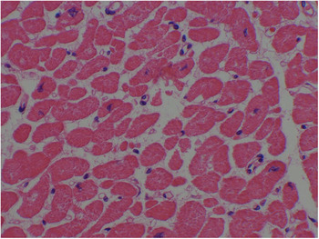
At autopsy, examination of the heart revealed massive cardiomegaly with a weight of 465 g, compared with an expected weight of 175 g (Fig 2). Marked left ventricular free wall and asymmetric septal hypertrophy reducing the left ventricular volume was present. The epicardial surface was covered by increased fatty tissue. The origin and course of the coronary arteries were normal. Histological analysis of the left ventricular myocardium demonstrated foci of myofibrillar disarray, proliferation of fibroblasts consistent with early fibrosis, and enlarged myofibres with heterogeneity of nuclear size including “box-car” nuclei (Fig 3). There was no evidence of myocarditis or findings of right ventricular dysplasia.
Gross evaluation revealed excessive epicardial fat with marked hypertrophy of the ventricles and papillary muscles. There was multifocal endocardial fibrosis, especially in the atria.

Histological evaluation demonstrated enlarged myofibres with hyperchromatic and pleomorphic nuclei. The interstitial tissue was expanded with an increased capillary network and occasional fibroblasts. No inflammatory infiltrates were noted (H & E stain, 40× objective).

Increased muscle mass, particularly of the upper body was present. Bilateral skin striae of the biceps and thighs were present. Bilateral adrenal atrophy with lipid depletion and reduction of the zona fasiculata was evident. No evidence of adrenal tumour was found. Fatty infiltration of the liver was noted. Secondary sexual precocity was present, with reduced gonadal size.
At the time when anabolic steroid use was suspected, urine was no longer available for chromatographic analysis for steroid metabolites.
Discussion
Anabolic androgenic steroids are synthetic derivatives of testosterone that promote the growth of skeletal muscles and development of male sexual characteristics.Reference Van Amsterdam, Opperhuizen and Hartgens 1 Commonly recognised cardiovascular side-effects of chronic anabolic steroid use include cardiac hypertrophy, atherosclerosis, and hypertension, with the rare occurrence of sudden death.Reference Dickerman, Schaller, Prather and McConathy 2 – Reference Nieminen, Ramo and Viitasalo 4 To our knowledge, this patient represents the first case of adolescent sudden cardiac arrest in the context of hypertrophic cardiomyopathy attributed to anabolic steroid use. Although we could not confirm the presence of anabolic steroids or their metabolites at the time of our patient’s death, the cardiac and systemic findings including adrenal atrophy, mature genitalia, muscular development, and skin striae are considered indicative of steroid use and are consistent with cases of steroid use in adult bodybuilders.Reference Dickerman, Schaller, Zachariah and McConathy 5 , Reference Achar, Rostamian and Narayan 6 In addition, familial cardiomyopathy is less likely in the absence of genetic or cardiac findings among family members. On the basis of previous health records, it was estimated that steroid use was likely initiated 10–12 months before cardiac arrest when the patient was 11 years old.
The use of steroids as performance-enhancing substances is estimated to occur in 0.7–2.2% of adolescent males in the United States.Reference Johnston, O’Malley, Miech, Bachman and Schulenberg 7 Many users are athletes for whom the possibility of improved body appearance and athletic performance outweighs the potential health consequences. It has been reported that up to 15% of adolescent males would consume a substance that would guarantee their fitness goals even if it may be harmful to their health, and up to 8% would continue to use even if it may shorten their lives.Reference Hoffman, Faigenbaum, Ratamess, Ross, Kang and Tenenbaum 8
Conclusion
Our patient represents the first case of adolescent cardiac arrest attributed to hypertrophic cardiomyopathy from anabolic steroid use, with documented ventricular fibrillation as the initiating arrhythmia. Improved education and awareness of the potentially fatal cardiac effects of anabolic steroid use of younger patients by practitioners is imperative.
Acknowledgement
None.
Financial Support
This research received no specific grant from any funding agency, commercial, or not-for-profit sectors.
Conflicts of Interest
None.