INTRODUCTION
Visceral leishmaniasis (VL), commonly known as kala-azar, is caused by obligate intracellular protozoan parasites, particularly by the species Leishmania donovani, Leishmania infantum and Leishmania chagasi. VL is a potentially fatal human disease with an estimated incidence of at least 0·2–0·4 million cases worldwide, causing 20 000–400 000 deaths each year (Alvar et al. Reference Alvar, Velez, Bern, Herrero, Desjeux, Cano, Jannin and den Boer2012). Moreover, VL promotes clinical progression of HIV and the development of AIDS-defining conditions, which may also increase the cumulative immunodeficiency against infections, disease severity and morbidity (Alvar et al. Reference Alvar, Aparicio, Aseffa, Den Boer, Canavate, Dedet, Gradoni, Ter Horst, Lopez-Velez and Moreno2008; Cota et al. Reference Cota, de Sousa and Rabello2011; Albuquerque et al. Reference Albuquerque, Mendonca, Cardoso, Baldacara, Borges, Borges Jda and Pranchevicius2014). The untreated disease in any age group can produce profound cachexia, thrombocytopaenia, susceptibility to secondary infections, ultimately resulting in death (Davidson, Reference Davidson1998; Osman et al. Reference Osman, Kager and Oskam2000; Da Luz et al. Reference Da Luz, Pimenta, Rabello and Schall2003; Davies et al. Reference Davies, Kaye, Croft and Sundar2003; Collin et al. Reference Collin, Davidson, Ritmeijer, Keus, Melaku, Kipngetich and Davies2004). Emergence of resistance is a serious problem in the control of VL against standard anti-leishmanials, such as pentavalent antimonials. In northern Bihar (India), a highly endemic region, up to 60% of VL patients do not respond to pentavalent antimony treatment (Sundar, Reference Sundar2001; Sundar et al. Reference Sundar, Pai, Kumar, Pathak-Tripathi, Gam, Ray and Kenney2001; Sundar and Rai, Reference Sundar and Rai2005). Human VL is characterized by a distinct humoral response and absence of parasite-specific cell-mediated immune response, associated with an inability to control infection. Leishmania donovani induces dynamic immune responses which, however, in patients with acute disease do not control the infection. However, patients recovered from VL have shown resistance to reinfection (Modabber, Reference Modabber1995), which indicates that development of protective immunity provides a rational basis for the development of vaccines that reveal potent cell-mediated immune responses. It has been well established that the ability to induce a pro-inflammatory response is essential for the control of the disease wherein the Th1 cytokines IL-2, IL-12 and IFN-γ play an important role (Melby et al. Reference Melby, Tryon, Chandrasekar and Freeman1998; Garg and Dube, Reference Garg and Dube2006). These cytokines direct the effector functions of macrophages and elicit a Th1 immune response (Peruhype-Magalhaes et al. Reference Peruhype-Magalhaes, Martins-Filho, Prata, Silva Lde, Rabello, Teixeira-Carvalho, Figueiredo, Guimaraes-Carvalho, Ferrari and Correa-Oliveira2005). These findings put forward that any intervention that facilitate shifting of the Th2-type immune response towards the Th1 type will have an impact on the treatment of VL. Therefore, the Th1 arm of the immune response could be exploited as vaccine candidates.
A comprehensive knowledge of antigens targeted during infection helps in the development of successful vaccine. Therefore, in recent years several workers explored and studied various antigens that predominantly stimulate Th1 responses in infected patients/rodents (Webb et al. Reference Webb, Kaufmann, Campos-Neto and Reed1996; Gurunathan et al. Reference Gurunathan, Sacks, Brown, Reiner, Charest, Glaichenhaus and Seder1997; Ahmed et al. Reference Ahmed, Bahloul, Robbana, Askri and Dellagi2004; Zadeh-Vakili et al. Reference Zadeh-Vakili, Taheri, Taslimi, Doustdari, Salmanian and Rafati2004; Bhowmick and Ali, Reference Bhowmick and Ali2009; Ghaffarifar et al. Reference Ghaffarifar, Jorjani, Sharifi, Dalimi, Hassan, Tabatabaie, Khoshzaban and Hezarjaribi2012; Khare et al. Reference Khare, Jaiswal, Tripathi, Joshi, Sundar and Dube2013). Given that the disease is related directly and exclusively to the propagation and resolution of amastigotes in macrophages of the vertebrate host, the antigenic molecules that are upregulated or selectively expressed in the amastigotes have the potential to be superior vaccine candidates. However, little is known about the possible protective antigens from the amastigote stage of the parasite. Still fewer studies look at amastigote-specific proteins including A2, cysteine proteinase, etc. as a suitable vaccine candidate (Rafati et al. Reference Rafati, Baba, Bakhshayesh and Vafa2000; Ghosh et al. Reference Ghosh, Zhang and Matlashewski2001; Coelho et al. Reference Coelho, Tavares, Carvalho, Chaves, Teixeira, Rodrigues, Charest, Matlashewski, Gazzinelli and Fernandes2003; Nylen et al. Reference Nylen, Maasho, McMahon-Pratt and Akuffo2004; Farajnia et al. Reference Farajnia, Mahboudi, Ajdari, Reiner, Kariminia and Alimohammadian2005; Zanin et al. Reference Zanin, Coelho, Tavares, Marques-da-Silva, Silva Costa, Rezende, Gazzinelli and Fernandes2007; Fernandes et al. Reference Fernandes, Costa, Coelho, Michalick, de Freitas, Melo, Luiz Tafuri, Resende Dde, Hermont, Abrantes Cde and Gazzinelli2008; Hsiao et al. Reference Hsiao, Yao, Storlie, Donelson and Wilson2008). Though availability of axenic cultures of several Leishmania strains (Pan, Reference Pan1984; Eperon and McMahon-Pratt, Reference Eperon and McMahon-Pratt1989; Bates, Reference Bates1993) allowed detailed studies of biological and immunological functions of amastigote proteins, numerous proteins were identified from axenic amastigotes (El Fakhry et al. Reference El Fakhry, Ouellette and Papadopoulou2002; Walker et al. Reference Walker, Acestor, Gongora, Quadroni, Segura, Fasel and Saravia2006), while the true amastigotes (from infected spleen) are still less characterized. There are several antigens expressed both in promastigotes and amastigotes, yet most of them are expressed preferentially in either stages of the parasite (Ghedin et al. Reference Ghedin, Zhang, Charest, Sundar, Kenney and Matlashewski1997; McMahon-Pratt et al. Reference McMahon-Pratt, Kima and Soong1998; Kar et al. Reference Kar, Soong, Colmenares, Goldsmith-Pestana and McMahon-Pratt2000).
On the basis of this rationale, our earlier studies led to the identification of two potent fractions (F2 and F3) in the range of 97–68 kDa and 68–43 kDa, respectively, inducing Th1-type cellular responses in cured Leishmania patients as well as in hamsters by fractionation and sub-fractionation of the soluble proteins from clinical isolates of L. donovani amastigotes (Kumari et al. Reference Kumari, Misra, Tandon, Samant, Sundar and Dube2012). Further, proteomic characterization of this sub-fraction led to the identification of numerous Th1-stimulatory proteins, in particular, the crucial stress-related proteins viz. cytosolic tryparedoxin peroxidase (cTryP) also known as thiol-specific antioxidant or TSA (Webb et al. Reference Webb, Campos-Neto, Ovendale, Martin, Stromberg, Badaro and Reed1998) and chaperonin TCP20 (TCP20) apart from several putative ones (communicated). cTryP is an essential enzyme (Castro et al. Reference Castro, Budde, Flohe, Hofmann, Lunsdorf, Wissing and Tomas2002; Wilkinson et al. Reference Wilkinson, Horn, Prathalingam and Kelly2003) in the kinetoplastid parasites for protection against oxidative stress (causing detoxification of peroxides) and also responsible for survival, drug response and virulence in Leishmania (Iyer et al. Reference Iyer, Kaprakkaden, Choudhary and Shaha2008; Romao et al. Reference Romao, Castro, Sousa, Carvalho and Tomas2009). cTryP has been found to express both in promastigote and amastigote forms of the parasite and conserve in most Leishmania species. This protein has been identified as immunogenic as well protective antigen in Leishmania major (Stober et al. Reference Stober, Lange, Roberts, Alcami and Blackwell2005, Reference Stober, Lange, Roberts, Gilmartin, Francis, Almeida, Peacock, McCann and Blackwell2006, Reference Stober, Lange, Roberts, Alcami and Blackwell2007; Fiorillo et al. Reference Fiorillo, Colotti, Boffi, Baiocco and Ilari2012). Moreover, cTryP antigen has been used as vaccine targets against L. infantum (Rodríguez-Cortés et al. Reference Rodríguez-Cortés, Ojeda, López-Fuertes, Timón, Altet, Solano-Gallego, Sánchez-Robert, Francino and Alberola2007; Carson et al. Reference Carson, Antoniou, Ruiz-Arguello, Alcami, Christodoulou, Messaritakis, Blackwell and Courtenay2009; Todoli et al. Reference Todoli, Solano-Gallego, de Juan, Morell, Nunez Mdel, Lasa, Gomez-Sebastian, Escribano, Alberola and Rodriguez-Cortes2010; Stober et al. Reference Stober, Jeronimo, Pontes, Miller and Blackwell2012). The other protein – chaperonin TCP20 – encodes a subunit of the cytosolic TCP-1chaperonin (chaperonin containing t-complex polypeptide-1) complex in eukaryotes. The human and yeast TCP20 genes encode representative members of a family of subunits of TriC (TCP ring complex), the TCP-l ring complex (Li et al. Reference Li, Lin, Frydman, Boal, Cardillo, Richard, Toth, Lichtman, Hartl, Sherman and Segel1994). Though the protein TCP20 has been reported to be involved in the folding and assembly of actin and tubulin structures (Frydman et al. Reference Frydman, Nimmesgern, Erdjument-Bromage, Wall, Tempst and Hartl1992; Gao et al. Reference Gao, Thomas, Chow, Lee and Cowan1992, Reference Gao, Vainberg, Chow and Cowan1993; Yaffe et al. Reference Yaffe, Farr, Miklos, Horwich, Sternlicht and Sternlicht1992; Sternlicht et al. Reference Sternlicht, Farr, Sternlicht, Driscoll, Willison and Yaffe1993; Li et al. Reference Li, Lin, Frydman, Boal, Cardillo, Richard, Toth, Lichtman, Hartl, Sherman and Segel1994; Miklos et al. Reference Miklos, Caplan, Mertens, Hynes, Pitluk, Kashi, Harrison-Lavoie, Stevenson, Brown and Barrell1994; Stoldt et al. Reference Stoldt, Rademacher, Kehren, Ernst, Pearce and Sherman1996), there has been no description of the protein in the case of either Leishmania or any pathogen that is important for causing human infections. Since, there has been no report of either of these proteins as vaccine candidates in the case of L. donovani, it was pertinent to assess their immunogenic and prophylactic potential against VL – a fatal infection. Therefore, the present study includes (1) molecular cloning and characterization of cTryP and TCP20; (2) evaluation of immunogenicity of both recombinant proteins using lymphocytes/PBMCs of cured Leishmania hamsters/patients and (3) evaluation of prophylactic efficacy of proteins against L. donovani infection in golden hamsters, which has been reported as a perfect experimental model for VL (Melby et al. Reference Melby, Tryon, Chandrasekar and Freeman1998).
MATERIALS AND METHODS
Ethics statement
Experiments on the animals (hamsters) were performed following the approval of the protocol and the guidelines of Institutional Animal Ethics Committee (IAEC) of the CDRI, which is adhered to the National Guideline of Committee for the Purpose of Control and Supervision on Experiments on Animals (CPCSEA) under the Ministry of Environment and Forest, Government of India. The approval reference number is 154/10/Para/IAEC dated 04·10·10. The protocol and study with patients was approved by the Ethics Committee of the Kala-Azar Medical Research Centre, Muzaffarpur (protocol # EC- KAMRC/Vaccine/VL/2007-01) and written informed consent was obtained from the patients before enrolment to this study. All the human subjects underwent clinical examination by a local physician for leishmanial and other possible infections.
Host and parasite
Laboratory-bred golden hamsters (Mesocricetus auratus, 45–50 g) of either sex (M/F), from the Institute's Animal House Facility were used as experimental host. They were housed in climatically controlled room and fed with standard rodent food pellet (Lipton India Ltd, Bombay) and water ad libitum. The L. donovani strain (2001) used in this study was isolated from patients at the Kala-Azar Medical Research Centre, Institute of Medical Sciences, Banaras Hindu University, Varanasi, India, and at its affiliated hospital at Muzaffarpur, Bihar, India, and cultured in vitro as described elsewhere (Garg et al. Reference Garg, Gupta, Tripathi, Naik, Sundar and Dube2005). Promastigotes were grown in complete RPMI medium at 26 °C (Sigma-Aldrich, USA) in 75 cm2 culture flasks (Nunc, Denmark) (Dube et al. Reference Dube, Singh and Sundar2005). The strain has also been maintained in hamsters through serial passage, i.e. from amastigote to amastigote to maintain its virulence (Dube et al. Reference Dube, Singh and Sundar2005).
Soluble L. donovani antigen from amastigote (AmSLD)
Soluble L. donovani amastigote antigen (AmSLD) was prepared as per the protocol described elsewhere (Kumari et al. Reference Kumari, Misra, Tandon, Samant, Sundar and Dube2012). Briefly, isolated splenic amastigotes were repeatedly washed in phosphate buffered saline (PBS) and subjected to ultrasonication followed by centrifugation at 40 000 g for 20 min at 4 °C. After assessing the protein contents by the Bradford method (Bradford, Reference Bradford1976), the antigen was distributed in small aliquots and stored at −80 °C.
Generation of recombinant proteins
Gene-specific primers were designed using L. donovani sequence available in the Gene bank database as follows: cTryP, 5′-GGATCCATGTCCTGCGGTGACGCCAAGATCAACTG-3′ as forward and 5′-GAATTCTTACTGCTTACTGAAGT ACCCCTCGACAGA-3′ as reverse; TCP20, 5′-GCTAGCATGTCGAGCCTGGCTTACATCAACCCTGGC as forward and 5′-AAGCTTCTACTGACCTCAGGAGCCGCGGCGCC as reverse, containing BamHI, EcoRI, NheI and HindIII sites (underlined), respectively. Both the genes were amplified using L. donovani amastigote genomic DNA as template and Taq DNA polymerase (Bangalore Genei, India) in a Thermocycler (Bio-Rad) and the amplified PCR product was cloned into the pTZ57R/T vector and then inserted in-frame with a 6xHis tag into the BamHI/EcoRI (LdcTryP) and NheI/HindIII (LdTCP20) site of the expression vector pET-28a (Novagen, USA) and overexpressed in Escherichia coli C41 (DE3) and Rosetta strain, respectively. Further, following IPTG (1 mm) induction, the overexpression of the recombinant proteins, rLdcTryP and rLdTCP20, were tested as reported earlier (Kushawaha et al. Reference Kushawaha, Gupta, Tripathi, Khare, Jaiswal, Sundar and Dube2012). Further, rLdcTryP was purified using Tris buffer (Kushawaha et al. Reference Kushawaha, Gupta, Tripathi, Khare, Jaiswal, Sundar and Dube2012) and rLdTCP20 was purified in denaturing condition as described by Khare et al. (Reference Khare, Jaiswal, Tripathi, Joshi, Sundar and Dube2013). The presence of endotoxin such as the lipopolysaccharide (LPS) content of the recombinant proteins was measured by the Limulus amoebocyte lysate test (QCL-1000H chromogenic kit, Lonza).
Confirmation of recombinant proteins with immunoblot analysis
Swiss mice were immunized with 25 μg of purified recombinant proteins (rLdcTryP and rLdTCP20) with Freund's complete adjuvant (FCA, Bangalore Genei, India) for primary immunization (day 0) followed by two booster doses of 25 μg of either rLdcTryP or rLdTCP20 in Freund's incomplete adjuvant (FIA) (day 14 and 21) for the generation of antibodies. Serum was obtained 7 days after the last booster and antibody titres were determined by ELISA; wherein, 96-well plates were coated with either rLdcTryP or rLdTCP20 overnight at 0·2 μg/100 μL well−1 in carbonate buffer. The serum obtained from mice was used as the primary antibody whereas goat anti-mice horseradish peroxidises (HRP) conjugate was taken as the secondary antibody. The binding of conjugate was visualized with 100 μL of O-phenylenediamine (OPD, Sigma-Aldrich) in citrate phosphate buffer (citric acid 0·1 m, Na2HPO4 0·2 m, pH 4·5). The reaction was stopped with 4 m H2SO4 and the absorbance was read using a spectrophotometer at 492 nm.
For immunoblots, AmSLD was separated on 12% SDS–PAGE and electrophoretically transferred onto nitrocellulose membrane (NCM, Millipore, USA) using a semi-dry blot apparatus (Hoefer Semiphor) (Towbin et al. Reference Towbin, Staehelin and Gordon1979). After overnight blocking in 5% skimmed milk, the membrane was incubated with antiserum to the recombinant protein at a dilution of 1:4000 for 2 h at room temperature. The membrane was washed 3 times with PBS containing 0·5% Tween 20 (PBS-T) and then incubated in goat anti-mice IgG HRP (Bangalore Genei, India) conjugate solution at a dilution of 1:10 000 for 1 h at room temperature. Finally, the blot was developed using diaminobenzidine (DAB, Sigma-Aldrich) imidazole and H2O2.
Verification of amastigote proteins by immunoblot analysis and immunolocalization
The whole cell lysate (WCL) of splenic amastigotes and promastigotes of L. donovani was prepared as described earlier (Kumari et al. Reference Kumari, Misra, Tandon, Samant, Sundar and Dube2012). Then the sample (25 μL, 300 μg/lane) was separated on two sets of 12% SDS–PAGE gel and then transformed onto NCM. Following transfer of protein, NCM was blocked overnight in 5% skimmed milk and then one NCM was probed with A2 antibody (Medimabs, Montreal, Canada) and followed by incubation with HRP-conjugated goat anti-mice antibodies (Bangalore Genei, India) at a dilution of 1:5000. Similarly the other blot was probed with an antiserum of either rLdcTryP or rLdTCP20, then incubated with HRP-conjugated goat anti-mice antibodies at a dilution of 1:10 000 for 1 h at room temperature. After washing with PBS-T (0·05% Tween-20), the protein bands were detected using Amersham ECL kit of enhanced chemiluminescence (Amersham Biosciences, Singapore). Briefly, NCM was incubated for 5 min with detection solutions (provided with ECL kit) in equal ratio.
For the immunolocalization experiment, L. donovani pure amastigotes were isolated from the spleen of infected hamsters by percoll gradients (Sigma-Aldrich, USA) as described elsewhere (Kumari et al. Reference Kumari, Misra, Tandon, Samant, Sundar and Dube2012). Isolated amastigotes were washed 3 times with PBS, allowed to adhere to poly-L-lysine (Sigma-Aldrich, USA)-coated cover slips followed by fixation with paraformaldehyde. Cells were then permeabilized with 0·2% (v/v) Triton X-100 and washed again with PBS. The fixed cells were incubated in PBS containing 3% BSA for 30 min and washed again. Then, the cells were incubated with primary antibodies (mice anti-rLdcTryP/anti-rLdTCP20) at a dilution 1:2000 for 1 h, rinsed with PBS and incubated again for 1 h with Fluorescein isothiocyanate (FITC)-conjugated goat anti-mice IgG. The coverslips were mounted on clean microslides using 10 μL of fluorescent mounting media (Calbiochem, Germany) and finally observed under a fluorescence microscope (Nikon 80i, Japan).
Treatment of L. donovani-infected hamsters and isolation of mononuclear cells from the lymph node
Approximately, 30 male hamsters were infected with 107 amastigotes intracardially (i.c) and checked for parasite burden as described elsewhere (Kumari et al. Reference Kumari, Samant, Misra, Khare, Sisodia, Shasany and Dube2008c ). Animals harbouring >20–30 amastigotes/100 macrophage cell nuclei were then treated with Miltefosine (SynphaBase, Switzerland) given orally at 40 mg kg−1 bodyweight for 5 days. After reassessment of splenic biopsy on day 30 post-treatment, mononuclear cells were separated from lymph nodes of cured, infected and normal hamsters as per the protocol of Garg et al. (Reference Garg, Gupta, Tripathi, Naik, Sundar and Dube2005).
Isolation of peripheral blood mononuclear cells (PBMCs) from different groups of human patients
The study groups for human samples belonged to four clinically well-characterized groups: (i) cured VL patients (age: 5–32 years); all the patients had received complete course of treatment with amphotericin B and recovered from VL and shown to have no parasites in splenic aspirates at least 6 months after treatment. Diagnosis was established using splenic aspirates and found negative at the time of study (n = 7; 5 male and 2 female); (ii) active VL patients (age: 8–49 years) showing clinical symptoms of VL and parasitologically confirmed using splenic aspirates and shown to have amastigote in Giemsa-stained slides (n = 8; 4 male and 4 female); (iii) endemic household contacts (age: 8–35 years) neither showed clinical symptoms nor received any treatment for VL (n = 7; 4 male and 3 female); (iv) normal healthy donors (age: 25–32 years) without any history of leishmaniasis (n = 9; 5 male and 4 female).
Heparinized venous blood (10 mL each) was collected from all study subjects and PBMCs were isolated from blood by density gradient centrifugation using histopaque (Histopaque 1077, Sigma-Aldrich, USA) as described by Garg et al. (Reference Garg, Gupta, Tripathi, Naik, Sundar and Dube2005).
Assessment of prophylactic efficacy of rLdcTryP and rLdTCP20 in hamsters to L. donovani challenge
Five groups of hamsters (15–20 animals per group) were made whereas groups 1–3 served as controls as described below and groups 4 and 5 as the main experimental groups: group 1, unvaccinated and unchallenged (normal control); group 2, unvaccinated and challenged (infected control); group 3, Bacillus Calmette-Guérin (BCG) alone; group 4, vaccinated with rLdcTryP+BCG and group 5, vaccinated with rLdTCP20 + BCG. The hamsters of groups 4 and 5 were vaccinated intradermally (i.d) on the back with either rLdcTryP or rLdTCP20 (50 μg/50 μL per animal) along with equal volume of BCG (0·1 mg/50 μL per animal) in emulsified form and group 3 was given BCG only. Similarly, 15 days later a booster dose of half of the amount of either rLdcTryP or rLdTCP20 along with BCG was given to groups 4 and 5, only BCG to group 3, and 50 μL of PBS was administered in group 1 animals which served as normal control.
Twenty-one days after the booster dose, the vaccinated and unvaccinated control groups were challenged via intracardiac (i.c.) route with 108 late logarithmic phase promastigotes of L. donovani. The prophylactic efficacy of rLdcTryP and rLdTCP20 was assessed in the spleen, liver and bone marrow of three vaccinated hamsters at different time intervals, i.e. on days 0, 45, 60, 90 and 120 post-challenge (p.c.). The criterion of prophylactic efficacy was the assessment of parasite load as the number of amastigotes/1000 cell nuclei in each organ in comparison to the unvaccinated controls and the percentage inhibition (PI) was calculated as described earlier (Garg et al. Reference Garg, Gupta, Tripathi, Hajela, Sundar, Naik and Dube2006):
Measurement of body, spleen and liver weight of vaccinated hamsters
The body weights as well as weight of the spleen and liver (on necropsy) of hamsters of all of the experimental groups were assessed at different time intervals, i.e. on days 0, 45, 60, 90, 120 and 180 p.c.
Immunological assays
Delayed-type hypersensitivity (DTH)
DTH was performed by injecting 50 μg/50 μL of AmSLD in PBS via i.d route into one footpad and PBS alone into another footpad, one of each of the vaccinated and unvaccinated controls. The response was evaluated 48 h later by measuring the difference in footpad swelling between the two with and without AmSLD for each animal (Bhowmick et al. Reference Bhowmick, Ravindran and Ali2007).
Lymphocyte proliferation assay (LTT)
Lymph node cells (1 × 10⁶ cells mL−1) of normal, infected (day 30 post-infection) and cured/vaccinated hamsters as well as the cured and infected patient's PBMC were cultured in 96-well flat bottom tissue culture plates (Nunc, Denmark). LTT assay was carried out as per the protocol described by Garg et al. (Reference Garg, Gupta, Tripathi, Naik, Sundar and Dube2005). Approximately 100 μL of predetermined concentration (10 μg mL−1) of mitogens phytohaemagglutinin (PHA Sigma, USA) for patient's PBMCs, concanavalin (Con A, Sigma, USA) for hamster lymphocytes, as well as antigens i.e. LdcTryP, LdTCP20 and AmSLD (10 μg mL−1 each) were added to the wells in triplicate. Wells without stimulants served as blank controls. Cultures were incubated at 37 °C in a CO2 incubator with 5% CO2 for 3 days in the case of the mitogens, and for 5 days in the case of the antigens (LdcTryP/LdTCP20/AmSLD). Eighteen hours prior to termination of experiment, 50 μL of XTT (Roche Diagnostics) was added to 100 μL of supernatants of each well and optical density at 480 nm was determined using 650 nm as the reference wavelength.
Estimation of Nitric oxide (NO) activity in macrophages of hamsters
Isolated lymphocytes from all the three study groups of hamster's, viz. normal, infected and cured, were suspended in cRPMI and plated at 105 cells well−1 and stimulated for 3 days in case of LPS and 5 days in case of rLdcTryP/LdTCP20 and AmSLD at a concentration of 10 μg mL−1. The presence of NO was assessed in the culture supernatants of cured hamster peritoneal macrophages after the exposure with supernatant of stimulated lymphocytes. The supernatants (100 μL) collected from macrophage cultures 24 h after incubation was mixed with an equal volume of Griess reagent (Sigma-Aldrich, USA) and left for 10 min at room temperature. The absorbance of the reaction was measured at 540 nm in an ELISA reader (Ding et al. Reference Ding, Nathan and Stuehr1988).
Assessment of cytokine levels-IFN-γ/IL-12/IL-10/IL-4 in PBMCs of cured/endemic patients
Further, levels of Th1/Th2-stimulatory cytokines were estimated in PBMCs from cured patients as well as in endemic contacts in response to recombinant proteins (rLdcTryP and rLdTCP20). Culture of PBMCs (1 × 10⁶ cells mL−1) was plated in 96-well culture plates and then AmSLD as well as rLdcTryP or rLdTCP20 were added at a concentration of 10 μg mL−1 in triplicate wells. The levels of IFN-γ, IL-12 as well as IL-10 and IL-4 were estimated using commercial ELISA kits according to the manufacturer's protocol (BD OptEIA kit, USA). The results were expressed as picograms of cytokine/mL, based on the standard curves generated using a recombinant cytokine provided in the kit.
Measurement of serum antibody response in vaccinated hamsters
The levels of anti-leishmanial antibody IgG1 and IgG2 in serum samples from hamsters of different groups were measured as described earlier (Samant et al. Reference Samant, Gupta, Kumari, Misra, Khare, Kushawaha, Sahasrabuddhe and Dube2009). The 96-well ELISA plates were coated either with rLdcTryP or rLdTCP20 (2 μg mL−1 in PBS) for overnight at 4 °C and blocked with 1·5% BSA at room temperature for 1 h. The sera were used at a dilution of 1:50 for 2 h at room temperature. Biotin-conjugated mouse anti-Armenian hamster IgG1 and mouse anti-Syrian hamster IgG2 (BD, Pharmingen, USA) were added for 1 h and the plates were further incubated for 1 h with HRP-conjugated streptavidin and finally developed using OPD (Sigma-Aldrich, USA) as substrate.
Evaluation of mRNA cytokines and inducible NO synthase (iNOS) in vaccinated hamsters by quantitative real time-PCR (qRT-PCR)
qRT-PCR was performed to assess the expression level of mRNAs for various cytokines and iNOS in splenic cells upon vaccination with rLdcTryP and rLdTCP20. The splenic tissues of hamsters of each experimental group at different time intervals were collected and total RNA was isolated using Tri-reagent (Invitrogen, USA). Following quantification using Gene-quanta (Bio-Rad, USA), 1 μg of total RNA was used for the synthesis of cDNA using a first-strand cDNA synthesis kit (Fermentas, USA).The primers for qRT-PCR were designed using Beacon Designer software (Bio-Rad, USA) on the basis of cytokines and iNOS mRNA sequences available on PubMed (Melby et al. Reference Melby, Tryon, Chandrasekar and Freeman1998). RT-PCR was performed as described earlier elsewhere (Samant et al. Reference Samant, Gupta, Kumari, Misra, Khare, Kushawaha, Sahasrabuddhe and Dube2009) using the iQ5 multicolour real-time PCR system (Bio-Rad, USA).
Post-challenge survival of vaccinated hamsters
Survival of hamsters belonging to groups 4 and 5 was checked till day 180 p.c. in comparison to normal hamsters (group 1). Animals in all of the groups were given proper care and were observed for their physical conditions until their survival period. Survivals of individual hamsters were recorded and the mean survival period was calculated.
Statistical analysis
Results were expressed as mean±s.d. Two sets of experiments were performed for vaccination studies and in each experiment 15–20 animals were used. The results (pooled data of two independent experiments) were analysed by one-way ANOVA test followed by Tukey's test using GraphPad Prism software program. One-way ANOVA statistical test was used to assess the significance of the differences among various groups and a P value of < 0·001 was considered highly significant.
RESULTS
Molecular characterization of recombinant proteins
Generation of rLdCTryP and rLdTCP20
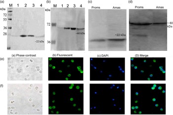
Both LdcTryP and LdTCP20 genes were successfully cloned first in pTZ57R/T (T/A) cloning vector and then into the pET28a vector and sequenced. The gene sequence was submitted to the National Centre for Biotechnology Information with accession number KC595509 and KC595510 (http://www.ncbi.nlm.nih.gov/nuccore/KC595509/KC595510). LdcTryP showed 99, 95 and 95% identity to those of L. infantum, L. major and Leishmania mexicana, respectively. Similarly, LdTCP20 showed, 99, 97 and 95% identity to those of L. infantum, L. major and L. mexicana, respectively. Further, 600 bp amplicon of LdcTryP gene encoded 200 amino acids with a predicted molecular mass of ~22 kDa, whereas 1617 bp amplicon of the LdTCP20 gene encoded 539 amino acids with a predicted molecular mass of ~60 kDa. Purification of rLdcTryP and LdTCP20 His-tagged was made by metal affinity chromatography and the recombinant proteins were used to raise polyclonal antibody in mice. Immunoblot analysis with mouse polyclonal antiserum to rLdcTryP and rLdTCP20 recognized a single band with molecular masses of ~22 kDa (Fig. 1a) and ~60 kDa (Fig. 1b) in amastigote L. donovani lysate, respectively.
Characterization of L. donovani cytosolic tryparedoxin peroxidase (LdcTryP) and chaperonin TCP20 (LdTCP20): (a) western blot with anti-rLdcTryP antibody; M: molecular weight (Mw) marker; lane1: uninduced E. coli WCL; lane 2: induced E. coli WCL, lane 3: purified protein; lane 4: amastigote-soluble L. donovani (AmSLD); (b) western blot with anti-rLdTCP20 antibody; M: Mw marker; lane1: uninduced E. coli WCL; lane 2: induced E. coli WCL, lane 3: purified protein; lane 4: AmSLD. Western blot analysis of cTryP and TCP20 protein expression in L. donovani amastigotes (Amas-lane 1) and promastigotes (Proms-lane 2) lysates using (c) anti-rLdcTryP antibody and (d) anti-rLdTCP20 antibody. Immunolocalization of LdcTryP (e) and LdTCP20 (f) in pure amastigotes by fluorescence microscopy using anti-LdcTryP antibody and secondary anti-mice FITC labelled IgG: Phase contrast (a), fluorescent (FITC) (b), DAPI (c), and FITC and DAPI merged image (d).

Confirmation of amastigote protein by immunoblot analysis using monoclonal A2 antibody and immunolocalization
Immunoblotting
Further, to ensure that the recombinant proteins we have generated in the present study are also expressing in amastigotes, a specific marker antibody A2, a stage-specific protein of amastigotes, was utilized (Charest et al. Reference Charest, Zhang and Matlashewski1996). Immunoblot analysis of WCL of splenic amastigotes and promastigotes using anti-rLdcTryP/anti-rLdTCP20 revealed the presence of these proteins in both stages of the parasite (Fig. 1c and d). Monoclonal antibody against amastigotes A2 served as control, detected in amastigotes lysate only (data not shown).
Immunolocalization
In addition, to know the sub-cellular location of these proteins in amastigotes, immunolocalization study was performed using pure splenic amastigote. cTryP was observed to be localized in whole of the cytoplasm of the cell wherein amastigotes showed bright green fluorescence (Fig. 1e). Similar was the case with chaperonin TCP20 (Fig. 1f).
rLdcTryP and rLdTCP20 induced LTT response and NO production in cured hamsters
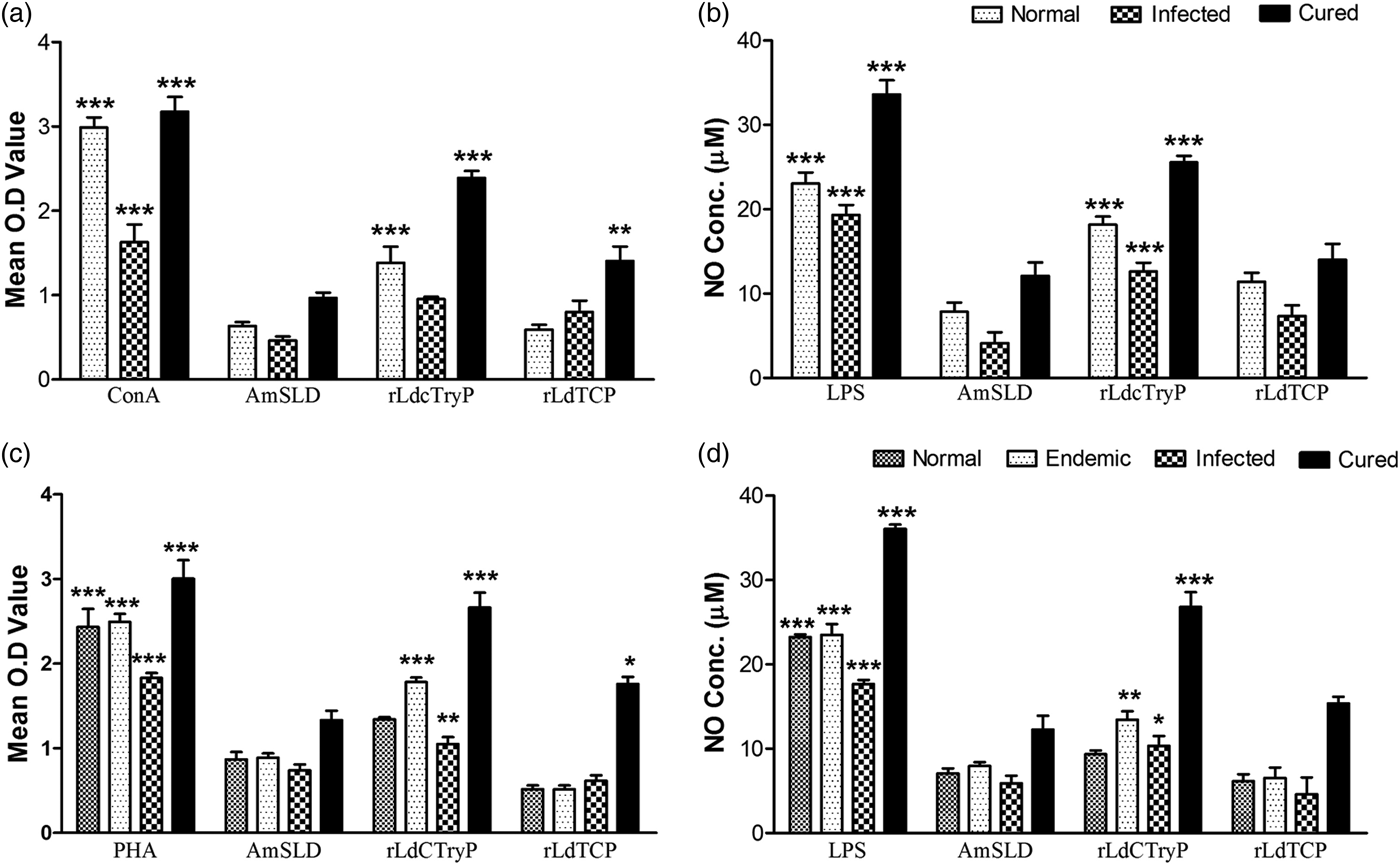
The cellular response of lymph node cells of cured hamsters was assessed by LTT simultaneously against Con A, AmSLD as well as test antigens (rLdcTryP and rLdTCP20) at a predetermined concentration of 10 μg mL−1. The LTT response of cured lymphocytes was compared with normal as well as L. donovani-infected lymphocytes, which were served as controls. The LTT response of Con A was observed better in all the experimental groups but was more significant in the cured group followed by normal group (Fig. 2a). The stimulation with rLdcTryP showed significantly higher proliferative response (~2·5-fold) in cured hamsters (O.D. 2·389 ± 0·067) than AmSLD (O.D. 0·965 ± 0·052). The difference was statistically significant (P < 0·001). On the other hand, in cured hamsters, rLdTCP20 stimulation showed inferior response (O.D. 1·581 ± 0·032) than rLdcTryP and ~1·5-fold when compared with AmSLD (P < 0·01) (Fig. 2a).
Cellular responses of rLdcTryP and rLdTCP20 in cured hamsters and patient lymphocytes: (a) LTT response of mononuclear cells from normal (n = 8), L. donovani-infected (n = 8) and treated (n = 8) hamsters in response to Con A, AmSLD, and rLdcTryP and rLdTCP20 at a concentration of 10 μg mL−1 each. Each bar represents the pooled data (mean ± s.d. value) of all the hamsters and represents the means of triplicate wells ± the s.d (b) NO production (μ m): peritoneal macrophages were stimulated with the supernatants of stimulated lymphocytes of normal/infected/cured hamsters (each group, n = 8) in response to rLdcTryP and rLdTCP20, AmSLD and LPS, respectively, at 10 μg mL−1 each. The estimation of NO production was done using Greiss reagent in supernatants collected from PBMCs, 24 h after incubation and O.D. was measured at 540 nm. (c) LTT response of PBMC from normal (n = 7), L. donovani-infected (n = 8), endemic (n = 7) and cured patients (n = 7) against PHA, AmSLD and rLdcTryP and rLdTCP20 stimulation at a concentration of 10 μg mL−1 each. Each bar represents the pooled data (mean ± s.d. value) of stimulated PBMCs of each group. (d) NO production (μ m): PBMCs from normal/infected/endemic/cured patients were stimulated with LPS, AmSLD as well as with rLdcTryP and rLdTCP20 at a concentration of 10 μg mL−1 each. Significance values indicate the difference between the AmSLD, and rLdcTryP and rLdTCP20 stimulation (*, P < 0·05, **, P < 0·01; ***, P < 0·001).

The NO-mediated macrophage effector mechanism also imparts cellular immune responses. Therefore, the production of NO in peritoneal macrophages of cured hamsters was estimated after 24 h of incubation in the presence of LPS, AmSLD and test antigens (rLdcTryP and rLdTCP20). NO production was recorded to be higher (~2·3-fold) upon stimulation with rLdcTryP (25·55 ± 0·753 μ m) as well as against rLdTCP20 (14·00 ± 1·683 μ m) as compared with AmSLD (12·128 ± 1·525 μ m) (Fig. 2b).
LTT and cytokine (IFN-γ, IL-12, IL-10 and IL-4) responses in PBMCs of cured Leishmania patients in response to recombinant proteins
Encouraged with the observations made with hamster's data, we further corroborated the proliferative responses and cytokine production in response to recombinant proteins in PBMCs of cured patients. The comparison was made with the PBMCs from patients with normal/L.donovani-infected/cured/endemic groups. All the individuals in each study group were found to have shown different responses. Endemic control as well as cured kala-azar patients groups has shown higher mean O.D. value against PHA (2·495 ± 0·081 and 3·005 ± 0·189, respectively) as compared with unstimulated control. Upon stimulation with rLdcTryP ~2-fold higher LTT response was exhibited in the cured group of patients (O.D. 2·663 ± 0·153) when compared with AmSLD (O.D. 1·337 ± 0·097) and the difference was highly significant (P < 0·001) (Fig. 2c). On contrary, rLdTCP20 stimulation induced lesser (~1·3-fold) LTT response in comparison to rLdcTryP but it was significant in comparison to AmSLD (P < 0·05). In a similar manner, rLdcTryP elicited better NO production (~1·8) in human PBMCs as compared with rLdTCP20 (Fig. 2d).
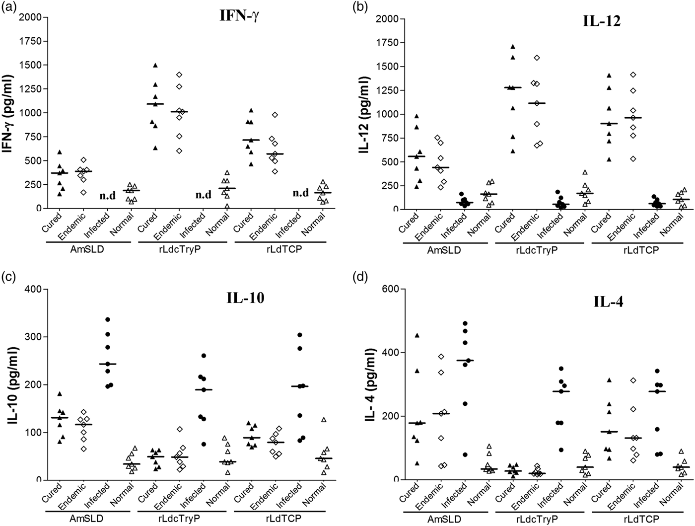
We further assessed the production of Th1/Th2 cytokine levels in PBMCs from cured patients in response to both test antigens (rLdcTryP and rLdTCP20). Individual donors in each study group were found to elicit different cellular responses. The level of IFN-γ and IL-12 cytokines, the signatures of Th1 type of immune response, was noticed to be higher in the supernatant from the samples of cured patients stimulated with rLdcTryP. The median level of IFN-γ and IL-12 in cured patients was 1092·47 pg mL−1 (range: 634·00–1499·46) and 1278·92 pg mL−1 (range: 613·92–1709·64), respectively, and in endemic contacts it was 1011·48 pg mL−1 (range: 602·35–1399·62) and 1115·89 pg mL−1 (range: 672·14–1592·68), respectively (Fig. 3a and b). However, the level of IFN-γ response with rLdTCP20 stimulation in cured and endemic contacts was 716·13 pg mL−1 (range: 465·66–1027·88 pg mL−1) and 571·34 pg mL−1 (range: 387·87–979·81 pg mL−1), respectively, whereas IL-12 response was 901·96 pg mL−1 (range: 529·10–1408·39 pg mL−1) in cured patients and 963·92 pg mL−1 (range: 533·92–1414·29 pg mL−1) in endemic contacts.
Cytokine production (a) IFN-γ, (b) IL-12, (c) IL-10 and (d) IL-4 in stimulated PBMCs from normal (n = 7), L. donovani infected (n = 7), cured VL patients (n = 7) and endemic controls (n = 7) in response to rLdcTryP, rLdTCP20 and AmSLD antigens, each data point represents one individual. The X-axis represents the group of individuals and the Y-axis corresponds to the values of respective cytokine concentrations in pg mL−1. The mean concentration of cytokine for each group is indicated by horizontal bars. Cytokine production was tested in triplicate in two independent experiments and the results were comparable (n.d, not detected).

Moreover, no detectable amount of IFN-γ was observed with the PBMCs of the L. donovani-infected patients against rLdcTryP and rLdTCP20 while very low level of IL-12 was detected. Conversely, the median level of IL-10 and IL-4 cytokine against rLdcTryP stimulation was observed quite low in cured PBMCs (49·49 pg mL−1 ranging from 24·10 to 63·30, and 27·41 pg mL−1 ranging from 11·07 to 47·15 pg mL−1, respectively) as well as in endemic contacts (48·56 pg mL−1 ranging from 22·58 to 106·90 and 19·85 pg mL−1 ranging from 16·54 to 43·66, respectively) (Fig. 3c and d). Similarly, the IL-10 and IL-4 level was also downregulated after stimulation with rLdTCP20. In contrast, these cytokines were upregulated in all the infected patients as compared with other groups (Fig. 3c and d).
Vaccination with rLdcTryP induced considerably good prophylactic efficacy in hamsters as compared with rLdcTCP20
The vaccinated hamsters were shown significant reduction in parasitic burden in the spleen, liver and bone marrow (~78%, in cTryP vaccinated; ~55 %, in TCP20 vaccinated) against infection of L. donovani on day 90 p.c (Fig. 4a, b and c). An increase in parasites in infected control and BCG groups was seen in Giemsa-stained splenic smears from days 45 to 90 p.c. In the rLdcTryP- and rLdTCP20-vaccinated group, parasite loads decreased onwards from day 45 to day 90 p.c. and a very low level of parasite load has been observed in rLdcTryP-vaccinated group (P < 0·001) followed by rLdTCP20-vaccinated groups. The hamsters vaccinated with rLdcTryP survived longer after the challenge of L. donovani and remained healthy up to 6–8 months post-infection (p.i.), while hamsters vaccinated with rLdTCP20 only survived up to 3–4 months p.i. On the other hand, there was progressive increase in parasite load in hamsters vaccinated with BCG (alone) and in infected controls and they succumbed to virulent L. donovani challenge within 2–3 months.
Prophylactic efficacy of rLdcTryP and rLdTCP20 in hamsters against L. donovani challenges: parasite burden (number of amastigotes/1000 macrophage) in the dab smears of (a) the spleen, (b) liver and (c) the bone marrow of hamsters (n = 3–4 animals from each group) on days 45, 60, 90, 120 and 180 p.c. Since the non-vaccinated challenged (infected control), the BCG vaccinated and challenged group died after day 90 (D 90) of the study period, their corresponding bars are not shown in a, b and c. Significance values indicate the difference between the rLdcTryP and rLdTCP20 vaccinated groups and infected group (*, P < 0·05; **, P < 0·01; and ***, P < 0·001).

Vaccination with rLdcTryP stimulates significant DTH, mitogenic and Leishmania-specific cellular responses in hamsters against L. donovani challenges
The cellular immune responses were generated following vaccination with rLdcTryP and rLdTCP20, and were characterized in hamsters. Though, DTH, an index of cell-mediated immunity (CMI) in vivo, and T cell stimulation with antigen in vitro is often related to successful vaccination. In the study we observed that the hamster's vaccinated with rLdcTryP + BCG elicited significantly stronger DTH response as compared with other control groups. However, the hamsters vaccinated with rLdTCP20+BCG displayed inferior response as compared with rLdcTryP. DTH response progressively increased after days 60–90 p.c. in hamsters vaccinated with rLdcTryP (P < 0·001) (Fig. 5a). The proliferative response of lymphocytes against ConA stimulation in rLdcTryP + BCG-vaccinated hamsters was observed to be higher throughout the entire p.c. period (Fig. 5b). Similarly the normal hamsters were shown to have much similar LTT response whereas it was lower in other control groups. In antigen-specific re-stimulation assays, rLdcTryP + BCG-vaccinated hamsters showed significant (P < 0·001) cellular immune response which was observed maximum on day 90 p.c. (Fig. 5c). On the other hand, lesser proliferative response was observed in hamsters vaccinated with rLdTCP20 + BCG (P < 0·01, Fig. 5c).
Prophylactic efficacy of rLdcTryP and rLdTCP20 in hamsters and challenged with L. donovani. DTH response (mm) to AmSLD as footpad swelling on days 45, 60 and 90 p.c (a); LTT response (mean O.D values) to ConA (b) and rLdcTryP and rLdTCP20 (c) stimulation-vaccinated hamsters, uninfected normal control, infected control, BCG-vaccinated hamsters (n = 3–4 from each groups) on days 45, 60 and 90 p.c.; NO production (μ m) to LPS (d), and rLdcTryP and rLdTCP20 (e), in the naive peritoneal macrophages co-incubated with supernatants of mononuclear cells isolated from vaccinated hamsters, uninfected normal control, infected control and BCG vaccinated hamsters (n = 3–4 from each groups) on days 45, 60 and 90 p.c. Significance values indicate the difference between the vaccinated groups and infected group (*, P < 0·05; **, P < 0·01; and ***, P < 0·001).

Therefore, NO is an important factor for controlling for leishmaniasis, macrophages isolated from naïve hamsters, when incubated with stimulated supernatants of mononuclear cells from rLdcTryP + BCG-vaccinated hamsters, induced significant (P < 0·001) amount of NO (24·27 ± 1·549 μ m) which was ~3-fold more than that of unvaccinated infected controls (7·82 ± 0·678) on day 90 p.c. (Fig. 5e). On the other hand, hamsters vaccinated with rLdTCP20 + BCG induced low levels of NO production on day 90 p.c. (16·15 ± 0·53 μ m, P < 0·01) (Fig. 5e).
Serological response (IgG1 and IgG2) in vaccinated hamsters
We measured the levels of IgG1 and IgG2 isotypes in the sera of control and vaccinated animals on day 45, 60 and 90 p.c. The hamsters vaccinated with rLdcTryP elicited higher levels of IgG2 antibody on days 60 and 90 p.c. as compared with other control groups (P < 0·001) (Fig. 6b). Moreover, the hamsters vaccinated with rLdcTryP elicited a lower ratio of IgG1/IgG2 (Fig. 6c). The hamsters vaccinated with rLdTCP20 elicited significant levels of IgG2 response (P < 0·01, day 60; P < 0·05, day 90) but it was inferior to rLdcTryP vaccination. In contrast, IgG1 levels were progressively decreased in the vaccinated group and increased in all the control groups (Fig. 6a). As a measure of CMI, the elevated level of IgG2 was consistent with the development of effective immune responses.
Ratio of IgG1:IgG2 antibody isotype in LdcTryP- and LdTCP20-vaccinated hamsters (n = 3–4 from each group): (a) IgG1, (b) IgG2 and (c) ratio of IgG1/IgG2 in rLdcTryP- and rLdTCP20-vaccinated hamsters in comparison to the unvaccinated infected control hamsters on days 45, 60 and 90 p.c. Significance values indicate the difference between the vaccinated group and unvaccinated infected group (*, P < 0·05; **, P < 0·01; and ***, P < 0·001).

rLdcTryP-vaccinated hamsters elicited prominent Th1-type cytokine responses, as evaluated by qRT-PCR
The expression level of Th1 and Th2 cytokines (IFN-γ, IL-12, TGF-β, IL-4, IL-10) and iNOS was quantified in hamsters vaccinated either with rLdcTryP + BCG or rLdTCP20 + BCG on days 45 and 90 p.c. and was compared with infected control groups. An elevated expression level of Th1 type of cytokines was observed on days 45 and 90 p.c. wherein, a significantly (P < 0·001) high level of IL-12 (~3·5-fold) was observed in rLdcTryP + BCG-vaccinated group on day 90 p.c. as compared with the infected control group whereas a moderate increase in expression levels of IL-12 mRNA transcripts was observed by hamsters vaccinated with rLdTCP20 + BCG on day 90 p.c. (P < 0·01) (Fig. 7b). In addition, a moderate expression level (~3-fold) of IFN-γ was noticed on day 45 p.c. (P < 0.01) in hamsters vaccinated with rLdcTryP + BCG which further increased up to ~4·7-fold (P < 0·001) on day 90 p.c. (Fig. 7c). In contrast the expression of IFN-γ in hamsters vaccinated with rLdTCP20 + BCG was lesser as compared with the rLdcTryP + BCG-vaccinated hamsters and significant only at day 90 (P < 0·05) (Fig. 7c). Moreover, the expression of iNOS transcripts was found upregulated (~4·2-fold) significantly (P < 0·001) on day 90 p.c. in hamsters vaccinated with rLdcTryP + BCG while the hamsters vaccinated with rLdTCP20 + BCG generated a lower response (Fig. 7a).
Analysis of mRNA expression profile of normal, infected control, BCG control and LdcTryP/LdTCP20-vaccinated hamsters (n = 3–4 from each groups) by qRT-PCR: Splenic inducible nitric oxide synthase (iNOS) (a) and cytokines, IL-12 (b), IFN-γ (c), IL-10 (d), IL-4 (e) and TGF-β (f); mRNA expression profile was assessed by qRT-PCR in all the experimental groups of hamsters on days 45 and 90 p.c. Data are presented as means ± s.d. and are representative of two independent experiments with similar results. Significance values indicate the difference between the vaccinated group and unvaccinated infected control group (*, P < 0·05; **, P < 0·01; and ***, P < 0·001).

Conversely, the expression of Th2-type cytokines (IL-10, TGF-β and IL-4) was downregulated in rLdcTryP- and rLdTCP20-vaccinated hamsters and upregulated in infected and BCG controls. The level of IL-10 was downregulated in both the vaccinated groups on day 90 (P < 0·001 in rLdcTryP + BCG-vaccinated group and P < 0·05 in rLdTCP20 + BCG vaccinated) (Fig. 7d). Similarly, the level of IL-4 m-RNA transcript was also downregulated on day 90 p.c. in both the vaccinated groups. Further, a maximum downregulation (3–4-fold, P < 0·001), in the expression levels of TGF-β, a Th1-deactivating cytokine, was observed in rLdcTryP + BCG-vaccinated groups as compared with the infected ones (Fig. 7f). While, rLdTCP20 + BCG-vaccinated groups have shown lesser level of downregulation of TGF-β transcript on day 90 p.c. (P < 0·01) (Fig. 7f). The upregulation of Th2 cytokines in BCG as well as infected controls are indicative of progressive VL while these cytokines were highly downregulated in vaccinated hamsters defining the situation in cured VL.
DISCUSSION
Immunoproteomic approaches were utilized for the identification of different antigenic Leishmania proteins in both the stages of L. infantum and L. donovani parasites (Vieira et al. Reference Vieira, Goldschmidt, Nashleanas, Pfeffer, Mak and Scott1996; Coelho et al. Reference Coelho, Oliveira, Valadares, Chavez-Fumagalli, Duarte, Lage, Soto, Santoro, Tavares, Fernandes and Coelho2012) to be targeted either for vaccine or for diagnostic purposes. In VL, most of the amastigote proteins were identified from amastigote-like stage but not from true splenic amastigotes. Recently, antigenic fractions from splenic amastigotes of L. donovani ranging from 97 to 68 kDa (F2) and 68 to 43 kDa (F3) were identified to be immunogenic as well as protective against L. donovani challenge in hamsters. The F2 fraction when subjected to proteomic characterization revealed the presence of several immunogenic proteins, which includes cTryP and chaperoninTCP20 (TCP20) apart from numerous hypothetical proteins (Ms communicated).
cTryP is mainly involved in the detoxification of peroxides and is also associated with survival and virulence of L. donovani parasites (Iyer et al. Reference Iyer, Kaprakkaden, Choudhary and Shaha2008). In addition, its role in metastasis in Leishmania guyanensis (Walker et al. Reference Walker, Acestor, Gongora, Quadroni, Segura, Fasel and Saravia2006) and in arsenite-resistant Leishmania amazonensis (Lin et al. Reference Lin, Hsu, Chiang and Lee2005) has been well documented. Apart from these, this protein has been also studied as a vaccine candidate in case of L. major (Stober et al. Reference Stober, Lange, Roberts, Alcami and Blackwell2005, Reference Stober, Lange, Roberts, Alcami and Blackwell2007), L. (vianna) panamensis (Jayakumar et al. Reference Jayakumar, Castilho, Park, Goldsmith-Pestana, Blackwell and McMahon-Pratt2011) (cutaneous forms) as well as L. infantum (Carson et al. Reference Carson, Antoniou, Ruiz-Arguello, Alcami, Christodoulou, Messaritakis, Blackwell and Courtenay2009) and L. infantum chagasi (Stober et al. Reference Stober, Jeronimo, Pontes, Miller and Blackwell2012) (zoonotic visceral forms). Recently, Singh et al. (Reference Singh, Stober, Singh, Blackwell and Sundar2012) studied some novel Leishmania antigens including cTryP on cured VL patient samples and inferred that these antigens can be taken forward as prophylactics against human leishmaniasis. The other protein – chaperonin TCP20 – is one of the key chaperonins essential in the folding of proteins to provide stability as well as protein conformations (Sternlicht et al. Reference Sternlicht, Farr, Sternlicht, Driscoll, Willison and Yaffe1993; Li et al. Reference Li, Lin, Frydman, Boal, Cardillo, Richard, Toth, Lichtman, Hartl, Sherman and Segel1994). Though, this protein has been identified as Th1 stimulatory for the first time in Leishmania amastigotes in our studies, this has also not been characterized in any of the protozoan parasite as yet. Therefore, in the present study we have carried out molecular and immunological characterization of both the proteins and assessed their prophylactic potentials against L. donovani infection.
Both the proteins – LdcTryP and LdTCP20 – were successfully cloned, overexpressed and purified. The immunolocalization studies carried out in purified splenic amastigotes revealed abundance of these proteins in the cytosolic region. Further, immunoblot study of L. donovani promastigote and amastigote lysate with anti-rLdcTryP and anti-LdTCP20 antibody revealed its presence in both the forms of Leishmania parasites. Since, cTryP was identified in a higher molecular weight range of 97 to 68 kDa in proteomics study as compared with its observed molecular mass could be attributed to the post-translational modifications that are widely prevalent in Leishmania (Gupta et al. Reference Gupta, Sisodia, Sinha, Hajela, Naik, Shasany and Dube2007; Kumari et al. Reference Kumari, Kumar, Samant, Sundar, Singh and Dube2008a , Reference Kumari, Samant, Khare, Sundar, Sinha and Dube b ; Kulkarni et al. Reference Kulkarni, Olson, Engman and McGwire2009).
It is well known that the development of cell-mediated immune response is a major factor responsible for healing of leishmaniasis, which includes Leishmania-specific lymphoproliferation and the stimulation of T-cells to produce various cytokines, including IFN-γ that contributes to killing of intracellular parasites. The earlier studies have documented that stimulation with Soluble L. donovani (SLD) as well as its sub-fractions, led to the generation of significant T-cell responses in cured hamsters as well as cured VL patients (Garg et al. Reference Garg, Gupta, Tripathi, Naik, Sundar and Dube2005; Kumari et al. Reference Kumari, Kumar, Samant, Sundar, Singh and Dube2008a , Reference Kumari, Samant, Khare, Sundar, Sinha and Dube b ). Further, it is also very well known that recovery from Leishmaniasis, mainly depends on the induction of dominant Th1-type of immune responses, which is characterized by enhanced induction of IFN-γ, IL-12 and increased expression of NO synthase (Liew, Reference Liew1991; MacMicking et al. Reference MacMicking, Xie and Nathan1997). Therefore, in the study we tested the immunogenicity of the recombinant proteins in vitro first in cured hamsters and then further validated in Leishmania patients, who were cured with amphotericin B. In the absence of commercially available cytokine reagents against hamsters, we studied the effect of rLdcTryP and rLdTCP20 on lymphocyte proliferation and nitrite production by peritoneal macrophages of hamsters. Therefore, when assessed for their immunogenicity in vitro, rLdcTryP exhibited superior cellular responses than rLdTCP20 when compared with AmSLD in cured hamster lymphocytes. Moreover, significant induction of NO either with rLdcTryP or rLdTCP20 supports the upregulation of iNOS by Th1-cell-associated cytokines. We further validated these findings on patient samples and made similar observations (proliferation of lymphocytes and NO production).
In addition, the cytokine responses in the patient samples were characterized following stimulation with both the recombinant proteins and we observed a significant upregulation of IFN-γ and IL-12 in response to rLdcTryP in endemic control and cured patients of VL. However, the cytokine response against the stimulation of rLdTCP20 was inferior to that of rLdcTryP. Further, since the protective immunity in L. donovani mainly depends on IL-12-driven Th1-type immune response and IFN-γ production (Murray et al. Reference Murray, Montelibano, Peterson and Sypek2000) and that IFN-γ-mediated killing of intracellular parasites occurs by activating the macrophages through the production of several mediators, principal among which is NO (Liew et al. Reference Liew, Li, Moss, Parkinson, Rogers and Moncada1991; Assreuy et al. Reference Assreuy, Cunha, Epperlein, Noronha-Dutra, O'Donnell, Liew and Moncada1994), the observation that rLdcTryP stimulated T-cells from cured/endemic individuals to proliferate and produce IFN-γ suggests that PBMCs response to rLdcTryP may be associated with protective immunity.
Further, prophylactic efficacy of rLdcTryP/rLdTCP20 was evaluated along with BCG in naïve hamsters against L. donovani challenges. The adjuvant property of BCG is well known (Khalil et al. Reference Khalil, El Hassan, Zijlstra, Mukhtar, Ghalib, Musa, Ibrahim, Kamil, Elsheikh, Babiker and Modabber2000; Misra et al. Reference Misra, Dube, Srivastava, Sharma, Srivastava, Katiyar and Naik2001; Armijos et al. Reference Armijos, Weigel, Calvopina, Hidalgo, Cevallos and Correa2004). Moreover, it has also been reported that BCG activates macrophages with induced NO release (MacMicking et al. Reference MacMicking, Xie and Nathan1997; Nozaki et al. Reference Nozaki, Hasegawa, Ichiyama, Nakashima and Shimokata1997) and elicits long-lasting cellular and humoral immune responses (Warren et al. Reference Warren, Vogel and Chedid1986). Interestingly, in the present study 3 months after the Leishmania challenge, significant reduction in parasite load (~78%, P < 0·001) was observed in rLdcTryP-vaccinated hamsters, which survived and remained healthy till the termination of the experiment on day 180 p.c., whereas rLdTCP20 provided lower prophylactic efficacy (~55%, P < 0·001) and survived up to 3–4 months only. Conversely, unvaccinated infected hamsters, succumbed to the lethal L. donovani challenge within 2–3 months p.c. Since, T-cell proliferation is impaired during active VL (Liew et al. Reference Liew, Millott, Parkinson, Palmer and Moncada1990a ) and the control of parasite growth and healing in VL accompanies with the development of strong cell-mediated immune response, such as T-cell proliferation, NO production and DTH responses (Melby et al. Reference Melby, Chandrasekar, Zhao and Coe2001; Afrin et al. Reference Afrin, Rajesh, Anam, Gopinath, Pal and Ali2002; Basu et al. Reference Basu, Bhaumik, Basu, Naskar, De and Roy2005), we further checked for T-cell proliferation in lymphocytes of hamsters vaccinated with rLdcTryP and rLdTCP20. There was significant LTT response in rLdcTryP + BCG-vaccinated hamsters and lesser LTT response in rLdTCP20+BCG-vaccinated hamsters was observed on day 90 p.c. as compared with unvaccinated infected control groups. Similarly, significant level of NO production was observed in rLdcTryP + BCG-vaccinated hamsters which supports the observation regarding the upregulation of iNOS by Th1-cell-associated cytokines, which, in turn, activates the macrophages to kill intracellular parasites (Armijos et al. Reference Armijos, Weigel, Calvopina, Hidalgo, Cevallos and Correa2004). Further, as earlier reports have shown that successful vaccination of humans and animals is often related to antigen-induced DTH responses in vivo (Howard and Liew, Reference Howard and Liew1984; Melby et al. Reference Melby, Tryon, Chandrasekar and Freeman1998), in the present study, hamsters vaccinated with rLdcTryP and rLdTCP20 have shown significant DTH response as compared with other control groups.
Apart from this, active VL is also associated with the production of high levels of the Leishmania-specific antibody, which is observed before detection of parasite-specific T-cell response (Ghose et al. Reference Ghose, Haldar, Pal, Mishra and Mishra1980). Evidence, as high level of IgG2 is a measure of cell-mediated immune response and it was observed significantly prominent in the vaccinated group in this study, is consistent with the development of effective immune responses (Samant et al. Reference Samant, Gupta, Kumari, Misra, Khare, Kushawaha, Sahasrabuddhe and Dube2009). Conversely, the level of anti-Leishmania IgG1 was elevated progressively in all the groups except the vaccinated one, which however concluded that protection against leishmaniasis is induced by T-cell response (Scott et al. Reference Scott, Pearce, Heath and Sher1987).
It has been well established that the resistance against leishmaniasis is conferred by Th1 cells while susceptibility is conferred by Th2 cells. Therefore, we further assess the expression of mRNA of these cytokines along with iNOS in the splenic cells of vaccinated hamsters through qRT-PCR. Interestingly, the expression of IFN-γ, a signature cytokine of the Th1-type immune response, was noticeably upregulated in vaccinated hamsters, which however along with TNF-α often reported to act in concert to activate iNOS for the production of NO, which is responsible for intracellular parasite killing (Liew et al. Reference Liew, Millott, Parkinson, Palmer and Moncada1990a ). In addition, it is also reported that TNF-α is one of the primary agents to stimulate macrophage to produce NO (Liew et al. Reference Liew, Millott and Schmidt1990b ). Moreover, in the present study, we observed downregulation of IL-12 in infected hamsters; in contrast, it was highly upregulated in vaccinated hamsters supporting the view that Th1 type of protective immunity is mediated by IL-12-dependent INF-γ production (Murray et al. Reference Murray, Montelibano, Peterson and Sypek2000). Conversely, the key Th2 cytokines IL-10 and IL-4, associated with an acute phase of VL were observed to be upregulated gradually in infected tissues (Melby et al. Reference Melby, Tryon, Chandrasekar and Freeman1998). Further, IL-4, IL-10 and TGF-β are major immunosuppressive cytokines reported in experimental and human VL (Melby et al. Reference Melby, Tryon, Chandrasekar and Freeman1998, Reference Melby, Chandrasekar, Zhao and Coe2001). We have also observed downregulation of IL-10, reported to downregulate IL-12 for disease progression (Caldas et al. Reference Caldas, Favali, Aquino, Vinhas, van Weyenbergh, Brodskyn, Costa, Barral-Netto and Barral2005), and IL-4, which is the marker for Th2 response (Sundar et al. Reference Sundar, Reed, Sharma, Mehrotra and Murray1997), in vaccinated hamsters as compared with infected control. TGF-β a notorious Th2 cytokine with diverse functions is known to be expressed at a moderate level even in normal hamsters (Melby et al. Reference Melby, Tryon, Chandrasekar and Freeman1998, Reference Melby, Chandrasekar, Zhao and Coe2001; Basu and Ray, Reference Basu and Ray2005) and it was apparently downregulated in vaccinated hamsters compared with the infected controls throughout the experiment.
In conclusion, both rLdcTryP and rLdTCP20 proteins, identified as Th1-stimulatory proteins in splenic amastigotes, were ascertained to be immunogenic and between the two, the former was found to be more potent. Moreover, our studies on successful vaccination with LdcTryP are consistent with various reports documented earlier against L. major (Stober et al. Reference Stober, Lange, Roberts, Alcami and Blackwell2005, Reference Stober, Lange, Roberts, Alcami and Blackwell2007) as well as L. infantum chagasi infection (Stober et al. Reference Stober, Lange, Roberts, Gilmartin, Francis, Almeida, Peacock, McCann and Blackwell2006, Reference Stober, Jeronimo, Pontes, Miller and Blackwell2012; Carson et al. Reference Carson, Antoniou, Ruiz-Arguello, Alcami, Christodoulou, Messaritakis, Blackwell and Courtenay2009). Further, we have reported for the first time the capability of rLdTCP20, a cytosolic chaperonin, to induce, though, a moderate CMI response in vitro but at the same time offered a considerable level of prophylactic efficacy. Since this protein is an important target, further attempts towards determination of immunodominant regions for designing fusion peptides may be taken up as for designing polyvalent synthetic and recombinant chimeric vaccines. An addition of promiscuous T-cell epitopes derived from the potential Th1-stimulatory proteins of L. donovani including rLdcTryP and rLdTCP20 may be required for effective intervention measures to control the disease in endemic areas.
ACKNOWLEDGEMENT
The authors thank the Director, Central Drug Research Institute (CDRI), India, for providing facilities for the experiments. This Ms has CSIR-CDRI communication No. 8857.
FINANCIAL SUPPORT
This work was supported by a grant from CSIR-CDRI's Supra-Institutional Network Project (SIP-0026).