Introduction
Starch, the major plant-energy-reserve polysaccharide, is one of the major nutrients in both human and animal diets (Gidenne et al., Reference Gidenne, Segura and Lapanouse2005). The α-amylase (α-1,4-glucan-4-glucanhydrolase, EC 3.2.1.1) and α-glucosidase (α-1,4-glucan-4-glucanhydrolase, EC 3.2.1.20) are among the digestive enzymes that degrade starch and related compounds. Many organisms living on a polysaccharide-rich diet depend on the effectiveness of amylases for their survival (Marchaiah and Vakil, Reference Marchaiah and Vakil1984; Strobl et al., Reference Strobl, Gomis-Rüth, Maskoss, Frank, Huber and Glocksuber1997), and several studies demonstrated their importance in carbohydrate metabolism in different organisms ranging from micro-organisms to higher organisms (Franco et al., Reference Franco, Rigden, Melo and Grossi-de-Sá2002; Kato et al., Reference Kato, Shimisu-Ibuka, Mura, Takeuch, Tokue and Arai2007).
In the insect kingdom, amylolytic enzymes are known to play an essential role in carbohydrate metabolism and energy production (Pelegrini et al., Reference Pelegrini, Murad, Grossi-de-Sá, Mello, Romeiro, Noronha, Caldas and Franco2006). Among the digestive enzymes so far studied, α-amylases were investigated in different insects such as the yellow mealworm Tenebrio molitor L. (Buonocore et al., Reference Buonocore, Poerio, Silvano and Tomasi1976), the house cricket Acheta domesticus L. (Teo and Woodring, Reference Teo and Woodring1994), the larger grain borer Prostephanus truncatus (Horn) (Mendiola-Olaya et al., Reference Mendiola-Olaya, Valencia-Jiménez, Valdes-Rodriguez, Delano-Frier and Branco-Labra2000), the apollofalter Parnassius appollo (L.) (Nakonieczny et al., Reference Nakonieczny, Michalczyk and Kedziorski2006), the lesser grain borer Rhyzopertha dominica (Fab.) (Cinco-Moroyoqui et al., Reference Cinco-Moroyoqui, Diaz-Malvaez, Alanis-Villa, Baron-Hoyos, Cardenas-Lopez, Cortez-Rocha and Wong-Corral2008), the long-horned beetle Morimus funereus Mulsant (Dojnov et al., Reference Dojnov, Bozic, Nenadovic, Ivanovic and Vujcic2008) and the American cockroach Periplaneta americana (L.) (Dué et al., Reference Dué, Kouadio, Kouakou, Dabonné, Niamké and Kouamé2008). It has been reported that insects are totally dependent on α-amylases for their survival (Sivakumar et al., Reference Sivakumar, Mohan, Franco and Thayumanavan2006). α-Glucosidase has been studied in the fruit fly Drosophila melanogaster Meigen (Tanamura et al., Reference Tanamura, Kitamura, Fukuda and Kikuchi1979), the European honey bee Apis mellifera L. (Takewaki et al., Reference Takewaki, Chiba, Kimura, Matsui and Koike1980; Nishimoto et al., Reference Nishimoto, Kubota, Tsuji, Mori, Kimura, Matsui and Chiba2001), P. americana (Kouamé et al., Reference Kouamé, Dué, Niamké, Kouamé and Kamenan2005) and the Asiatic honey bee Apis cerana japonica Fab. (Wongchawalit et al., Reference Wongchawalit, Yamanoto, Nakai, Kim, Sato, Nishimoto, Okuyama, Mori, Saji, Chanchao, Wongsiri, Surarit, Svasti, Chiba and Kimura2006). Studies on biochemical characteristics of the digestive enzymes from insects are useful since such investigations give new opportinuties for development of control protocols for insect pests.
The giant cricket Brachytrupes membranaceus is abundant in West Africa, particularly in forests and savannas (Chiffaud-Mestre and Gillon, Reference Chiffaud-Mestre and Gillon1985). This cricket, which can ingest mainly roots among varied plant materials, is well known to be the most popular edible insect species in West Africa and recognized as a rich source of nutrients (Dunkel, Reference Dunkel1996). To our knowledge, no study on the digestive enzymes of B. membranaceus has been reported.
To provide data for a better understanding of the digestive physiology of this insect, the present work aimed to study the enzymatic profiles and biochemical characteristics of amylase and α-glucosidase activities in its digestive tract.
Materials and methods
Chemicals
The soluble starch p-nitrophenyl-α-d-glucopyranoside (α-pNPG) and bovine serum albumin were purchased from Sigma Aldrich Ltd (St Louis, MO, USA), and silica gel for thin layer chromatography (TLC) was purchased from Merck Inc. (Darmstadt, Germany). All other chemicals and reagents were of analytical grade.
Insects
Specimens of adult B. membranaceus were collected in September 2008 in the savannas of the Lamto Ecological Station (6°13′N/5°1′W) in Côte d'Ivoire. The station is located 170 km north-west of Abidjan on the eastern bank of the Bandama River. All adult crickets had body weights ranging from 5 to 7 g with a medium size between 5 and 6 cm from the head to the abdominal apex. After sampling, the insects were transported to the laboratory. These insects (an equal proportion for each sex) were kept alive in our laboratory without food for 48 h, after which they were stored at − 20 °C until analyses.
Enzyme source preparation
Defrozen crickets that were previously washed in distilled water were dissected and the guts were isolated, then 10 g of guts were ground and homogenized in 100 ml of 0.9% NaCl (w/v) with a microcrusher ULTRA-TURRAX® (type TP 10/18). After centrifugation at 4 °C for 20 min at 5000 rpm, the supernatant was kept at 4 °C and used as crude enzymatic extract.
Amylase and α-glucosidase assays
The amylase assays were performed as described below (Bernfeld, Reference Bernfeld, Colowick and Kaplan1955). Briefly, the enzyme extract (50 μl) was incubated with 0.5% starch solution (80 μl) in 100 mm phosphate buffer at pH 7.0 (170 μl) at 37 °C for 30 min. The amount of reducing sugars produced was determined by the dinitrosalicylic acid method with maltose as the standard. One unit of the enzyme activity (UI) was defined as the amount of enzyme that liberated 1 μmol of maltose equivalent per minute under the above conditions. Specific activity was expressed as units per mg of protein. The activity of α-glucosidase was measured by the release of p-nitrophenol from α-pNPG. The standard reaction mixture containing 75 μl of 5 mm α-pNPG in 125 μl of 100 mm phosphate buffer at pH 7.0 and 50 μl of crude enzymatic extract was incubated at 37 °C for 10 min. The reaction was stopped by the addition of 2 ml of 2% Na2CO3 (w/v). Absorbance of the reaction mixture was measured at 410 nm using a spectrometer. One unit of α-glucosidase activity (UI) was defined as the amount of enzyme liberating 1 μmol of p-nitrophenol per minute under the above conditions. Specific activity was expressed as units per mg of protein.
Amylase and α-glucosidase profiles in the crude enzymatic extract
The profiles of amylase and α-glucosidase activities in the crude extract were evaluated using a zymogram. An aliquot of crude enzymatic extract was loaded onto 10% acrylamide gel prepared according to Laemmli (Reference Laemmli1970) without sodium dodecyl sulphate (SDS). The crude extract aliquot was previously combined with sample buffer without 2-mercaptoethanol and was not heat treated. For amylase, after electrophoresis, the gel was submerged in 50 ml of a 1% soluble starch solution for 1 h. The gel was briefly rinsed in assay buffer to remove excess starch, and activity bands of amylase were developed by adding 50 ml 0.014 m KI–0.01 m I2 solution. For α-glucosidase, after electrophoresis, activity staining was done by immersing the gel in 5 mm α-pNPG containing 100 mm phosphate buffer at pH 7.0 for 1 h, and the activity band was developed by adding 50 ml of 2% Na2CO3.
Protein concentration
The concentration of protein in the crude enzymatic extract was measured according to the Lowry method using bovine serum albumin as standard (Lowry et al., Reference Lowry, Rosebrough, Farr and Randall1951).
pH and temperature optima
Effect of pH on the amylase and α-glucosidase activities was determined by measuring the hydrolysis of soluble starch and α-pNPG, respectively, in a series of phosphate buffers (100 mm) at various pH values ranging from 5.6 to 8.0. Different enzyme activities were measured at 37 °C under the standard test conditions as described above. The effect of temperature on the amylase and α-glucosidase activities was followed in 100 mm phosphate buffer at pH 7.0 and at temperatures ranging from 30 to 80 °C using 0.5% starch (w/v) and 5 mm α-pNPG, respectively, under the standard test conditions.
Stabilities of enzyme activities at pH and temperature
The stability of amylase and α-glucosidase activities of the enzyme extracts was followed at pH 5.6–8.0 in 100 mm phosphate buffer. After 2 h of incubation at 37 °C, the aliquots were taken and immediately assayed for residual amylase and α-glucosidase activities. The thermal denaturation of the amylase and α-glucosidase activities was analysed at various temperatures (30–80 °C) for 5 min. The thermal inactivation of the two activities was analysed with the enzyme extracts preincubated at 37 and 55 °C (amylase) or 40 °C (α-glucosidase) for 3 h. Aliquots were withdrawn at regular intervals of 10 min and immediately cooled in ice-cold water. Residual activities, determined in both cases at 37 °C under the standard test conditions, are expressed as percentage activity of zero-time control of untreated enzyme.
Effects of some chemical agents
The effects of various compounds (ions, ethylenediaminetetraacetic acid (EDTA), dithionitrobenzoic acid (DTNB) and 4-chloromercuribenzoic acid (pCMB)) as possible activators or inhibitors of the enzyme activities were studied. The enzyme extracts were preincubated at 37 °C for 30 min with each compound, and then the activity was assayed. The substrate (0.5% starch or 5 mm α-pNPG) was added to the medium and incubated at 37 °C for 30 min (amylase) or 10 min (α-glucosidase). The residual activities were assayed under the standard conditions.
TLC analysis of starch hydrolysis by amylolytic enzymes from the digestive tract of B. membranaceus
Hydrolysis of soluble starch by amylolytic enzymes from the digestive tract of B. membranaceus was tested with 50 μl of crude enzymatic extract mixed in 170 ml of 100 mm phosphate buffer at pH 6.6 and 80 μl of 0.5% starch at 37 °C for 1 and 2 h. At each time, the aliquots (5 μl) were removed and spotted onto a TLC plate. Hydrolysed products were separated using butanol–acetic acid–water (9:3.75:2.25, v/v/v) and developed using naphthoresorcinol in ethanol and H2SO4 20% (v/v). The sugar spots were visualized keeping the plate at 110 °C for 5 min.
Results
Specific activities and profiles of amylase and α-glucosidase in the crude enzymatic extract
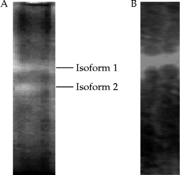
The values of amylase- and α-glucosidase-specific activities measured in the digestive tract of the cricket B. membranaceus were 3.31 and 13.02 UI/mg, respectively. The profiles of amylase and α-glucosidase activities in crude enzymatic extract evaluated using zymogram showed two activity bands corresponding to two isoforms for amylase and one activity band for α-glucosidase (Fig. 1).
Amylase and α-glucosidase profiles of the crude enzymatic extract from the digestive tract of Brachytrupes membranaceus obtained using zymogram analysis after electrophoresis without SDS and 2-mercaptoethanol. Sample was loaded onto 10% gels. A, amylase profile; B, α-glucosidase profile.

Effect of pH and temperature on amylase and α-glucosidase activities and stabilities
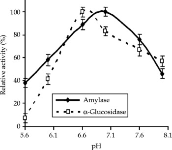
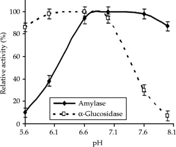
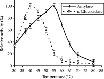
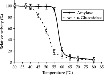
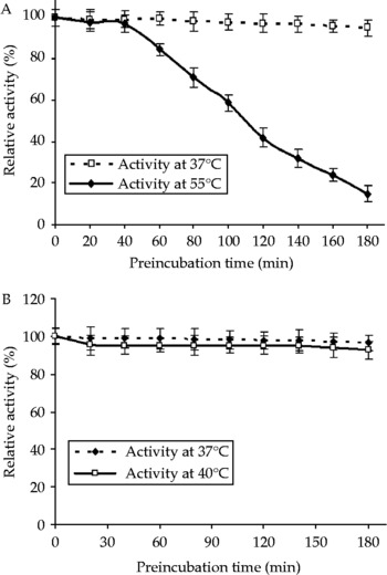
The findings of the optimal conditions (pH and temperature) for the digestive enzyme activities are presented in Figs 2 and 3. The activity of α-amylase displayed optimum pH at 6.6 (Fig. 2) and was stable at pH 6.6–7.6 (Fig. 3). For α-glucosidase activity, the optimum pH was 7.0 (Fig. 2) and the enzyme was stable at pH 6.0–7.0 (Fig. 3). With regard to temperature, optimum temperature and enzyme stabilities are presented in Figs 4–6. The α-amylase exhibited optimum activity at 55 °C (Fig. 4) and was fairly stable at temperatures up to 55 °C (Fig. 5). Above 55 °C, its activity declined rapidly as the temperature increased. α-Glucosidase activity had an optimum temperature of 40 °C (Fig. 4), and the enzyme lost more than 80% of its original activity at 55 °C (Fig. 5). The thermal inactivation studies indicated that at their optimum pH, both crude enzymes remained fully stable during 180 min at 37 °C (Fig. 6A and B). At its optimum temperature (55 °C), the crude amylase remained 84% at 60 min and lost 85% of its original activity, respectively, after 180 min (Fig. 6A). Crude α-glucosidase retained 96% of its original activity at its optimum temperature (40 °C) after 180 min (Fig. 6B).
Effect of pH on amylase and α-glucosidase activities from the digestive tract of Brachytrupes membranaceus. The experiments were carried out at 37 °C with soluble starch (amylase) and p-nitrophenyl-α-d-glucopyranoside (α-glucosidase) as substrates in a series of phosphate buffer ranging from pH 5.6 to 8.0. The enzyme activities were measured at 37 °C under the standard test conditions. Values are the average ( ± SD) of three independent determinations.

Effect of pH on amylase and α-glucosidase stabilities from the digestive tract of Brachytrupes membranaceus. The stability of each enzyme activity was followed at pH ranging from 5.6 to 8.0 in 100 mm phosphate buffer. After 2 h of incubation at 37 °C, aliquots were taken and immediately assayed for residual amylase and α-glucosidase activities. Values are the average ( ± SD) of three independent determinations.

Effect of temperature on amylase and α-glucosidase activities from the digestive tract of Brachytrupes membranaceus. The effect of temperature on each enyme activity was followed in 100 mm phosphate buffer at pH 7.0 at temperatures ranging from 30 to 80 °C using soluble starch (amylase) and α-p-nitrophenyl-α-d-glucopyranoside (α-glucosidase) as subtrates under the standard test conditions. Values are the average ( ± SD) of three independent determinations.

Thermal denaturation of amylase and α-glucosidase activities from the digestive tract of Brachytrupes membranaceus. The enzyme extracts were preincubated at each temperature for 5 min. The residual activity of each enzyme was measured at 37 °C under the standard test conditions. Values are the average ( ± SD) of three independent determinations.

Thermal inactivation of amylase and α-glucosidase activities from the digestive tract of Brachytrupes membranaceus. A, amylase activity; B, α-glucosidase activity. The thermal inactivation of each enzyme activity was analysed with enzyme extracts that were preincubated at 37 and 55 °C (amylase) or 40 °C (α-glucosidase) for 3 h. Aliquots were withdrawn at regular intervals of 10 min and immediately cooled in ice-cold water. Residual activities determined at 37 °C under the standard test conditions are expressed as percentage activity of zero-time control of untreated enzyme. Values are the average ( ± SD) of three independent determinations.

Effects of chemical agents
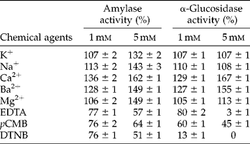
Results on the effects of chemical agents are presented in Table 1. The activities of the enzymes were activated by 1 and 5 mm of Ca2+ and Ba2+. At 1 mm concentration, K+, Na+ and Mg2+ had no significant effects on the activities of both enzymes, whereas at 5 mm, α-amylase activity was activated by these ions. However, at 5 mm, the same ions had no significant effect on α-glucosidase activity. Both enzyme activities that were studied were inhibited by EDTA, DTNB and pCMB.
Effect of some chemical agents on amylase and α-glucosidase activities of enzyme extracts from the digestive tract of Brachytrupes membranaceus+
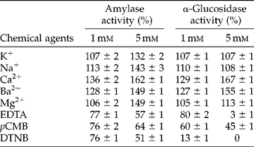
The relative activity is expressed as a percentage of the control without the chemical agent. Values are the average ( ± SD) of three independent determinations. The used ions are under chloride form.
Products formed by amylolytic enzymes from the digestive tract of B. membranaceus from soluble starch
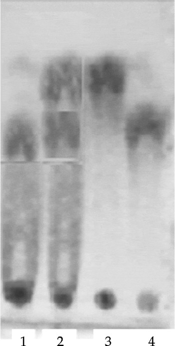
The action of amylolytic enzymes in the crude enzymatic extract on soluble starch was examined by TLC analysis (Fig. 7). After 1 h, maltose and a small amount of glucose were the products of hydrolysis. After 2 h, the amount of glucose increased, and the essential end products were maltose and glucose.
TLC analysis of the products of hydrolysis obtained from 0.5% soluble starch by crude enzymatic extract from the digestive tract of Brachytrupes membranaceus. These experiments were carried out at 37 °C for 1 and 2 h using 100 mm phosphate buffer at pH 6.6. At each time, aliquots (5 μl) were removed and spotted onto a TLC plate. Hydrolysed products were separated using butanol–acetic acid–water (9:3.75:2.25, v/v/v) and developed using naphthoresorcinol in ethanol and H2SO4 20% (v/v). The sugar spots were visualized keeping the plate at 110 °C for 5 min. Lane 1: hydrolytic products after 1 h; lane 2: hydrolytic products after 2 h; lane 3: glucose; lane 4: maltose.

Discussion
This study shows that crude enzymatic extracts obtained from the digestive tract of B. membranaceus contain amylase and α-glucosidase activities. These results indicate that amylase and α-glucosidase play a fundamental role in energy production of this insect. According to our findings, the activity of amylase measured in B. membranaceus was lower compared with both the values of 24 and 9.5 UI/mg obtained, respectively, for R. dominica and M. funereus (Cinco-Moroyoqui et al., Reference Cinco-Moroyoqui, Diaz-Malvaez, Alanis-Villa, Baron-Hoyos, Cardenas-Lopez, Cortez-Rocha and Wong-Corral2008; Dojnov et al., Reference Dojnov, Bozic, Nenadovic, Ivanovic and Vujcic2008). However, amylase activity in the giant cricket was higher than those investigated in P. truncatus (2.6 UI/mg) (Mendiola-Olaya et al., Reference Mendiola-Olaya, Valencia-Jiménez, Valdes-Rodriguez, Delano-Frier and Branco-Labra2000) and P. americana (1.89 UI/mg) (Dué et al., Reference Dué, Kouadio, Kouakou, Dabonné, Niamké and Kouamé2008). For α-glucosidase activity, the value obtained from the giant cricket was higher than the activity of the same enzyme measured in P. americana (0.57 UI/mg) (Kouamé et al., Reference Kouamé, Dué, Niamké, Kouamé and Kamenan2005) and A. cerana japonica (0.13 UI/mg) (Wongchawalit et al., Reference Wongchawalit, Yamanoto, Nakai, Kim, Sato, Nishimoto, Okuyama, Mori, Saji, Chanchao, Wongsiri, Surarit, Svasti, Chiba and Kimura2006). It is important to note that the variations of enzyme activities reported in the literature largely depend on the assay procedure and tissue on which the enzymatic analyses were done. However, several authors argue for genetic, species or physiological considerations to explain insect digestive physiology (Mandal et al., Reference Mandal, Choudhuri and Choudhuri1983; Nijhout, Reference Nijhout2003; Kunieda et al., Reference Kunieda, Fujiyuki, Kucharski, Foret, Ament, Toth, Ohashi, Takeuchi, Kamikouchi, Kage, Morioka, Beye, Kubo, Robinson and Maleszka2006). The zymogram analysis showed the presence of two isoforms of amylase in the digestive tract of B. membranaceus. The existence of multiple isoforms of α-amylase has been described for other insects. Two major isoforms of α-amylase have been reported in P. truncatus (Mendiola-Olaya et al., Reference Mendiola-Olaya, Valencia-Jiménez, Valdes-Rodriguez, Delano-Frier and Branco-Labra2000) and the bruchid Zabrotes subfasciatus (Bohemann) (Lemos et al., Reference Lemos, Campos, Silva and Xavier-Filho1990), while several isoforms of α-amylase have been identified in the cowpea bruchid Callosobruchus maculatus (Fab.) (Campos et al., Reference Campos, Xavier-Filho, Silva and Ary1989), R. dominica (Baker, Reference Baker1991; Cinco-Moroyoqui et al., Reference Cinco-Moroyoqui, Diaz-Malvaez, Alanis-Villa, Baron-Hoyos, Cardenas-Lopez, Cortez-Rocha and Wong-Corral2008) and M. funerus (Dojnov et al., Reference Dojnov, Bozic, Nenadovic, Ivanovic and Vujcic2008). According to Dojnov et al. (Reference Dojnov, Bozic, Nenadovic, Ivanovic and Vujcic2008), this existence of multiple forms of α-amylase in insects could be explained by the fact that different insects use a sophisticated system involving these isoforms to hydrolyse starch to get round the inhibition by α-amylase natural inhibitors. Furthermore, many insect species depend to a large extent on the effectiveness of their amylases for survival (Buonocore et al., Reference Buonocore, Poerio, Silvano and Tomasi1976; Mendiola-Olaya et al., Reference Mendiola-Olaya, Valencia-Jiménez, Valdes-Rodriguez, Delano-Frier and Branco-Labra2000; Sivakumar et al., Reference Sivakumar, Mohan, Franco and Thayumanavan2006). On the other hand, the digestive tract of B. membranaceus contains one form of α-glucosidase, while those of D. melanogaster and A. cerana japonica each contain three isoforms (Tanamura et al., Reference Tanamura, Kitamura, Fukuda and Kikuchi1979; Wongchawalit et al., Reference Wongchawalit, Yamanoto, Nakai, Kim, Sato, Nishimoto, Okuyama, Mori, Saji, Chanchao, Wongsiri, Surarit, Svasti, Chiba and Kimura2006).
Amylase from the digestive tract of B. membranaceus was more active in the pH ranges (6.6–7.6) with optimum pH of 6.6. This optimum pH value was similar to that obtained by Cinco-Moroyoqui et al. (Reference Cinco-Moroyoqui, Diaz-Malvaez, Alanis-Villa, Baron-Hoyos, Cardenas-Lopez, Cortez-Rocha and Wong-Corral2008) for the three isoamylases from R. dominica that showed optimum pH between 6.5 and 7.0. However, the optimum pH value was different for the amylases from T. molitor larvae (pH 5.8) (Buonocore et al., Reference Buonocore, Poerio, Silvano and Tomasi1976), P. truncatus (pH 6.0) (Mendiola-Olaya et al., Reference Mendiola-Olaya, Valencia-Jiménez, Valdes-Rodriguez, Delano-Frier and Branco-Labra2000), M. funereus larvae (pH 5.2) (Dojnov et al., Reference Dojnov, Bozic, Nenadovic, Ivanovic and Vujcic2008) and P. americana (pH 5.6) (Dué et al., Reference Dué, Kouadio, Kouakou, Dabonné, Niamké and Kouamé2008). α-Glucosidase showed maximum activity in the pH range 6.0–7.0 with optimum pH of 7.0. These results were different from the values of optimum pH of α-glucosidase studied in P. americana (pH 5.0) (Kouamé et al., Reference Kouamé, Dué, Niamké, Kouamé and Kamenan2005) and A. cerana japonica (pH 5.0) (Wongchawalit et al., Reference Wongchawalit, Yamanoto, Nakai, Kim, Sato, Nishimoto, Okuyama, Mori, Saji, Chanchao, Wongsiri, Surarit, Svasti, Chiba and Kimura2006). The optimum value of pH of digestive α-glucosidase in the Sunn pest Eurygaster integriceps Puton varied from 4 to 6 (Mehrabadi et al., Reference Mehrabadi, Bandani, Saadati and Sultan2009). According to the findings reported in the literature, amylases of certain insects showed high activities at alkaline pH, while others are well adapted to acidic physiological environment (Applebaum and Konijn, Reference Applebaum and Konijn1965; Podoler and Applebaum, Reference Podoler and Applebaum1971; Buonocore et al., Reference Buonocore, Poerio, Silvano and Tomasi1976; Sivakumar et al., Reference Sivakumar, Mohan, Franco and Thayumanavan2006). This is true for insects like T. molitor, the larva of which contains a single acidic α-amylase with an optimum pH of 5.8 for the hydrolysis of starch (Strobl et al., Reference Strobl, Gomis-Rüth, Maskoss, Frank, Huber and Glocksuber1997). The enzyme of this larva is accordingly well adapted to its physiological environment in its midgut, where a slightly acid pH is prevalent. The optimum temperature of amylase from B. membranaceus was 55 °C, similar to that in P. americana (Dué et al., Reference Dué, Kouadio, Kouakou, Dabonné, Niamké and Kouamé2008). However, it was generally higher than those reported for amylases from insects such as P. truncatus (30 °C) (Mendiola-Olaya et al., Reference Mendiola-Olaya, Valencia-Jiménez, Valdes-Rodriguez, Delano-Frier and Branco-Labra2000), Z. subfasciatus (20–30 °C) (Pelegrini et al., Reference Pelegrini, Murad, Grossi-de-Sá, Mello, Romeiro, Noronha, Caldas and Franco2006) and M. funereus larvae (45 °C) (Dojnov et al., Reference Dojnov, Bozic, Nenadovic, Ivanovic and Vujcic2008). In B. membranaceus, α-glucosidase was optimally active at 40 °C, lower than for P. americana (50 °C) (Kouamé et al., Reference Kouamé, Dué, Niamké, Kouamé and Kamenan2005) and A. cerana japonica (up above 40 °C) (Wongchawalit et al., Reference Wongchawalit, Yamanoto, Nakai, Kim, Sato, Nishimoto, Okuyama, Mori, Saji, Chanchao, Wongsiri, Surarit, Svasti, Chiba and Kimura2006).
At 55 °C, the crude amylase remained at 84% of its original activity after 60 min. This behaviour was similar to the three isoamylases from R. dominica which retained around 85% of their activity after 60 min at 50 °C (Cinco-Moroyoqui et al., Reference Cinco-Moroyoqui, Diaz-Malvaez, Alanis-Villa, Baron-Hoyos, Cardenas-Lopez, Cortez-Rocha and Wong-Corral2008). Moreover, amylase from B. membranaceus was more stable than that reported from P. truncatus, which has high thermal instability above 40 °C (Mendiola-Olaya et al., Reference Mendiola-Olaya, Valencia-Jiménez, Valdes-Rodriguez, Delano-Frier and Branco-Labra2000). Crude α-glucosidase retained 96% of its original activity at 40 °C after 180 min, which is similar in A. melifera (Takewaki et al., Reference Takewaki, Chiba, Kimura, Matsui and Koike1980) and A. cerana japonica (Wongchawalit et al., Reference Wongchawalit, Yamanoto, Nakai, Kim, Sato, Nishimoto, Okuyama, Mori, Saji, Chanchao, Wongsiri, Surarit, Svasti, Chiba and Kimura2006), which were also stable at 40 °C.
According to Terra et al. (Reference Terra, Ferreira, Jordao, Dillon, Lehane and Billingsley1996), Pelegrini et al. (Reference Pelegrini, Murad, Grossi-de-Sá, Mello, Romeiro, Noronha, Caldas and Franco2006) and Dojnov et al. (Reference Dojnov, Bozic, Nenadovic, Ivanovic and Vujcic2008), α-amylases from insects are known to be Ca2+ dependent. This observation could explain the result obtained for Ca2+ which was an activator for B. membranaceus. Dué et al. (Reference Dué, Kouadio, Kouakou, Dabonné, Niamké and Kouamé2008) have also reported the same result with amylase in P. americana. Moreover, the important inhibition of the amylase activity by a chelating reagent such as EDTA and its activation by the metal ion Mg2+ suggest a metalloprotein for this enzyme, as shown for the α-amylase of the large roundworm Ascaris suum (Goeze), a nematode in pigs (Zoltowska, Reference Zoltowska2001). In addition, the important inhibition of both enzymes by sulphydryl reagents as DTNB and pCMB suggests the existence of such essential sulphhydryl groups in the molecules of these enzymes.
The analysis of hydrolytic products after soluble starch hydrolysis by amylolytic enzymes in crude enzymatic extract from the digestive tract of B. membranaceus by TLC revealed that maltose and glucose were the major products. From a physiological point of view, this result confirms that the cricket utilizes starch as one of the main sources of energy. Our findings are similar to those reported for α-glucosidase and α-amylase from P. americana (Kouamé et al., Reference Kouamé, Dué, Niamké, Kouamé and Kamenan2005; Dué et al., Reference Dué, Kouadio, Kouakou, Dabonné, Niamké and Kouamé2008).
Conclusion
Our findings showed that amylase and α-glucosidase activities from the digestive tract of the giant cricket B. membranaceus had many biochemical characteristics similar to those reported for α-amylases and α-glucosidases from other insects. These amylolytic enzymes play a fundamental role in energy production for this insect. The present study also demonstrated that amylolytic enzyme activities from the digestive tract of B. membranaceus can be assayed in optimal conditions with pH 6.6 and 55 °C for α-amylases, and with pH 7.0 and 40 °C for α-glucosidase. To complete the biochemical characteristics of these enzymes, further work on the purification, SDS-polyacrylamide gel electrophoresis analysis and kinetic properties will be necessary for a detailed study of these two digestive enzymes.
Acknowledgements
We are grateful to Professor Mamadou Dagnogo, director of the Laboratoire de Biologie et de Cytologie, UFR des Sciences de la Nature, for advice and contribution on identification of the specimens of B. membranaceus. We are also grateful to students for their help during the insect collection.