Introduction
Cystic echinococcosis is a zoonotic infection caused by the larval form of Echinococcus granulosus sensu lato (s.l.) and is an important public health and veterinary problem in humans and various animal species worldwide. The life cycle of this parasite includes dogs and other carnivorous animals as definitive hosts and livestock such as sheep, cattle, goats, and ungulates such as horses and donkeys as intermediate hosts. However, hydatid cyst infections in ungulates are reported relatively rarely, and epidemiological and molecular data in these animals are limited (Borhani et al. Reference Borhani, Fathi, Darabi, Jalousian, Simsek, Ahmed, Kesik, Hosseini, Romig, Harandi and Mobedi2021).
In the current taxonomy of the genus Echinococcus, five species are reported: E. granulosus sensu lato, E. multilocularis, E. oligarthrus, E. vogeli, and E. shiquicus. The E. granulosus s.l. complex includes E. granulosus sensu stricto (s.s.) (G1/G3), E. equinus (G4), E. ortleppi (G5), E. canadensis (G6/7 and G8-G10), and E. felidis (Vuitton et al. Reference Vuitton, McManus, Rogan, Romig, Gottstein, Naidich, Tuxun, Wen and Menezes da Silva2020).
The prevalence of hydatid cysts in ungulates varies depending on regional conditions, environmental factors, the size of the animal population, and local sanitation conditions. Echinococcosis is particularly prevalent in rural and livestock-raising areas. Equids may become infected, particularly in environments where exposure to infected dogs is possible. Studies show that the prevalence of cysts in these animals can range from 5 to 30%, although there may be regional variations. The prevalence of hydatid cysts in ungulates varies in different geographical regions, and molecular characterisation of the disease is important to accurately identify the type of parasite. The use of molecular techniques helps to identify and manage infections more accurately (Borhani et al. Reference Borhani, Fathi, Darabi, Jalousian, Simsek, Ahmed, Kesik, Hosseini, Romig, Harandi and Mobedi2021; Gessese Reference Gessese2020; Varcasia et al. Reference Varcasia, Garippa, Pipia, Scala, Brianti, Giannetto, Battelli, Poglayen and Micagni2008). Although cases of E. equinus are rare in Türkiye, molecular evidence for the presence of this parasite in various animals has been obtained in recent years. In particular, E. equinus infections were reported in a mule in 2014, a donkey in 2015, and a human in 2021 (Kesik et al. Reference Kesik, Kilinc, Simsek and Gul2019; Macin et al. Reference Macin, Orsten, Samadzade, Colak, Cebeci and Fındık2021; Simsek and Cevik Reference Simsek and Cevik2014; Simsek et al. Reference Simsek, Roinioti and Eroksuz2015). These findings indicate that E. equinus is present in different hosts in Türkiye and carries a potential zoonotic risk.
This study aimed to investigate the presence of hydatid cysts in equine necropsies conducted at Firat University, perform gross and histopathological examinations, conduct molecular characterization of the detected cysts using PCR amplification and sequencing, and carry out phylogenetic analyses to determine genetic variations and evolutionary relationships.
Materials and methods
A retrospective analysis of 52 equine necropsies conducted at the Firat University, Faculty of Veterinary Medicine, Pathology Department, between 2020 and 2025 was performed to evaluate the presence of hydatid cysts. Germinal membranes of individual hydatid cysts identified during this period were excised and preserved in 70% ethanol for molecular analysis. Three cases – one Arabian horse (21-year-old) and two donkeys (22-year-old and 23-year-old) – exhibited gross and microscopic features consistent with hydatid cysts. Tissue samples of liver and lung were routinely processed.
Genomic DNA isolation
For molecular analysis, small tissue fragments from the germinal membrane of each hydatid cyst were washed with 1X PBS (pH 7.4) to eliminate residual ethanol. Genomic DNA (gDNA) was isolated from individual cyst samples using the Hibrigen Tissue Kit (Hibrigen, Ankara, Türkiye), following the manufacturer’s instructions with slight modifications including extended overnight incubation at 65°C. The germinal membranes were homogenized, and 400 μL of lysis buffer along with 20 μL of Proteinase K was added to each sample tube. After vortexing, the samples were incubated overnight at 65°C in a water bath. The next day, DNA extraction was completed according to the kit’s protocol, and the gDNA was stored at -20°C until further use.
PCR amplification
PCR amplification was carried out targeting the mitochondrial cytochrome oxidase subunit 1 (mt-CO1) gene, producing an amplicon of 875 base pairs. The primers used for amplification were previously described by Nakao et al. (2000) and included Forward-CO1 (5’-TTGAATTTGCCACGTTTGAATGC-3’) and Reverse-CO1 (5’-GAACCTAACGACATAACATAATGA-3’). The PCR reaction mixture was prepared with 5 μL of 10X PCR buffer, 5 μL of 25 mM MgCl₂, 400 μM of each dNTP’s, 20 pmol of each primer, 0.2 μL (1.25 IU) of Taq DNA polymerase (Hibrigen, Ankara, Türkiye), and 28.8 μL of sterile distilled water. Approximately 50–100 ng/μL of gDNA was used per PCR reaction. A previously sequenced gDNA confirmed as E. granulosus s.s. served as the positive control, while distilled water was used as the negative control. Amplification was performed using a thermal cycler (SensoQuest, GmbH, Germany) under the following cycling conditions: an initial denaturation at 94°C for 10 minutes, followed by 30 cycles of denaturation at 94°C for 30 seconds, annealing at 52°C for 45 seconds, extension at 72°C for 1 minute, and a final extension at 72°C for 10 minutes (Kesik et al. Reference Kesik, Celik, Simsek, Kilinc, Ahmed and Gul2021). The PCR products were analyzed by electrophoresis on a 1.4% agarose gel stained with ethidium bromide. The amplified bands were excised and purified using the QIAquick Gel Extraction Kit (Qiagen). The purified DNA fragments were then commercially sequenced by BM Labosis (Ankara, Türkiye) using a unidirectional Forward-CO1 primer. All sequences generated in this study were submitted to the NCBI database.
Alignment and phylogenetic analysis of sequence data
The sequencing data were examined using FinchTV 1.4.0 software (Geospiza Inc., Seattle, Washington, USA) (http://www.geospiza.com). Sequence trimming was conducted through the BLAST search tool (http://www.ncbi.nlm.nih.gov/BLAST/) by comparing the obtained sequences with previously documented ones. The refined sequences were then imported into MEGA-X software, aligned with the reference sequence (NC_020374), and assessed for nucleotide variations. For phylogenetic analysis, reference sequences were retrieved from the NCBI PubMed database. Multiple sequence alignment was carried out using ClustalW, integrated into MEGA-X (Kumar et al. Reference Kumar, Stecher and Tamura2016). The TN93 + I model was selected as the most appropriate substitution model within MEGA-X, and a phylogenetic tree was constructed using the Maximum Likelihood (ML) method. Branch reliability was evaluated through bootstrap analysis with 1,000 replicates (Kumar et al. Reference Kumar, Stecher and Tamura2016).
Results
Pathological and histopathological findings
Based on the case histories, two animals (donkeys) had sustained severe traumatic injuries from traffic accidents and were referred to the Pathology Department of the Faculty of Veterinary Medicine for postmortem examination. The third animal (horse), evaluated in the Surgery Department, was euthanized due to a bone fracture. Apart from cachexia, bone fractures, and subcutaneous hemorrhages, no significant additional pathological findings were noted. In the first donkey, the liver contained multifocal cysts ranging from 2 mm to 4 mm in diameter, filled with either caseified or calcified material or clear fluid (Figure 1A). Similarly, the second donkey exhibited multifocal serous cysts in the liver, though bigger in size (2 mm–4 cm in diameter) (Figure 1B). In contrast, the horse had focal cysts localized within the pulmonary tissue.
Gross appearance of the hydatid cyst (arrows) in livers. Scale bars: 5 cm. 1A: Donkey-1, multifocal subcapsular miliary hepatic hydatid cysts (2–4 mm diameter), 1B: Donkey-2, multifocal subcapsular hepatic hydatid cysts of varying sizes (indicated by arrow and arrowhead) with partial caseation.

Histopathological analysis revealed hydatid cysts containing various numbers of scolices in both the liver and lungs, along with caseified and calcified cysts (Figure 2A–C). The cysts were lined by a thin syncytial germinal layer, from which brood capsules originated (Figure 2A). The inner portion of the fibrous capsule was relatively acellular and was composed of mature collagenous connective tissue. Beneath this layer, the acellular, lamellar hyaline outer layer of the hydatid cyst wall was observed. Fertile cysts were surrounded by granulation tissue containing mononuclear cells and eosinophils (Figure 2D). Some hydatid cysts exhibited degenerative changes, characterized by structural collapse, caseification, or calcification (Figure 2C).
Histological appearence of hydatid cysts and inflammatory reaction in cyst wall, 2A: Protoscolex (arrows) and three-layered cyst wall, including germinal layer (1), laminated layer (2), and adventitial layer (3), 2B: Protoscoleces (arrowheads) and germinal layer (arrow), 2C: Hydatid cyst with calcification (arrow), 2D: Cyst wall composed of fibrous connective tissue with infiltration of mononuclear cells (asterisk) and eosinophil leukocytes (arrow).

Molecular findings and alignment
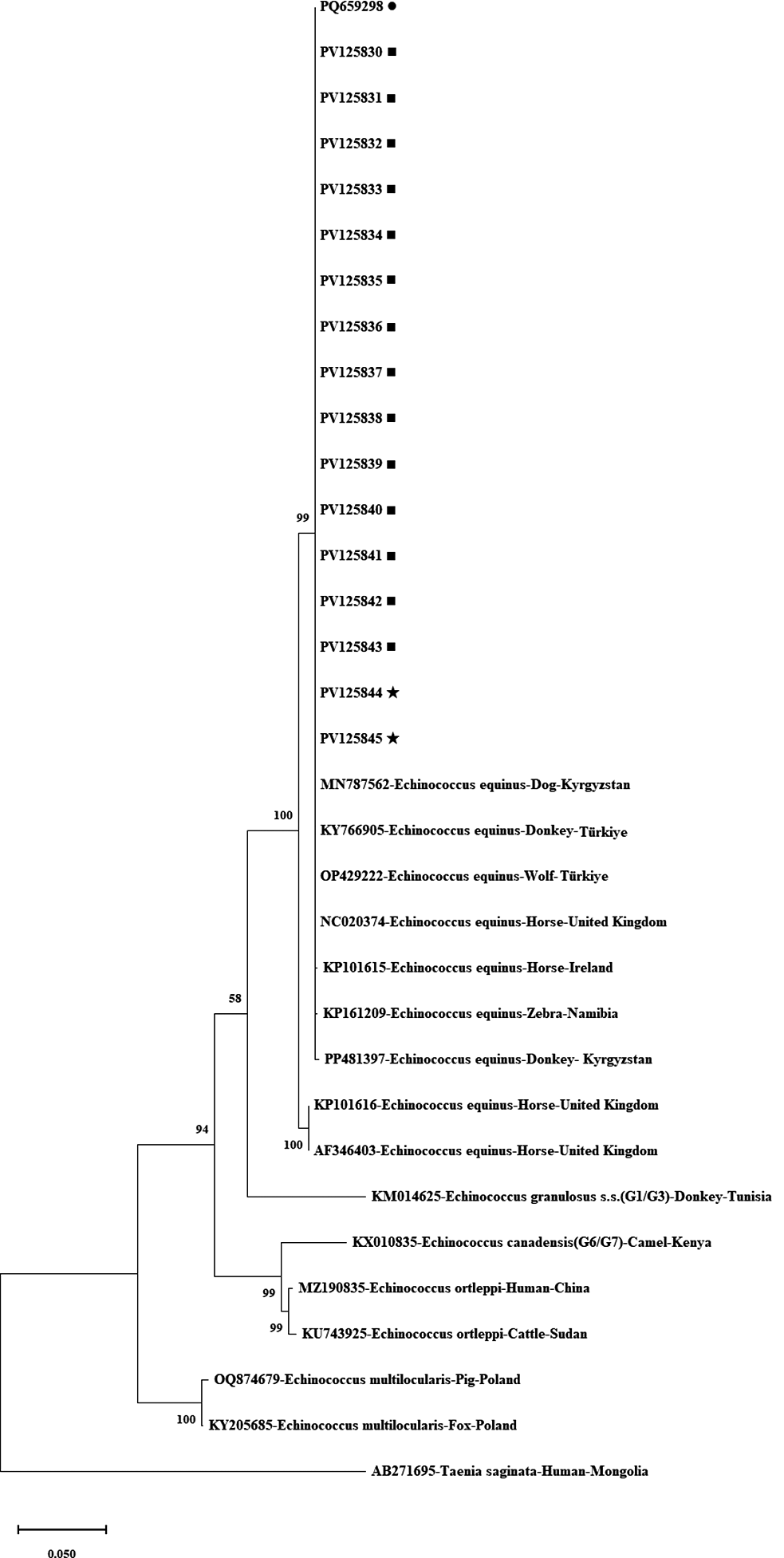
Amplification of the mitochondrial cytochrome oxidase subunit 1 (mt-CO1) gene yielded a distinct 875 bp product, which was successfully detected in all analyzed samples. The resulting nucleotide sequences were compared with previously published reference sequences and subsequently trimmed, yielding a final sequence length of 800 bp. Although three animals tested positive, multiple cysts (up to 14 from one donkey) were sequenced to assess intra-host variation, resulting in a total of 17 high-quality sequences. BLAST analysis confirmed that all sequences corresponded to E. equinus. The sequences generated in this study have been submitted to the NCBI database with the following accession numbers: PQ659298, PV125830–PV125843, and PV125844–PV125845. A phylogenetic tree illustrating the alignment of E. granulosus s.l. mt-CO1 gene sequences is shown in Figure 3.
Phylogenetic confirmation of E. equinus isolates using partial mt-CO1 (800 bp) sequence. It was created based on the TN93 + I model with the Maximum Likelihood method in MEGA-X, and the reliability of the tree was ensured by 1,000 bootstrap tests. The GenBank accession numbers and the species name were given for each reference sequences as follows: MN787562, KY766905, OP429222, NC_020374, KP101615, KP161209, PP481397, KP101616, AF346403 (E. equinus), KM014625 (E. granulosus s.s.), KX010835 (E. canadensis, G6/G7), MZ190835, KU743925 (E. ortleppi), OQ874679, KY205685 (E. multilocularis), AB271695 (T. saginata), •: Current study, the sequence of a cyst obtained from a donkey (E. equinus, PQ659298), ■: Current study, the sequence of 14 individual cysts obtained from a donkey (E. equinus, PV125830- PV125843),★: Current study, sequences of two cysts from a horse (E. equinus, PV125844-PV125845).

Discussion
The present study provides valuable insights into the occurrence and molecular characterization of E. equinus in equids from Türkiye. Hydatid cysts were identified in both horses and a donkey, with molecular analysis confirming the presence of E. equinus. These findings contribute to the growing body of literature on equine echinococcosis and highlight the importance of further epidemiological studies to better understand the disease’s prevalence and transmission dynamics.
Hydatid cysts in equids have been previously documented in various regions, with E. equinus being the primary species infecting horses and donkeys (Alvarez Rojas et al. Reference Alvarez Rojas, Romig and Lightowlers2014; Kesik et al. Reference Kesik, Kilinc, Simsek and Gul2019; Moro and Schantz Reference Moro and Schantz2009; Simsek and Cevik Reference Simsek and Cevik2014; Simsek et al. Reference Simsek, Roinioti and Eroksuz2015). Unlike E. granulosus s.s., which is a significant zoonotic agent, E. equinus has historically been considered non-zoonotic (Thompson Reference Thompson2017). However, Macin et al. (Reference Macin, Orsten, Samadzade, Colak, Cebeci and Fındık2021) reported the first confirmed case of E. equinus infection in a human host in Türkiye, challenging the long-standing assumption of its non-zoonotic nature. This finding raises critical questions about the actual zoonotic potential of E. equinus, particularly in regions where close interactions occur between equids and dogs, the latter being the definitive host. While such cases remain rare, their emergence underscores the importance of integrating molecular surveillance and public health awareness into echinococcosis control programs. Further studies are essential to determine whether these human cases represent isolated spillovers or a broader, previously underrecognized transmission cycle. This species is primarily maintained in an equine-canine cycle, where dogs act as definitive hosts, excreting infective eggs into the environment (Cardona and Carmena Reference Cardona and Carmena2013). The identification of E. equinus in this study aligns with previous findings from Europe and other regions where equine echinococcosis has been reported (Blutke et al. Reference Blutke, Hamel, Hüttner, Gehlen, Romig, Pfister and Hermanns2010; Boufana et al. Reference Boufana, Lett, Lahmar, Buishi, Bodell, Varcasia, Casulli, Beeching, Campbell, Terlizzo, McManus and Craig2015; Kesik et al. Reference Kesik, Kilinc, Simsek and Gul2019; Romig et al. Reference Romig, Deplazes, Jenkins, Giraudoux, Massolo, Craig, Wassermann, Takahashi and de la Rue2017; Simsek et al. Reference Simsek, Roinioti and Eroksuz2015).
Pathological examination revealed hepatic and pulmonary involvement, with hydatid cysts demonstrating varying degrees of calcification and degeneration. These findings are consistent with previous studies reporting that horses develop thick-walled cysts with limited fertility, potentially acting as dead-end hosts for the parasite (Eckert et al. Reference Eckert, Deplazes and Craig2001). The presence of fertile cysts in equids may suggest a potential role in maintaining the life cycle of E. equinus in endemic regions, particularly where equids and dogs coexist. However, the pathogenic significance of E. equinus in equids remains poorly understood, necessitating further investigations (Jenkins et al. Reference Jenkins, Romig and Thompson2005).
Molecular characterization using the mt-CO1 gene confirmed the identity of E. equinus in all positive cases. The obtained sequences exhibited high similarity to previously published E. equinus sequences, reinforcing the genetic consistency across global isolates. The phylogenetic analysis further confirmed the close relationship between the detected strains and reference sequences from other geographical regions. The use of molecular tools in this study underscores the importance of genetic characterization in distinguishing E. equinus from other Echinococcus species, particularly in endemic areas (Snábel et al. Reference Snábel, D’Amelio, Mathiopoulos, Turcekova and Dubinsky2016).
Despite the relatively low number of positive cases in this study, the detection of E. equinus in necropsied equids suggests that the parasite may be more prevalent than currently recognized. Given the detection of fertile cysts and the limited available data, future studies should include systematic molecular surveillance of both equids and dogs in endemic areas to better understand transmission dynamics. Further sero-epidemiological studies and molecular surveillance are needed to better assess the extent of equine echinococcosis and its potential impact on animal health.
In conclusion, this study provides the molecular confirmation of E. equinus in equids from Türkiye, contributing to the limited but growing knowledge of this parasite’s distribution. Future research should focus on elucidating the epidemiology, pathology, and transmission dynamics of E. equinus, particularly in regions where canine definitive hosts are prevalent. Enhanced surveillance and molecular studies will be crucial for a comprehensive understanding of equine echinococcosis and its implications for veterinary and public health.
Acknowledgements
Not applicable.
Author contributions
FC, BK, EC, and MU: Data acquisition, material examination, analysis, and manuscript preparation; FC, BK: Conceptualization and data analysis; YE and SS: Critical manuscript analysis.
Data availability
All the parasite specimens are deposited at the Molecular Parasitology Laboratory, Firat University Veterinary Faculty, Elazig, Türkiye, and can be verified after sending a request to the corresponding author. DNA sequence data were deposited in GenBank.
Competing interests
The authors declare no competing interests.
Ethical standard
Not applicable.
Consent for publication
Not applicable.