Introduction
The pig is faced with many new challenges during the post-weaning period such as a change in diet, removal from the sow and other littermates and a new environment (Pluske et al., Reference Pluske, Hampson and Williams1997). These stressors can lead to an intestinal imbalance (Estrada et al., Reference Estrada, Drew and Van Kessel2000), which has been associated with reduced growth rates, changes in gut morphology and microbial population numbers and an increased susceptibility to scouring and disease (Hampson, Reference Hampson1986; Estrada et al., Reference Estrada, Drew and Van Kessel2000; Drew et al., Reference Drew, Van Kessel, Estrada, Ekpe and Zijlstra2002). Recently, seaweeds and seaweed extracts have been investigated as a potential feed additive in pig diets due to their wide range of biological actions (Grinstead et al., Reference Grinstead, Tokach, Dritz, Goodband and Nelssen2000; Gardiner et al., Reference Gardiner, Campbell, O’Doherty, Pierse, Lynch, Leonard, Stanton, Ross and Lawlor2008). Seaweeds are rich in polysaccharides that are resistant to digestion and absorption in the small intestine but are completely or partially fermented in the large intestine by the microbiota present (Deville et al., Reference Deville, Damas, Forget, Dandrifosse and Peulen2004). The seaweed extracts are a potential source of soluble dietary fibres such as laminarins and fucoidans (Michel et al., Reference Michel, Lahaye, Bonnet, Mabeau and Barry1996).
The chemical structure of laminarin consists mainly of a linear β-(1→3)-linked glucan with some random β-(1→6)-linked side-chains depending on the variety of seaweed (Yun et al., Reference Yun, Estrada, Van Kessel, Park and Laarveld2003; Brown and Gordon, Reference Brown and Gordon2005). Water-soluble β-glucans such as those derived from Laminaria digitata contain small levels of β-(1→6)-linked side-chains while water-insoluble β-glucans such as those derived from Laminaria hyperborea only contain linear β-(1→3)-linked residues (Read et al., Reference Read, Currie and Bacic1996). β-glucans have been shown to exhibit prebiotic properties by increasing the bifidobacteria and lactobacilli spp. in the large intestine (Mathers and Annison, Reference Mathers and Annison1993; Jaskari et al., Reference Jaskari, Kontula, Siitonen, Jousimies-Somer, Mattila-Sandholm and Poutanen1998), thus having a positive effect on gut health (De lange, Reference De Lange2000). However, Michel et al. (Reference Michel, Bernard, Lahaye, Formaglio, Kaeffer, Quemener, Berot, Yvin, Blottiere and Cherbut1999) and Deville et al. (Reference Deville, Gharbi, Dandrifosse and Peulen2007) reported that β-glucans with β-(1→6) linkages did not exhibit prebiotic properties as such but are known to have many benefits including protection against fungal, bacterial and viral infections. They have the ability to stimulate the host immune system by stimulating the production of cytokines and chemokines and activate leukocytes such as macrophages and neutrophils (Yun et al., Reference Yun, Estrada, Van Kessel, Park and Laarveld2003; Brown and Gordon, Reference Brown and Gordon2005).
Fucoidans are sulphated polysaccharides extracted from the cell wall of various species of brown seaweeds (Berteau and Mulloy, Reference Berteau and Mulloy2003). They are known to have antibacterial properties as they have been shown to have the ability to inhibit the attachment of certain bacterial species to porcine gastric mucin in vitro (Shibata et al., Reference Shibata, Limuro, Uchiya, Kawamori, Nagaoka, Ueyama, Hashimoto, Yokokura, Sugimura and Wakabayashi2003) as well as having antiviral and anti-inflammatory properties (Marais and Joseleau, Reference Marais and Joseleau2001).
The objective of this experiment was to evaluate the effects of dietary supplementation with extracts containing both laminarin and fucoidan from two different varieties of brown seaweed, L. hyperborea and L. digitata on gut morphology, selected intestinal microbiota populations, nutrient digestibilities, volatile fatty acid (VFA) concentrations and the immune status of the weaned pig. It is hypothesised that the inclusion of L. hyperborea or L. digitata seaweed extracts will enhance selected indices of gut health in the newly weaned pig due to the biological properties of laminarins and fucoidans. This should result in an altered population of selected microbiota in the gastro-intestinal tract as well as improving the pig’s immune status due to the immune-enhancing effects of laminarin. It is also hypothesised that the combination of seaweed extracts will act synergistically in the gastro-intestinal tract.
Material and methods
All procedures described in this experiment were conducted under experimental licence from the Irish Department of Health in accordance with the Cruelty to Animals Act 1876 and the European Communities (Amendments of the Cruelty to Animals Act, 1976) Regulations, 1994.
Experimental design and dietary treatments
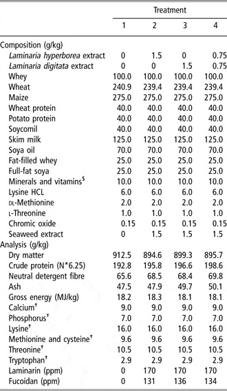
The experiment was designed as a complete randomised design comprising of four dietary treatments. The dietary treatments were as follows: (T1) basal diet (control); (T2) basal diet and L. hyperborea seaweed extract at 1.5 g/kg; (T3) basal diet and L. digitata seaweed extract at 1.5 g/kg; and (T4) basal diet and an equal proportion of L. hyperborea and L. digitata seaweed extract at 1.5 g/kg. The L. hyperborea-supplemented diet contained 170 ppm laminarin and 131 ppm fucoidan. The L. digitata-supplemented diet contained 170 ppm laminarin and 136 ppm fucoidan. The diet supplemented with the combination of L. hyperborea and L. digitata contained 170 ppm laminarin and 134 ppm fucoidan. The seaweed extracts were supplied by Bioatlantis Ltd (Tralee, County Kerry, Ireland). The diets were fed for 7 days ad libitum; after which time the pigs were humanely sacrificed. Diets were formulated to have similar digestible energy (DE) (16 MJ/kg) and ileal digestible lysine (14 g/kg) contents. The amino acid requirements were met relative to lysine (NRC, 1998). Chromium III oxide (Cr2O3) was added to the diets at the time of milling to give a concentration of 150 ppm in the diet for the determination of the nutrient digestibilities. The ingredient composition and chemical analysis of the dietary treatments are presented in Table 1.
Diet composition and chemical analysis of experimental starter diets (as fed)

$Provided per kg of complete diet: 3 mg retinol, 0.05 mg cholecalciferol, 40 mg alpha-tocopherol, 90 mg copper as copper II sulphate, 100 mg zinc as zinc oxide, 0.3 mg selenium as sodium selenite, 25 mg manganese as manganous oxide and 0.2 mg iodine as calcium iodate on a calcium sulphate/calcium carbonate carrier.
†Calculated from proximate analysis (Ministry of Agriculture, Fisheries and Food, 1991).
Animals and management
Twenty-eight piglets were selected from a commercial pig unit at 24 days of age, with an average weaning weight of 6.5 kg (s.d. 1.4 kg) and were blocked on the basis of weaning weight and litter of origin and within each block were randomly assigned to one of the four dietary treatments. The piglets were individually housed in fully slated pens (1.7 m × 1.2 m). They were individually fed and had ad libitum access to feed and water. The house temperature was thermostatically controlled at 30°C throughout the experiment. The piglets were weighed at the beginning of the experiment (day 0) and at the end of the experiment (day 7). Feed was available up to the final weighing and all remaining feed in the trough was weighed back in order to calculate the food conversion ratio. Feed samples were taken for chemical analysis at the end of the 7-day period. Faecal grab samples were taken every day from all pens during the 7-day period and were combined for subsequent nutrient analysis. After collection, the faeces samples were dried at 100°C for 48 h.
Chemical analysis
The feed and faeces samples were analysed for ash, dry matter, gross energy, neutral detergent fibre (NDF), nitrogen and chromium concentration. The feed and faeces samples were milled through a 1-mm screen (Christy and Norris Ltd, Ipswich, UK). The ash content of the feed was determined after ignition of a known weight of feed in a muffle furnace (Nabertherm, Bremen, Germany) at 500°C for 4 h (Association of Official Analytical Chemists (AOAC), 1995). The dry matter of both the feed and faeces samples was determined after drying overnight (minimum of 16 h) at 103°C. Gross energy was measured using a Parr 1201 oxygen bomb calorimeter (Parr, Moline, IL, USA). The NDF was determined using a Fibretec extraction unit (Tecator, Hoganans, Sweden) according to the method of Van Soest et al. (Reference Van Soest, Robertson and Lewis1991). The nitrogen content was determined as Kjeldahl N X 6.25 (AOAC, 1995) using the LECO FP 528 instrument (LECO Instruments, UK Ltd, Newby Road, Hazel Grove, Stockport, Cheshire, UK). The chromium concentration was determined according to Williams et al. (Reference Williams, David and Iismaa1962). The total laminarin content of the diets was analysed using a Megazyme kit (Megazyme, Bray Business Park, Bray, Wicklow, Ireland). The fucoidan levels were measured according to the method of Usov et al. (Reference Usov, Smirnova and Klochkova2001).
Gut morphological analysis
The piglets were humanely sacrificed on day 7 by lethal injection with Euthatal (pentobarbitone sodium BP) at a rate of 1 ml per 1.4 kg bodyweight. The digestive tract was removed and sections of the duodenum (10 cm from the stomach), the jejunum (60 cm from the stomach) and the ileum (15 cm from the caecum) were cut and fixed in 10% phosphate-buffered formalin. The preserved segments were prepared using standard paraffin-embedding techniques. The samples were sectioned at 5 μm thickness and stained with haemotoxylin and eosin. Villous height and crypt depth were measured on the stained sections (4× objective) using a light microscope fitted with an image analyser (Image Pro Plus; Media Cybernetics, Bethesda, MD, USA). The measurements of 20 villi and crypts were taken for each segment. The villous height was measured from the crypt–villous junction to the tip of the villous. The crypt depth was measured from the crypt–villous junction to the base. The results were expressed as the mean villous height or crypt depth in μm.
Microbial analysis
Digesta samples (approximately 10 ± 1 g) were removed from the caecum and colon of each animal immediately after slaughter and stored in sterile containers (Sarstedt, Wexford, Ireland) on ice and transported to the laboratory shortly afterwards. A 1.0 g sample was removed from each digesta sample, serially diluted (1 : 10) in 9.0 ml aliquots of maximum recovery diluent (MRD, Oxoid, Basingstoke, UK) and spread plated (0.1 ml aliquots) onto selective agars. Enterobacteria, bifidobacteria and lactobacilli species were isolated according to the methods described by Pierce et al. (Reference Pierce, Callan, McCarthy and O’ Doherty2005) and the colonies of each were counted and the numbers of bacteria were expressed per gram of wet digesta.
Volatile fatty acid analysis
Samples of digesta from individual pigs were taken from the caecum and colon to measure the VFA concentration and molar proportions of VFAs. VFA concentrations in the digesta were determined using gas liquid chromatography according to the method described by Pierce et al. (Reference Pierce, Callan, McCarthy and O’ Doherty2005).
Ammonia concentration
Digesta samples from the caecum and colon were collected to determine the concentration of ammonia-nitrogen (NH3-N) using the microdiffusion method of Conway (Reference Conway1957).
Blood analysis
Blood samples were collected on day 0 (am) prior to commencing feeding and on day 6 of the experiment (am). The blood was taken from the jugular vein of each piglet. Ten millilitres of blood in total was collected into vacutainers containing heparin anticoagulant (Becton Dickinson, Ireland). Two millilitres of blood was used to determine the total number of leukocytes and the proportions of lymphocytes, monocytes and neutrophils in the whole blood via flow cytometry, while the remaining 8 ml of blood was used for the isolation of peripheral blood mononuclear cells (PBMCs) for use in gene expression studies.
Phagocytosis estimation of blood cells by flow cytometry
The phagocytotic capacity of the white blood cells was measured using the PHAGOTEST® kit (Orpegen Pharma, Heidelberg, Germany). This procedure was performed following the guidelines of the manufacturer. For the Phagotest assay, the samples were measured using a Dako Cyan-ADP flow cytometer (Dako, Glostrup, Denmark). Ten thousand leukocytes were analysed from each blood sample on the flow cytometer. All analysis of the samples was carried out with Summit v. 4.2 software (Dako). The results of the phagotest assay were expressed as the total number of phagocytosing cells per cell type.
Preparation of peripheral blood mononuclear cells for cytokine gene expression analysis
Approximately 8 ml of whole blood was collected from each piglet into sterile vacutainers containing lithium heparin as an anticoagulant. PBMCs were separated as follows: 50 ml of lysis solution containing 155 mM Ammonium Chloride, 1 mM EDTA and 10 mM of Potassium Hydrogen Chloride was added to each falcon conical centrifuge tube (Becton Dickinson, Ireland). The blood for each piglet was then distributed evenly between two falcon tubes by pouring approximately 4 ml of the whole blood into each tube, followed by a gentle mixing by inversion. The tubes were then incubated for 10 min at 37°C, followed by centrifugation at 350 × g for 15 min. The red blood cells were then poured off. The remaining pellet was resuspended in 50 ml of Hanks Buffered Salt Solution (HBSS) (Gibco/Invitrogen, Carsbad, CA, USA, Cat No: 14175), and mixed gently by inversion. The solution was centrifuged at 225 × g for 6 min. The HBSS was then poured off and the pellet was resuspended. The two falcon tubes for each individual piglet were then pooled together. Approximately 10 ml of HBSS was then added and the tube was mixed gently by inversion. This was then centrifuged at 225 × g for 6 min. The HBSS was then poured off. Finally the pellet was resuspended in 1 ml of Tri-Reagent (Sigma-Aldrich, St Louis, MO, USA), flash frozen in liquid nitrogen and stored at −80°C.
RNA extraction and concentration determination
RNA was extracted from the PBMCs of individual piglets using Tri-Reagent (Sigma-Aldrich). The procedure was as follows: 200 μl of chloroform was added to each PBMC sample and centrifuged at 1200 × g for 15 min at 4°C. The upper phase containing the RNA was removed to a fresh centrifuge tube and 500 μl of isopropanol was added to precipitate the RNA. This was incubated at room temperature for 10 min and then centrifuged at 1200 × g for 10 min at 4°C. The isopropanol was poured off and the pellet was washed in 1 ml of cold ethanol and centrifuged. The ethanol was poured off and the remaining pellet was air-dried. RNAsed-free water (52 μl) was then added followed by incubation in a water bath. Eight microlitres of 10× RQ1 Dnase buffer and 20 μl of RQ1 Dnase were added and then incubated at 37°C for 30 min. The quality (RNA Integrity Number: RIN Value) and quantity of extracted total RNA were estimated by UV spectrophotometry and using the Bioanalyzer 2100 RNA 6000 Nanochip (Aligent Technologies, Walbronn, Germany).
cDNA synthesis and real-time polymerase chain reaction
One microgram of total RNA extracted from the PBMCs of the pigs was used for cDNA synthesis, which was carried out using Superscript™ II First-Strand synthesis for real-time polymerase chain reaction (RT-PCR) (Invitrogen Life Technologies, Carlsbad, CA, USA) and random hexamers as the primers following the manufacturers instructions. RT-PCR was then carried out on the cDNA using the ABI PRISM 7900HT sequence detection system, using 384-well plates (Applied Biosystems, Foster City, CA, USA). All samples were prepared in duplicate using SYBR Green PCR Master Mix (Applied Biosystems), cDNA as template and specific primers for the genes selected. The thermal cycling conditions involved an initial denaturation step at 95°C for 10 min, 40 cycles at 95°C for 15 s and finally 65°C for 1 min.
Experimental and reference genes
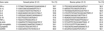
Primers for the porcine experimental genes interferon (IFN)-γ, Interleukin-1α (IL-1α), IL-4, IL-6, IL-8, IL-10, tumour necrosis factor (TNF)-α, and the reference genes β-actin, β2-microglobin, Cyclophilin and glyceraldehyde-3-phosphate dehydrogenase (GAPDH) were designed using Primer Express™ software version 3.0 (Applied Biosystems) and were synthesised by MWG Biotech (Milton Keynes, UK). Sequence data for the primers used for RT-PCR are listed in Table 2. Analysis of the reference or ‘housekeeping’ genes was carried out using geNorm version 3.5. Based on the geNorm analysis, GAPDH and Cyclophilin were used as the reference genes and a normalisation factor was calculated. The cycle threshold (Ct) values for each experimental gene were then normalised relative to the normalisation factor and the results were expressed relative to the control treatment on day 0.
Porcine oligonucleotide primers used for real time PCR

Statistical analysis
Data from the experiment were analysed using the General Linear Model (GLM) procedure of the Statistical Analysis Systems Institute (1985). For the performance data, the statistical model used included the effects of treatment and block with liveweight at weaning used as a covariate. The statistical model for analysing villous height, crypt depth and villous height : crypt depth ratio included the effects of treatment and block with feed intake included as a covariate in the model. In the analysis of the immune parameters, values at day 0 were used as a covariate in the model. A contrast statement was used to compare T1 (control diet) v. T2 (L. hyperborea-supplemented diet), T1 v. T3 (L. digitata-supplemented diet) and T1 v. T4 (combination of both L. hyperborea and L. digitata). The individual pig served as the experimental unit. All results are presented in the tables as least square means ± standard error of the means.
Results
Animal performance and nutrient digestibilities
The supplementation of neither L. hyperborea nor L. digitata seaweed extracts alone or in combination had any significant effect on feed intake (272, 306.3, 291.9, 276.1 g/day; s.e. 16.1), average daily gain (246.1, 256.8, 246.5, 269 g/day; s.e. 28) or food conversion ratio (1.307, 1.215, 1.238, 1.104; s.e. 0.145 for T1 control, T2 L. hyperborea, T3 L. digitata or T4 combination of L. hyperborea and L. digitata, respectively) compared to the control treatment (P > 0.05).
Also, the supplementation of L. hyperborea or L. digitata seaweed extracts alone or in combination had no significant effect on apparent nutrient digestibility (dry matter digestibility (85.30, 82.59, 82.96, 81.99; s.e. 1.248); NDF (35.68, 37.77, 35.92, 34.43; s.e. 5.60); nitrogen digestibility (79.50, 77.85, 79.83, 78.18; s.e. 1.828) or ash digestibility (57.59, 53.26, 51.55, 52.88; s.e. 2.20) for T1 control, T2 L. hyperborea, T3 L. digitata or T4 combination of L. hyperborea and L. digitata, respectively) compared to the control treatment (P > 0.05).
Gut morphology
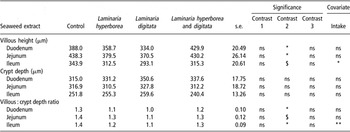
There was no effect of L. hyperborea seaweed extract or the combination of L. hyperborea and L. digitata seaweed extracts on villous height, crypt depth and the villous height : crypt depth ratio in the duodenum, jejunum or the ileum (P > 0.05). Pigs offered the L. digitata seaweed extract had a reduced villous height in the duodenum, jejunum (P < 0.05) and a numerical tendency towards a reduction in the ileum (P = 0.08) compared to the control treatment. Subsequently, a lower villous height : crypt depth ratio in the duodenum and ileum (P < 0.05) was observed with the supplementation of L. digitata seaweed extract (Table 3).
Effect of seaweed extract on villous height, crypt depth and the villous height : crypt depth ratio in the gastro intestinal tract of the weanling pig (least-square means and s.e.)

Probability of significance: *P < 0.05, **P < 0.01, ns = non-significant (P > 0.05).
$ = Approaching significance (P = 0.1).
Contrast 1: Control v. Laminaria hyperborea.
Contrast 2: Control v. Laminaria digitata.
Contrast 3: Control v. Laminaria hyperborea and Laminaria digitata.
Microbiology
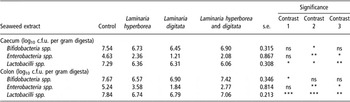
Pigs offered diets containing L. hyperborea seaweed extract had a lower population of bifidobacteria in the colon (P < 0.05), and a reduction in the population of lactobacilli in the caecum (P < 0.05) and colon (P < 0.001) when compared to the control treatment. The inclusion of L. digitata seaweed extract was associated with lower populations of enterobacteria in the caecum and colon (P < 0.01), bifidobacteria in the caecum (P < 0.05), and lactobacilli in the caecum (P < 0.05) and colon (P < 0.001) compared to the control treatment. Pigs offered the combination of L. hyperborea and L. digitata seaweed extracts also had a significantly lower population of enterobacteria spp. (P < 0.05) and lactobacilli spp. (P < 0.01) in the caecum and colon compared to the control treatment (Table 4).
Effect of seaweed extract on selected microbial populations in the caecum and colon of the weanling pig (least-square means and s.e.)

Probability of significance: *P < 0.05, **P < 0.01, ***P < 0.001, ns = non-significant (P > 0.05).
Contrast 1: Control v. Laminaria hyperborea.
Contrast 2: Control v. Laminaria digitata.
Contrast 3: Control v. Laminaria hyperborea and Laminaria digitata.
Volatile fatty acids and ammonia
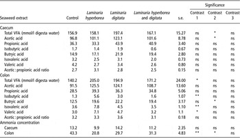
In the caecum, there was a significantly higher total VFA concentration and proportion of acetic acid in piglets offered diets supplemented with L. digitata seaweed extract compared to the control treatment (P < 0.05).
In the colon, pigs offered L. hyperborea-supplemented diets had a higher total VFA concentration compared to the control treatment (P < 0.05). Pigs offered diets supplemented with L. hyperborea seaweed extract had a significantly higher proportion of valeric acid (P < 0.05), isobutyric acid (P < 0.01) and isovaleric acid (P < 0.01) compared to the control treatment. The supplementation of L. digitata seaweed extract in pig diets resulted in a significantly higher molar proportion of butyric acid compared to the control treatment (P < 0.05). The ammonia concentration in the colon was significantly reduced with the supplementation of L. hyperborea seaweed extract (P < 0.01) and L. digitata seaweed extract (P < 0.05) to the diet (Table 5).
Effect of seaweed extract on the total volatile fatty acid (VFA) concentrations, the proportions of VFAs and the ammonia concentrations in the caecum and colon of the weanling pig (least-square means and s.e.)

Probability of significance: *P < 0.05, **P < 0.01, ns = non-significant (P > 0.05).
Contrast 1: Control v. Laminaria hyperborea.
Contrast 2: Control v. Laminaria digitata.
Contrast 3: Control v. Laminaria hyperborea and Laminaria digitata.
Phagocytotic capacity of the whole blood
The supplementation of L. hyperborea or L. digitata seaweed extracts alone or in combination had no significant effect on the total number of neutrophils or lymphocytes compared to the control treatment (P > 0.05). The inclusion of L. hyperborea seaweed extract resulted in an increase in the total number of monocytes compared to the control treatment (P < 0.05). There was no significant effect of L. hyperborea or L. digitata seaweed extract or the combination of both on the phagocytotic capacity of leukocytes or on the individual populations of leukocytes, i.e. neutrophils, monocytes or lymphocytes when compared to the control treatment (P > 0.05).
Cytokine gene expression analysis
The supplementation of L. hyperborea or L. digitata seaweed extracts alone or in combination had no significant effect on the expression of the cytokines IFN-γ, IL-1α, IL-4, IL-6, IL-10 or TNF-α. The inclusion of the combination of L. hyperborea and L. digitata seaweed extract resulted in an increase in the expression of IL-8 mRNA when compared to the control treatment (P < 0.05).
Discussion
The hypothesis of the current experiment is that the inclusion of L. hyperborea and L. digitata seaweed extracts either alone or in combination would enhance selected indices of gastro-intestinal health and immune status in the newly weaned piglet. In this experiment, the performance levels were similar between the control group and all of the seaweed extract-supplemented groups. Only marginal effects were observed on gut morphology and on the peripheral blood immune response. However, a significant reduction in the enterobacteria, bifidobacteria and lactobacilli populations were observed in the caecum and colon of seaweed extract-supplemented groups. The performance and general health of the pigs in this experiment were considered good and this may explain the lack of responses to seaweed extract inclusion. Under these conditions, little or no response to feed additives such as prebiotics can be expected (Pierce et al., Reference Pierce, Callan, McCarthy and O’ Doherty2005).
The results of this experiment suggest that the inclusion of seaweed extracts in the diet of weaned pigs caused a significant reduction in microbial populations in the large intestine. Within the caecum and colon, bifidobacteria and lactobacilli populations were reduced. Similarly, the enterobacteria population was significantly reduced in the caecum and colon. These results are comparable to those obtained by Gardiner et al. (2008) who demonstrated that the inclusion of an Ascophyllum nodosum seaweed extract had an inhibitory effect on the enterobacteria and bifidobacteria population within the caecum and colon of grower-finisher pigs. We propose that there are two possible explanations for this observation. Firstly, this reduction might be due to the fact that certain algal fibres present in the seaweed are not a suitable substrate for fermentation by the microbial populations in the large intestine. Goni et al. (Reference Goni, Gudiel-Urbano, Bravo and Saura-Calixto2001) suggested that algal fibres such as fucoidans were resistant to bacterial metabolism. Fucoidans from different species of brown algae have been shown to inhibit the attachment of certain bacterial species in the porcine gastro-intestinal tract as well as preventing the binding of enterococci and streptococci species to the extracellular matrix proteins of animal cells (Shibata et al., Reference Shibata, Limuro, Uchiya, Kawamori, Nagaoka, Ueyama, Hashimoto, Yokokura, Sugimura and Wakabayashi2003), indicating that the fucoidans may be acting as a powerful antimicrobial in reducing the microbial population. Secondly, the non-selective reduction in the microbial populations may be due to the agglutination properties of the extract. Yeast-derived β-glucans, which have a similar structure to algal β-glucan, have the ability to agglutinate certain bacterial species, which in turn would inhibit their attachment to epithelial cells, thus preventing them from colonising the mucosal surface (Mirelman et al., Reference Mirelman, Altmann and Eshdat1980; Kogan and Kocher, Reference Kogan and Kocher2007).
In the current study, the highest concentration of total VFA was produced from the L. digitata-supplemented treatment in the caecum, while in the colon the L. hyperborea-supplemented treatment had the highest production of total VFA. This result suggests that L. digitata had the highest fermentation rate in the caecum while L. hyperborea was being fermented in the colon. L. hyperborea is mainly insoluble in water due to the small amount of β-(1-6) branching on its structure (Annan et al., Reference Annan, Hirst and Manners1965). As a result, this insoluble fraction may be passed into the colon where it is fermented by the microbial population. The inclusion of the L. digitata extract resulted in a higher proportion of butyric acid in the caecum and colon compared to the control treatment. This increase in butyric acid as well as the reduction in the enterobacteria population indicates the potential benefits of the seaweed extract inclusion to the weaned pig. Butyrate is quickly metabolised by colonocytes (Roediger, Reference Roediger1982) and accounts for about 70% of the total energy consumption of the colon (Smith and German, Reference Smith and German1995). It is not unreasonable to expect that the L. digitata extract would have an important role to play in the prevention of diarrhoea in piglets (Williams et al., Reference Williams, Verstegen and Tamminga2001).
In the current experiment, pigs supplemented with L. digitata seaweed extract had significantly shorter villi in the duodenum and jejunum, when compared with the control treatment. Subsequently, a reduction in the villous height : crypt depth ratio in the duodenum, jejunum and ileum was observed. However, there was no reduction in performance in terms of daily gain, food intake or food conversion ratio in this treatment group in comparison to the control group. Previous studies have identified that a reduction in villous height can have detrimental effects on the absorptive capacity of nutrients, electrolytes and water in the small intestine and this has been shown to increase the incidence of post-weaning diarrhoea (Nabuurs and Hoogendoorn, Reference Nabuurs and Hoogendoorn1993). However, no incidence of diarrhoea was observed in the current experiment. This is probably due to the fact that the reduction in villous height was less (control 388 μm v. L. digitata 334 μm) in our study in comparison to the pigs with post-weaning diarrhoea (control 350 μm v. post-weaning diarrhoea group 200 μm) in the study of Nabuurs and Hoogendoorn (Reference Nabuurs and Hoogendoorn1993). Similar results were reported by Grinstead et al. (Reference Grinstead, Tokach, Dritz, Goodband and Nelssen2000), who found no difference in weaned pig performance when the diet was supplemented with Spirulina platensis seaweed extract. There has been very little data published on the effects of seaweed extracts on gut morphology in pigs or other species, so it is unclear as to the exact cause of the reduction in the villous height and subsequently the villous height: crypt depth ratio. There are a number of possible explanations for this reduction; firstly dietary fibres such as mixed linked β-glucans from cereal or algal sources are both soluble and viscous and can cause an increase in the viscosity of the intestinal contents (Montagne et al., Reference Montagne, Pluske and Hampson2003) and high viscous diets have been shown to decrease villous height in newly weaned pigs (McDonald et al., Reference McDonald, Pethick, Mullan and Hampson2001). Another possible explanation may be due to the presence of small amounts of components within the L. digitata seaweed extract such as tannins, which may not be fully removed during processing. This observation is supported by the data of Gardiner et al. (2008) who reported a reduced performance in grower-finisher pigs fed a diet supplemented with Ascophyllum nodosum seaweed extract, which included high levels of tannins. Tannins are anti-nutritional factors that are known to have effects on the gut structure and function in pigs (Pluske et al., Reference Pluske, Hampson and Williams1997).
In the current study it was found that seaweed extract supplementation had only marginal effects on immune parameters. No significant change in the expression of IFN-γ, IL-1α, IL-4, IL-6, IL-10 or TNF-α mRNA was found, which is similar to work carried out by Li et al. (Reference Li, Li, Xing, Cheng and Lai2006), who found no differences in the expression of IL-6, IL-10 and TNF-α with the inclusion of yeast-derived β-glucans. However, an increase in the expression of IL-8 mRNA was observed with the inclusion of the combination of L. hyperborea and L. digitata seaweed extract. Il-8 is a chemokine that is involved in the recruitment and activation of neutrophils from the blood to the site of infection. The reason for this increase in IL-8 expression with the combination of seaweed extracts is unclear as individually the seaweed extracts do not increase the expression of IL-8. The combination of seaweeds may be acting synergistically to induce IL-8 expression due to the combination of insoluble β-glucans derived from L. hyperborea and soluble β-glucans derived from L. digitata. An increase in total monocyte number was observed in pigs supplemented with L. hyperborea seaweed extract. Deliberate pathogen infection studies may be required to fully explore the potential of seaweed extracts to control pathogens or improve performance in post-weaned pigs.
In conclusion, when individually fed, the seaweed extracts had no significant effect on the pig’s immune function; however, in combination a marginal response was observed, indicating that the structure and chemical composition of components of the seaweed extract may influence IL-8 expression. The reduction in the enterobacteria, bifidobacteria and lactobacilli populations examined indicates that by modifying the diet with seaweed extracts such as L. hyperborea or L. digitata the composition of the gastro-intestinal microbiota can be modified without disrupting animal performance. Further studies will explore the potential of these diets to prevent the negative impact of a pathogen challenge.