

Parkinson’s disease (PD) is a neurodegenerative disorder, mainly defined as the degeneration of pigmented cells in the substantia nigra and the consequent dysfunction of the nigrostriatal system. Cognitive deficits have traditionally been seen as an executive dysfunction secondary to frontostriatal system affectation. In this schema, this executive dysfunction is responsible for other cognitive disturbances such as deficits in memory or linguistic functions. Although this type of description is frequent, recent evidence has questioned this interpretation (Barone et al., Reference Barone, Aarsland, Burn, Emre, Kulisevsky and Weintraub2011).
As regards declarative memory, alterations in learning and free delayed recall without deficits in cued recall have been reported. This pattern of affectation has been related to a deficit in the use of self-elaborated encoding and recall strategies. Thus, the aforementioned deficit has been considered as being indicative of frontal dysfunction due to frontostriatal circuitry impairment (Stefanova, Kostic, Ziropadja, Ocic, & Markovic, Reference Stefanova, Kostic, Ziropadja, Ocic and Markovic2001; Tröster & Fields, 1995). However, some recent findings indicate that declarative memory deficits are not confined to poor learning strategies or impaired free recall. In fact, patients can continue to show altered execution even after the use of external cues (semantic keys, recognition) (Beyer et al., Reference Beyer, Bronnick, Hwang, Bergsland, Tysnes, Larsen and Apostolova2013; Higginson, Wheelock, Carroll, & Sigvardt, Reference Higginson, Wheelock, Carroll and Sigvardt2005; Whittington, Podd, & Stewart-Williams, Reference Whittington, Podd and Stewart-Williams2006). On the other hand, a poor performance in WCST is well documented, which is one of the most commonly used instruments to assess executive function (Lees & Smith, Reference Lees and Smith1983; Liozidou, Potagas, Papageorgiou, & Zolonis, Reference Liozidou, Potagas, Papageorgiou and Zalonis2012; Muslimovic, Post, Speelman, & Schmand, Reference Muslimovic, Post, Speelman and Schmand2007; Witt, Nuhsman, & Deuschl, Reference Witt, Nuhsman and Deuschl2002), but none of the other tests used to measure these functions has yielded conclusive evidence. This is particularly relevant in the case of verbal fluency tests as, despite being one of the measures most strongly linked to prefrontal activity, there are as many studies showing altered performance in patients (Muslimovic et al., Reference Muslimovic, Post, Speelman and Schmand2007; Obeso, Casabona, Bringas, Alvarez, & Jahanshahi, Reference Obeso, Casabona, Bringas, Alvarez and Jahanshahi2012; Signorini & Volpato, Reference Signorini and Volpato2006; Uc et al., Reference Uc, Rizzo, Anderson, Qian, Rodnitzky and Dawson2005) as those in which no differences are observed (Brand et al., Reference Brand, Labudda, Kalbe, Hilker, Emmans, Fuchs and Markowitscha2004; Piatt, Fields, Paolo, Koller, & Troster, Reference Piatt, Fields, Paolo, Koller and Troster1999; Schneider, Reference Schneider2007). With regard to language, some authors have reported that deficits in comprehension may occur in complex sentences and that they are mainly due to limitations in executive resources such as working memory (Grossman, Reference Grossman1999; Grossman, Carvell, Stern, Gollomp, & Hurtig, Reference Grossman, Carvell, Stern, Gollomp and Hurtig1992; Hochstadt, Nakano, Lieberman, & Friedman, Reference Hochstadt, Nakano, Lieberman and Friedman2006). However, other results have questioned this assumption: alterations in sentence comprehension are not limited to those of greater complexity, but they are also found in simple sentences. In addition, these deficits are present in patients without alteration of working memory (Skeel et al., Reference Skeel, Crosson, Nadeau, Algina, Bauer and Fennell2001).
Contributions from neuroimaging studies also suggest that cognitive deficits in PD are not explained by frontostriatal system affectation alone. These findings demonstrate that, even in nondemented cases, patients may present hippocampal, frontal and parietal atrophy related to alterations in different cognitive functions (Beyer et al., Reference Beyer, Bronnick, Hwang, Bergsland, Tysnes, Larsen and Apostolova2013; Jokinen et al., Reference Jokinen, Bruck, Aalto, Forsback, Parkkola and Rinne2009; Pereira et al., Reference Pereira, Junque, Marti, Ramirez-Ruiz, Bargallo and Tolosa2009). Furthermore, the development of dementia in PD involves structural changes in limbic areas and extensive atrophy of different cortical regions (Ibarretxe-Bilbao, Tolosa, Junque, & Marti, Reference Ibarretxe-Bilbao, Tolosa, Junque and Marti2009).
Many of the findings obtained so far are based on studies of isolated cognitive functions which do not allow in-depth study of the relationships among these different functions or their components and do not provide a holistic impression of these deficits. Taking this situation into account, the aim of this work is to study cognitive functioning in PD patients over a wide range of cognitive domains and the main components of these.
Method
Participants
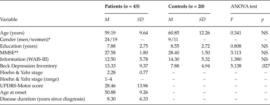
The study included 63 participants: 43 patients with idiopathic PD and 20 healthy and neurologically normal controls. Patients were recruited from the Hospital Universitario Ntra. Sra. Candelaria (HUNSC, Tenerife), where they were evaluated at the movement disorder unit using the Hoehn and Yahr Scale (Hoehn & Yahr, Reference Hoehn and Yahr1967) and UPDRS (Fahn & Elton, Reference Fahn, Elton, Fahn, Marsden, Goldstein and Calne1987). All the patients met the clinical criteria for the diagnosis of PD (Hughes, Daniel, Kilford, & Lees, Reference Hughes, Daniel, Kilford and Lees1992). Those who met the criteria of the Movement Disorder Society (MDS) for the diagnosis of dementia associated with PD (PD-D) were excluded (Emre et al., Reference Emre, Aarsland, Brown, Burn, Duyckaerts, Mizuno and Dubois2007). The diagnostic criteria for PD-D were operationalized according to the level 1 algorithm proposed by the MDS Task Force (Dubois et al., Reference Dubois, Burn, Goetz, Aarsland, Brown, Broe and Emre2007). Patients and controls were matched in age, years of study, gender, manual preference and estimated IQ (information WAIS-III) (Wechsler, Reference Wechsler1997a). The Beck Depression Inventory was administered for the assessment of mood state (Beck, Ward, Mendelson, Mock, & Erbaugh, Reference Beck, Ward, Mendelson, Mock and Erbaugh1961).
Both groups of participants were informed about the aims of the investigation and participated voluntarily. All gave their informed consent. The data was obtained in accordance with the regulations of the Ethics Committees of the Universidad de La Laguna and in compliance with the Helsinki Declaration for Human Research.
Procedure
An extensive assessment protocol was designed in order to assess a wide range of cognitive domains and their main components. Tests were selected in such a way that no or only limited movements had to be carried out by the patient. The neuropsychological tests which were administered are described below:
Processing speed, attention and working memory
-Reaction Time test: This system permits the dissociation of decision and motor times. Decision time (DT) is a cognitive measure of information processing speed. Motor time (MT) reflects motor and coordination deficits (Schuhfried, Reference Schuhfried1992).
-Paced Auditory Serial Addition Test (PASAT): adaptation of original version (Gronwall, Reference Gronwall1977). Stimuli are presented with a temporal interval of 3 seconds. The subject must say if every number heard is higher or lower than the previous one.
-Digit Span and Spatial Span (WMS-III) (Wechsler, Reference Wechsler1997b).
Visuospatial, visuoperceptive and visuoconstructive functions
-Judgment of Line Orientation Test (JLOT, 15 items simplified version) (Benton, Hamsher, Varney, & Spreen, Reference Benton, Hamsher, Varney and Spreen1983).
-Facial Recognition Test (FRT, simplified version) (Benton et al., Reference Benton, Hamsher, Varney and Spreen1983).
-Block design subtest: Three designs of four cubes (5, 7, 9 designs) and three designs of nine cubes (10, 11, 12 designs) were selected, measured according to standard procedure, except for time bonuses (Wechsler, Reference Wechsler1997a).
Executive functions
-Stroop Color and Word Test (Golden, 1978): This Stroop Test version includes an index to assess the interference related to the word–color conflict by comparing the subject’s performance in the third sheet (Word–Color), with the same subject’s performance in the other two neutral conditions (Word and Color sheets).
-Wisconsin Card Sorting Test (Heaton, Reference Heaton1981).
-Verbal fluency tasks: These tasks consist of asking the participants to rapidly generate words beginning by a given letter (phonemic fluency —FAS), to generate only animals (semantic fluency) (Benton & Hamsher, Reference Benton and Hamsher1989), and to rapidly generate verbs (action fluency) (Piatt, Fields, Paolo, & Troster, Reference Piatt, Fields, Paolo, Koller and Troster1999).
Learning and memory
-California Verbal Learning Test (CVLT) (Delis, Kramer, Kaplan, & Ober, Reference Delis, Kramer, Kaplan and Ober1987). The test includes learning over a five-trial presentation of a 16-word list, free and cued delayed recall and recognition.
-8/30 Spatial Recall Test (8/30 SRT): 7/24 SRT adaptation (Barbizet & Cany, Reference Barbizet and Cany1968). Subjects must learn the spatial location of eight black circles displayed in a matrix from 6 x 5 boxes. When the sheet is removed the subject must place eight circles in the corresponding locations on an empty matrix. The test includes five trials of learning and two trials of delayed recall (short and long term).
Linguistic functions
-Actions and Noun Naming Test: a naming task of pictorial visual stimuli. The Noun Naming test consists of 40 stimuli taken from the work of Cuetos and colleagues (Cuetos, Ellis, & Álvarez, Reference Cuetos, Ellis and Álvarez1999). The Action Naming task consists of 20 stimuli (Druks & Masterson, Reference Druks and Masterson2000).
-Sentence Comprehension Test: based on studies by Grossman and colleagues (1992, 1999) and Skeel and colleagues (2001). The test consists of auditory stimuli- 20 sentences presented on a PC, each followed by a question to assess their understanding. The level of syntactic complexity was manipulated as follows: 10 were simple sentences (The bellboy greeted the slim receptionist. Who greeted? or Who was greeted?) and 10 complex sentences with subordinate clause, in which the subject of the main clause is in turn the subject of the relative clause (The girl who pinched her cousin was naughty. Who pinched? or Who was pinched?
Data Analysis
Statistical comparisons were conducted using one-way analyses of variance (ANOVA). Correlational analyses were performed using Pearson´s correlation coefficient and linear regression analysis in order to examine the relation between cognitive functions. The level of statistical significance was established as p < .05. All the analyses were performed with SPSS-PC software version 15.0 for Windows.
Results
Processing speed, attention and working memory
In terms of reaction times, patients showed poorer performance in Motor Time, compared to controls, F(1, 60) = 6.68, p = .012. No difference was found in Decision Time, F(1, 60) = 1.52, p = .22. The PD group had significantly lower scores than controls in both the direct and inverse subtest in Spatial Span (direct: F(1, 61) = 7.61, p = .008; inverse: F(1, 61) = 4.57, p = .037). There were no differences between controls and patients in PASAT, F(1, 61) = 0.01, p = .94, and Digit Span (direct: F(1, 61) = 0.08, p = .78; inverse: F(1, 61) = 1.88, p = .18).
Visuospatial, visuoperceptive and visuoconstructive functions
The PD group performed significantly worse than the control group in the JLOT, F(1, 60) = 7.69, p = .007, on the FRT, F(1, 60) = 12.47, p = .001, and Block Design subtest, F(1, 59) = 6.07, p = .017.
Executive functions
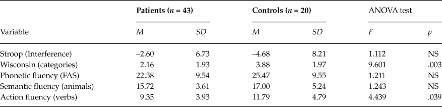
The patients showed a poorer performance compared to controls in the Wisconsin categories and Action Fluency tests. No differences between groups were found in either Stroop interference or in Phonetic and Semantic Fluency (Table 2).
Performance in executive functions

Note: M: mean. SD: standard deviation. NS: not significant.
Learning and memory
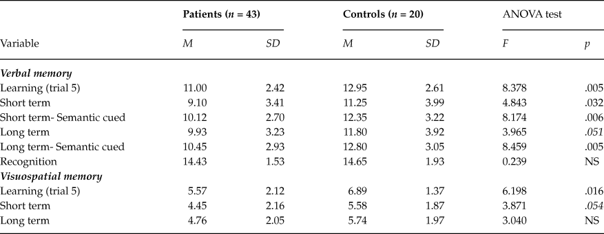
As for verbal memory, patients showed a lower performance in learning as well as in free and cued short-term memory. A marginally significant difference was obtained between groups in free long-term memory. In addition, patients performed significantly worse than the control group in cued recall. In terms of visual memory, the PD group performed significantly worse in visuospatial learning compared to the control group. However, patients and controls did not differ in any of the visual recall measures (Table 3).
Performance in verbal memory and visuospatial memory

Note: M: mean. SD: standard deviation. NS: not significant.
In order to control for the possible effects of executive functioning on memory performance participant`s scores on WCST and Action fluency were included as covariates and reanalyzed group differences. The ANCOVA showed that WCST performance only had a significant effect as a covariate on the free verbal short-term memory, F(1, 59) = 5.247, p = .026, and visuospatial learning, F(1, 59) = 4.333, p = .042. Nonetheless, WCST performance did not account for all the variance in these measures and significant between-groups differences remained (free verbal short-term memory, F(1, 59) = 4.691, p = .005, visuospatial learning, F(1, 59) = 4.744, p = .005).
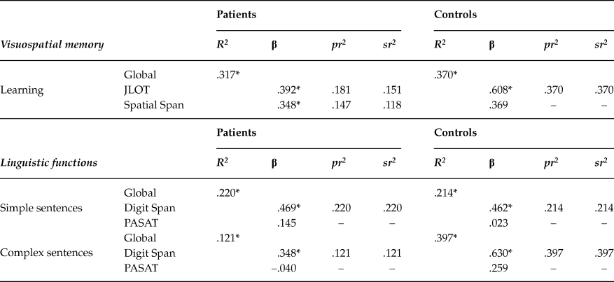
Visual memory performance can be conditioned by spatial perception (JLOT) and spatial working memory (Spatial Span). A linear regression analysis was performed to determine the nature of these relationships. The results showed that JLOT and Spatial Span accounted for 32% of visuospatial learning variance in the PD group, F(2, 40) = 8.80, p = .001. In the controls, 37% of visuospatial learning variance was explained by JLOT, F(1, 18) = 9.99, p = .001 (Table 4).
Regression analysis for memory and linguistic functions

Note: *p < .05.
Linguistic functions
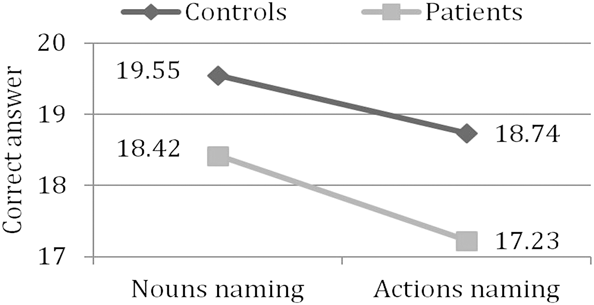
Correct responses in the naming task were analyzed using repeated measures, with one within-subject factor (task, 2 levels) and one between-subject factor (group, 2 levels). There was a significant main effect of group, F(1, 60) = 9.02, p < .001, and task, F(1, 60) = 17.59, p < .001, but no significant interaction between the two factors, F(1,60) = 0.60, p = .44. Patients performed poorly compared to controls in naming nouns and actions. Both patients and the control group subjects performed worse in the naming of actions (Figure 1). Analysis of errors showed that patients made more mistakes in all types of errors (perceptive, F(1, 60) = 12.36, p = .001, semantic, F(1, 60) = 10.19, p = .002, and mixed errors, F(1, 60) = 5,71, p = .02).
Performance in Actions and Nouns Naming Test.

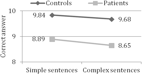
Performance in sentence comprehension was analyzed using repeated measures, with one within-subject factor (complexity, 2 levels) and one between-subject factor (group, 2 levels). There was a significant main effect of group, F(1, 60) = 7.77, p = .007, but no significant main effect of complexity, F(1, 60) = 2.89, p = .09, or significant interaction between the two factors, F(1, 60) = 0.35, p = .56. The PD group performed poorly on simple and complex sentences (Figure 2).
Performance in Sentences Comprehension Test.

An ANCOVA was performed in order to statistically control for the possible effects of executive functions on linguistic functions. Group differences in the linguistic tests were reanalyzed with participant’s scores on the WCST and Action Fluency tests included as covariates. WCST performance had a significant effect as a covariate on Naming Nouns, F(1, 60) = 8.623, p = .005, Naming Actions, F(1, 60) = 8.639, p = .005, Simple Sentences, F(1, 60) = 5.927, p = .018, and Complex Sentences, F(1, 60) = 7.206, p = .01. Nonetheless, significant between-group differences remained in all cases (Naming Nouns, F(1, 60) = 9.682, p < .001, Naming Actions, F(1, 60) = 7.837, p < .001, Simple Sentences, F(1, 60) = 5.625, p = .002, and Complex Sentences, F(1, 60) = 6.686, p = .001).
Performance in sentence comprehension can be conditioned by attention (PASAT) and verbal working memory (Verbal Span). A linear regression analysis was carried out to determine the nature of these relationships. Results in the PD group showed that Verbal Span explained 22% of variance in Simple Sentences and 12.1% of the variance in Complex Sentences, F(1, 41) = 11.56, p = .002 and F(1, 41) = 5.65, p = .022 respectively. In the control group, Verbal Span accounted for 21% of the variance in Simple Sentences whereas PASAT explained 40% of the variance in Complex Sentences, F(1, 18) = 4.62, p = .046 and F(1, 18) = 11.19, p = .004 respectively (Table 4).
Cognitive performance and depression
Significantly higher scores on the BDI were obtained in patients than in controls (table 1). BDI was considered as a covariate in statistical analyses in order to control for the possible effects of depression on cognitive functions. Depression was not associated with any of the analyzed variables, with the exception of Noun Naming (r = –.31; p = .04). Although depression had a significant effect as a covariate, F(1, 60) = 6.01, p = .017, between-group differences remained significant, F(1, 60) = 7.22, p = .009.
Demographic data and clinical characteristics

Note: M: mean. SD: standard deviation. NS: not significant. *number of men and women; **correction by age and educational level (Blesa et al., Reference Blesa, Pujol, Aguilar, Santacruz, Bertran-Serra and Hernández2001).
Discussion
The aim of this study was to examine the nature of the cognitive deficits associated with PD in depth. The results confirm the presence of motor slowing, along with alterations in visuospatial perception, visuoperceptive processing, visuoconstructive functions and spatial working memory.
In terms of executive functions, patients presented poor performance in the concept formation, set shifting and verb retrieval (WCST, Action Fluency), a result which coincides with those observed in other studies (Lees & Smith, Reference Lees and Smith1983; Signorini & Volpato, Reference Signorini and Volpato2006). However, performance was normal in usual measures of verbal fluency (Phonetic and Semantic) and in response inhibition. Consequently, generalized impairment of prefrontal functions was not observed. It is worth mentioning that there was normal achievement in one of the most studied tests -Phonetic Fluency- which has been strongly associated with executive functioning.
With regard to verbal declarative memory, patients showed a deficient performance in learning and delayed recall, both in free and cued tests (semantic cues). Verbal memory alterations in PD have traditionally been interpreted as deficits in the use of self-elaborated encoding strategies and information recovery (Caballol, Marti, & Tolosa, Reference Caballol, Marti and Tolosa2007; Pillon, Deweer, Agid, & Dubois, Reference Pillon, Deweer, Agid and Dubois1993). However, the results here do not support this hypothesis but agree with the findings of some recent investigations in which memory deficits persist even when external cues facilitating memory are provided (Higginson et al., Reference Higginson, Wheelock, Carroll and Sigvardt2005).
As regards visual declarative memory, patients presented deficits in visuospatial learning, although no alteration in delayed memory was observed. The extent to which alterations in spatial working memory and visuospatial perception could predict visuospatial learning performance was analyzed to examine these visual memory deficits more closely. These variables had considerable predictive power (patients: 32%; control group subjects: 37%), so it can be assumed that they play a relevant role in explaining visuospatial learning impairment. These results reinforce previous findings (Galtier, Nieto, Barroso, & Lorenzo, Reference Galtier, Nieto, Barroso and Lorenzo2009).
In relation to linguistic functions, we assessed production through a visual Noun and Action Naming test which indicated deficient performance among patients was assessed. No differences have been reported in previous studies between naming nouns and actions in the control group and the lower performance for naming of actions in the patients. This finding has been interpreted as evidence for frontal impairment in PD (Cotelli et al., Reference Cotelli, Borroni, Manenti, Zanetti, Arevalo, Cappa and Padovani2007; Rodriguez-Ferreiro, Menendez, Ribacoba, & Cuetos, Reference Rodriguez-Ferreiro, Menendez, Ribacoba and Cuetos2009). In the present study, however, both the patients and control group performed worse in Action Naming, which brings into question whether this naming deficit is related to frontal dysfunction. On the other hand, it has been suggested that visuosperceptive deficit is a relevant factor in explaining alterations in the naming of pictorial material (Bertella et al., Reference Bertella, Albani, Greco, Priano, Mauro, Marchi and Semenza2002). In the particular case of this study, the lack of predominance of perceptive errors would indicate that naming deficits among patients cannot be attributed to perceptive dysfunction.
Language comprehension was assessed with a Simple and Complex Sentence test, with patients’ performance being altered in both types of sentence. The results of this study are consistent with those obtained by Skeel and colleagues (2001), where deficits are not limited to the comprehension of more complex sentences. Alterations in comprehension have been related to attentional difficulties, processing speed and working memory, and have been considered a consequence of executive resource limitations (Grossman et al., Reference Grossman, Carvell, Stern, Gollomp and Hurtig1992; Hochstadt et al., Reference Hochstadt, Nakano, Lieberman and Friedman2006). Nevertheless, in the present study, patients did not show deficits in either attentional functions or in verbal working memory. Moreover, these variables did not have a greater predictive power in patients (12–22%) compared to the control group (21–40%), or a greater weight in complex sentences (12%), compared to simple ones (22%). If alterations in comprehension were a consequence of executive resource limitation, a greater involvement of working memory would be expected and the predictive power of working memory would be related to the level of sentence complexity. Thus, the results here do not indicate that comprehension deficits can be interpreted as a result of executive dysfunction.
In sum, the results of this work shed more light on the nature of cognitive deficits in PD questioning the idea that the neuropsychological profile associated with PD is exclusively a consequence of frontostriatal connection impairment. A generalized impairment of executive functions is not observed in the present study and the deficits observed in memory and language domains cannot be attributed to predominantly frontal impairment. The cognitive deficits observed are indicative of frontal dysfunction, but also of alterations in temporal and parietal functions and so one could consider that they cannot be reduced to an effect of dysfunction of frontostriatal circuits. In this sense, the findings of the present study are coherent with those from neuroimaging studies (Apostolova et al., Reference Apostolova, Alves, Hwang, Babakchanian, Bronnick, Larsen and M. K2012; Ibarretxe-Bilbao et al., Reference Ibarretxe-Bilbao, Tolosa, Junque and Marti2009) and the contributions of Braak and colleagues (Braak, Rüb, & Del Tredici, Reference Braak, Rüb and Del Tredici2006), which demonstrate that PD progression involves structural and metabolic changes that, after basal ganglia impairment, progressively affect medial temporal structures, limbic areas and other regions of the cerebral cortex. Further studies are needed to understand the cognitive heterogeneity of PD examining a wide range of cognitive domains and their relationship with neuroimaging data.
This research was supported by a ULL-CajaCanarias grant.