Implications
We here present a clinical scoring system for piglets that can be used to identify litters or herds with a high prevalence of clinical signs of inflammation and necrotic lesions, at the tail, ears, claws and nipples in this age group. The prevalence of inflammatory lesions is associated with the animals’ genetic background and with the practice of tail docking, being higher in piglets with undocked tails. These lesions, collectively described as swine inflammatory and necrosis syndrome (SINS) are impairing animal welfare, but clearly differ from earlier described clinical conditions of fattening pigs.
Introduction
Ensuring animal welfare is one of the major challenges in modern pig farming. Animal welfare is influenced by multiple factors such as animal genetics, farm management, stocking density and animal handling, housing facilities including climate control, nutrition at different stages of life and the stimulation of natural (species-specific) behavioural traits, for example, by the application of roughage and/or pen enrichment materials (e.g. see, Van de Weerd and Day, Reference Van de Weerd and Day2009; Nannoni et al., Reference Nannoni, Sardi, Vitali, Trevisi, Ferrari, Barone, Bacci, Barbieri and Martelli2016; Nasirahmadi et al., Reference Nasirahmadi, Edwards, Metheson and Sturm2017). In fattening pigs, welfare scoring schemes comprise various behavioural parameters such as alertness, general behaviour (restlessness and aggressiveness) and the use of enrichment (percentage of pigs actively using enrichment materials) (e.g. see, Studnitz et al., Reference Studnitz, Jensen and Pedersen2007; Casal-Plana et al., Reference Casal-Plana, Manteca, Dalmau and Fabrega2017; Winfield et al., Reference Winfield, Macnamara, Macnamara, Hall, Ralph, O´Shea and Cronin2017; Lingling et al., Reference Lingling, Bo, Huizhi, Schinckel, Tingting, Qingpo, Yuan and Feilong2018). Impairment of animal welfare can also be associated with suboptimal health conditions such as an increased prevalence of lameness, loss of tail integrity and tail lesions, or any other signs of inflammation, and the number of animals requiring treatment/hospitalization (European Food Safety Authority (EFSA) 2012 and 2014).
Tail lesions induced by tail biting are of special interest, not only due to their severe impact on animal welfare, but also in the light of the demands to ban tail docking in the EU (EU directive 2008/120/EC). Field observations and scientific studies have shown that discontinuing tail docking under current practical conditions can increase the prevalence of tail biting (Valros et al., Reference Valros, Ahlström, Rintala, Häkkinen and Saloniemi2004; Lahrmann et al., Reference Lahrmann, Busch, D’Eath, Forkman and Hansen2017). While various factors may induce tail biting (EFSA, 2007; D’ Eath et al., Reference D’ Eath, Arnott, Turner, Jensen, Lahrmann, Busch, Niemi, Lawrence and Sandøe2014), earlier studies focussed on tail-biting as a behavioural disorder attributable to the barren environment and housing conditions. However, even under extensive outdoor conditions as they are the common practice in Switzerland, a prevalence of tail biting between 14% and 20% was recorded in 2004 (Walker and Bilkei, Reference Walker and Bilkei2006). This suggests that three causalities have to be distinguished: tail biting as a behavioural disorder (also denoted primary biting; Taylor et al., Reference Taylor, Main, Mendl and Edwards2010), and secondary biting of pre-damaged tails, where the victim tolerates interactions with pen mates, due to a primary inflammatory process resulting in ischemic tail lesions. Moreover, tail necrosis without biting can be observed. Currently, the cause of such secondary tail biting is controversially discussed (EFSA, 2007; D’ Eath et al., Reference D’ Eath, Arnott, Turner, Jensen, Lahrmann, Busch, Niemi, Lawrence and Sandøe2014), but the implementation of the non-docking policy requires further understanding of the reasons for tail lesions that are observed in modern pig production.
In addition to tail lesions, inflammation and necrosis of the ears of pigs kept in production systems have been described (Pringle et al., Reference Pringle, Backhans, Otman, Sjölund and Fellström2009; Pejsak et al., Reference Pejsak, Markowska-Daniel, Pomorska-Mól, Porowski and Kołacz2011; Papatsiros, Reference Papatsiros2012; Weissenbacher-Lang et al., Reference Van Limbergen, Devreese, Croubels, Broekaert, Michiels, De Saeger and Dominiek Maes2012; Park et al., Reference Park, Friendschip, Poliak, DeKay and Slavic2013). For example, Pejsak et al. (Reference Pejsak, Markowska-Daniel, Pomorska-Mól, Porowski and Kołacz2011) described an ear necrosis syndrome in weaners and fatteners and discussed the possible association with (sub-) clinical infections with Mycoplasma suis as well as Staphylococcus hyicus and Streptococcus suis. Park et al. (Reference Park, Friendschip, Poliak, DeKay and Slavic2013) associated ear necrosis with environmental factors and infections with Staphylococcus aureus and Staphylococcus hyicus. In these previous investigations it remained, however, unclear, whether the pathogens were causative or secondary invaders. Papatsiros (Reference Papatsiros2012) associated ear necrosis (porcine necrotic ear syndrome) in weaners with Porcine Cirocvirus Type 2 (PCV-2) infections and recommended to intensify vaccination control. Earlier, Pringle et al. (Reference Pringle, Backhans, Otman, Sjölund and Fellström2009) associated necrotic ear lesions with Treponema socranskii. In contrast, Weissenbacher-Lang et al. (Reference Weissenbacher-Lang, Voglmayr, Waxenecker, Hofstetter, Weissenböck, Hoelzle, Hoelzle, Welle, Ogris, Bruns and Ritzmann2012) described a comparable syndrome, denoted PENS (porcine ear necrosis syndrome) in 5 to 10-week-old pigs. The authors investigated the prevalence of infectious agents in the herd, such as Streptococci, Staphylococci and Mycoplasmata, but could not detect any of these agents in the affected animals. The authors proposed that other (non-infectious) causes, such as mycotoxin exposure or stress factors, should to be considered as primary cause of PENS. Indeed, recent investigations showed that exposure of pregnant sows to the mycotoxin DON (deoxynivalenon) resulted in typical necrotic tail lesions even in neonatal piglets (Van Limbergen et al., Reference Van Limbergen, Devreese, Croubels, Broekaert, Michiels, De Saeger and Dominiek Maes2017).
The current study was designed to achieve a first indication of the prevalence of such inflammatory reactions in piglets from conventional pig farms. The decision to focus on young piglets was made in consideration of the fact that some common stressors such as high stocking density, crowding and poor climate control, pen construction (flooring) probably are less prominent risk factors at this early age. For the assessment of clinically visible alterations, such as inflammatory reactions and necrotic lesions on the tails, ears, coronary bands, heels and teats of suckling piglets a scoring system was developed and applied in a cohort consisting of more than 4700 piglets from 19 individual herds, which had been randomly selected in one geographic region.
Material and methods
Ethical statement
This study was supported by Thuringian piglet producers in Germany and conducted in agreement with the responsible authorities (Landesamt für Verbraucherschutz, Thuringia, Germany; AZ: 22.2684-04-07-001/16).
Herds and animals
Between October 2015 and May 2016, data from 4725 piglets from 382 litters in 19 herds were collected. These numbers were not obtained by sample size calculations. Rather, they resulted from herd sizes of the farms that were willing to participate in the investigation. The herds were randomly selected, but all had reported a high prevalence of tail lesions, since they had started to phase-out tail docking from the beginning of 2015. Litters (12.4 ± 1.7 piglets (mean ±SD)) were monitored at an age of 5.5 ± 2.7 days to allow a statement on the early suckling piglet phase. The herds were visited at a time when most litters were about 4 to 7 days old. Younger and older litters, if any, were also examined. Each litter was monitored only once. Each herd had one of four different genetic lines of sows (G1, 6 herds, 123 litters; G2, 5 herds, 99 litters; G3, 6 herds, 120 litters; G4, 2 herds, 40 litters). These were typical production lines of various breeding companies (not further specified). The herds used Pietrain (13 herds, 263 litters) or Duroc boars (6 herds, 119 litters) to produce hybrid fatteners. Piglets were traditionally tail-docked by cutting-off approximately one-half (13 herds, 224 litters with 2807 piglets) or one-third (tip-docking: 6 herds, 148 litters with 1824 piglets). The selection was made by the farmer. The selected litters per herd were kept in the same unit in direct proximity to each other to avoid a housing bias. At the time of the experiment, four out of the 19 herds additionally produced piglets without tail docking (n = 94). These herds simultaneously used litters tail-docked by one-half, tail-docked by one-third and litters without tail-docking. Because of this relatively small number of litters with piglets without tail docking, these animals were excluded from all analyses except the direct comparison between the three degrees of docking.
Housing and husbandry
Herds had on average 1000 sows, producing with 1- or 3-week rhythms. Sows were vaccinated against porcine parvo virus and erysipelothrix in each herd. Most herds vaccinated piglets also against Mycoplasma hyopneumoniae and Porcine circovirus type 2. Sows and litters were kept in farrowing pens (2.4 × 2.0 m) with farrowing crates (2.0 × 0.7 m) during the entire lactation period. Sows were stabled about 1 week before farrowing. They did not receive any nesting material. The lactation period varied between herds from 21 to 28 days. The temperature in the farrowing rooms was set at 18°C to 22°C, with supplemental heat pads and heat lamps provided for the younger piglets (for up to 10 days). The pens were provided with upper-floor ventilation and slatted (polyvinylchloride) floors with only minor differences due to different suppliers, without straw or any other rooting material. The lactation diet (dry or liquid) was formulated to meet or exceed standard requirements for all nutrients, and in most cases obtained from commercial feed mills. Sows were not additionally supplemented with roughage. Water was supplied by so-called mother–child troughs or by individual troughs for sows and piglets. Litters were cross-fostered to 13/14 piglets no later than 48 h after birth. Tail docking, teeth grinding and surgical castration of male piglets after the application of an analgesic (0.2 ml meloxicam i.m.; Metacam™, Boehringer, Ingelheim) was performed at an age of 2 to 3 days. Iron (Fe3+-hydroxide-dextran complex) was routinely administered to all piglets according to the specific product information.
Clinical scores
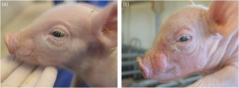
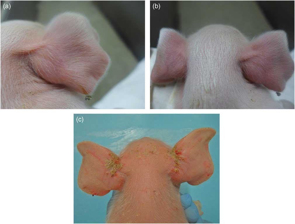
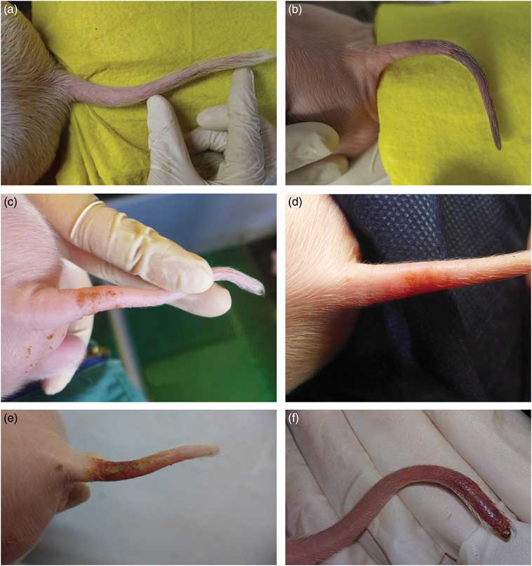
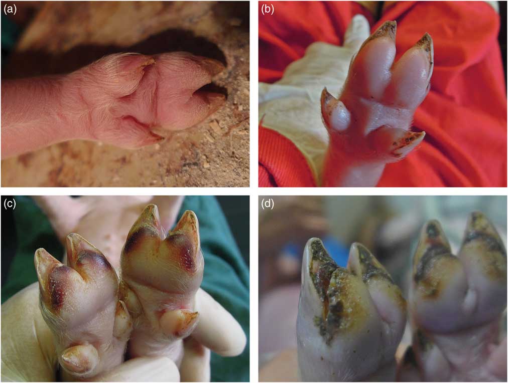
All litters were scored by a standardized clinical scoring system for the number and percentage of piglets that showed signs of inflammation and necrosis of the tail, inflammation of the ear base, inflammation of the heels, inflammation of the coronary band, inflammation/necrosis of the teats, inflammation of the umbilicus, swelling of the vulva and facial oedema and/or injuries. The scoring systems for the piglets was binary (0 = normal; 1 = affected). Examples of typical lesions are shown in Figures 1 to 8. Grades of clinical signs were not further differentiated. All examinations in any of the herds were conducted by the same two persons after a thorough introduction and training programme. All piglets were validated by both persons together; therefore there were no inter observer effects. The observers were blinded to the hypotheses of the study. All litters were scored only once in the pre-weaning period at an age of the piglets of 5.5 ± 2.7 days (Mean ± SD).
Faces were scored as follows: 0, pale and pink skin, with bristles and without any deposit or efflorescence (a); 1, presence of oedema, exudation and/or erosions (b). As soon as erosions were visible, face biting between piglets could not be ruled out.

Ear bases of piglets were scored as follows: 0, pale pink, with bristles and without any deposit or efflorescence (a); 1, presence of swelling, erythema (b), exudation, encrustation or necrosis (c).

Tail bases and tail tips of piglets were scored as follows: 0, pale pink, with bristles and without any lesions, deposit or efflorescence (a); 1, if there were loss of bristles, swelling and discolouration (b), swelling, redness, erosion or coating (c), signs of inflammation at the tail base (d, e), encrustation and/or necrosis at the tail tip (f). Within the scope of the study, inflammation/necrosis of the base or tip of the tail were aggregated to the parameter inflammation/necrosis of the tail. There were no signs of bitten tails or mechanical destruction of tails.

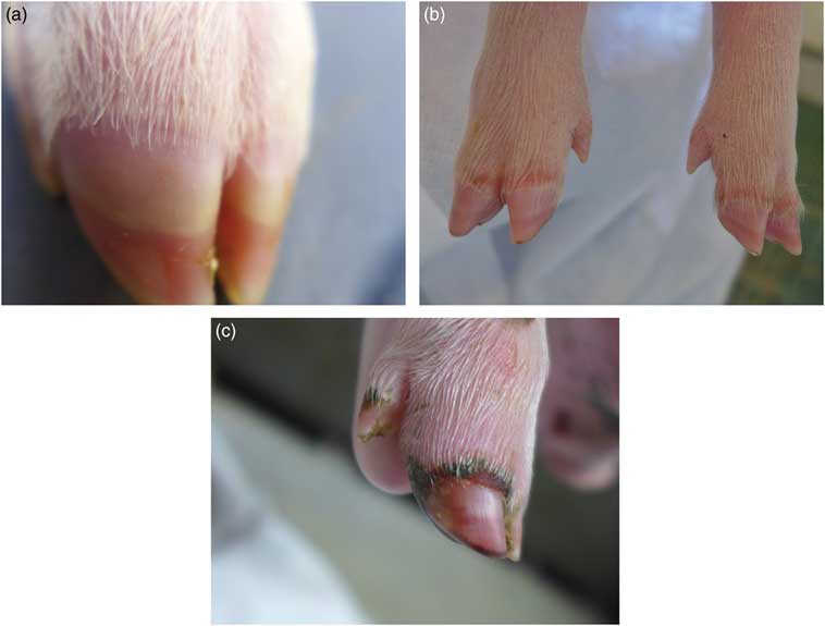
Heels of piglets were scored as follows: 0, not swollen and free from cracks and signs of bleeding (a); 1, presence of swelling (b), internal bleeding (c), cracks and separation from sole (d). Moreover, the dew claws could be involved.

Coronary bands of piglets were scored as follows: 0, not swollen and free from any lesions (a); 1, presence of clinical signs of inflammation like swelling, redness or exudation (b), encrustation or necrosis (c). Moreover, the dew claws could be involved.

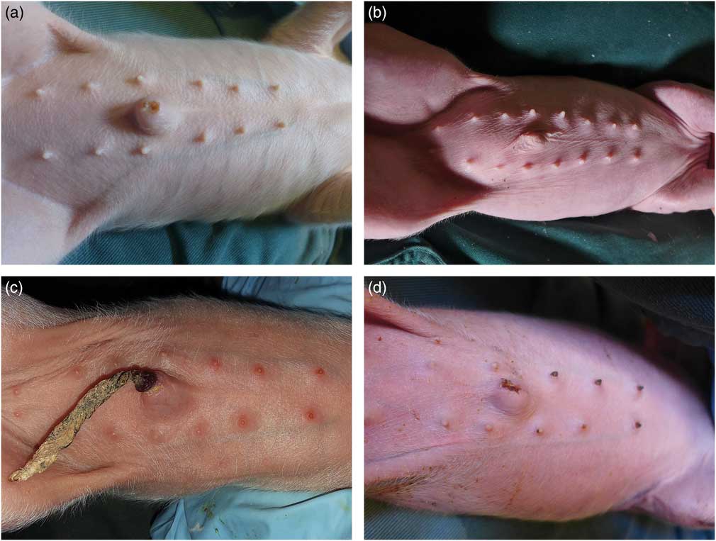
Teats of the piglets were scored as follows: 0, not swollen and free from any deposit or efflorescence (a); 1, presence of swelling (b), redness (c) or necrosis (d).

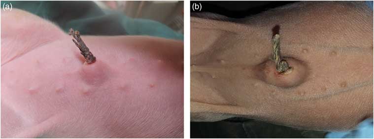
The umbilicus of each piglet was scored as follows: 0, not swollen and free from any signs of inflammation (a); 1, presence of swelling or redness (b).

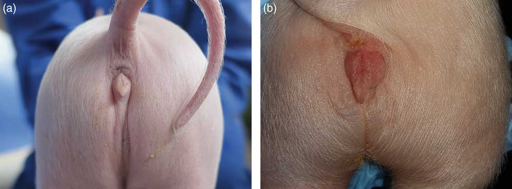
The vulva of female piglets was scored as follows: 0, not swollen and free from any signs of inflammation (a); 1, presence of swelling and redness (b).

Statistics
The statistical evaluation was conducted on data from 19 herds, including 382 litters and 4725 piglets. Analysis of traits was performed using the IBM SPSS (Version 23) software package. Piglet data were aggregated by litter to provide the percentage of piglets per litter with alterations in the form of inflammation/necrosis of the tail, ear base, heels, coronary bands, teats and umbilicus, vulva, and face. The statistical unit was the litter (n = 382). General data analysis was performed using the descriptive function, based on the mean, standard deviation, minimum and maximum. Effects of sows’ and boars’ genetics were tested using the following generalised mixed linear model, leading to least square means:
 $$\mu {\rm {\equals}mean {\plus}}H\left( {S{\times}B} \right)_{I} {\rm {\plus}}S_{j} {\rm {\plus}}B_{k} {\rm {\plus} }D_{l} {\rm {\plus} }A_{m} {\rm {\plus}}e_{{ijklmn}} $$
where mean is the mean for the trait, H(S × B)i = random effect of herds within genetics (boar after sow); S j the fixed effect of sow’s genetics, B k the fixed effect of boar’s genetics, D l the fixed effect of degree of docking, A m the age of piglets as covariate, e ijklmn the error.
Residuals were tested and found to be distributed approximately normally. Correlations were analysed as Spearman correlations. Multiple testing was adjusted by Bonferroni correction. Levels of significance was P <0.05.
The effects of intact tails (non-tail docked piglets) on the outcome analysis were estimated for the following data subset out of four herds with 83 litters (531 piglets in 45 litters with tails docked half; 337 piglets in 28 litters with 1/3-docked tails; 94 piglets in 10 litters without tail docking, respectively). The percentage of piglets with inflammation/necrosis of the tail was analysed by univariate ANOVA, with the herd as a random effect and the piglets’ age as a covariate. Data are presented as means and standard error. The coefficient of determination for this model was 27%.
The effect of the piglet’s age was estimated as a random factor in ANOVA.
Results
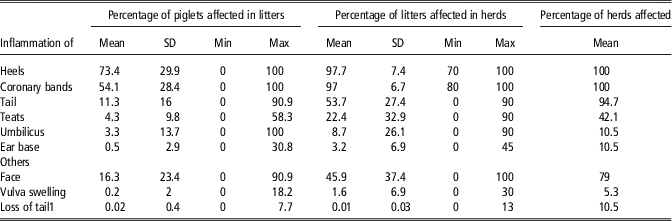
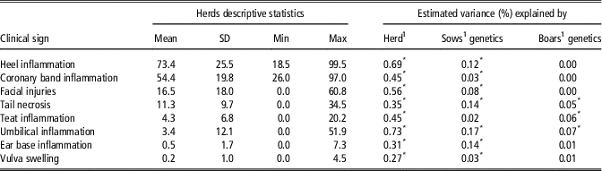
Table 1 shows the descriptive statistics for piglets, litters and herds. Piglets were affected by inflammation of the heels, coronary bands and docked tails in 73.4%, 54.1% and 11.3% of cases. The other clinical findings were visible at lower rates. There was a clear variation between litters, ranging from 0% to 100% of piglets affected. Almost all litters were affected by inflammation of the heels and coronary bands and more than 50% of the litters had at least one piglet with tail inflammation. These three dominant clinical findings were present in almost all herds (94.7% to 100%). Within herds, zero to 34.5% of the piglets could be affected by tail inflammation on distinct herds (Table 2). The herd effect was statistically significant for all clinical findings, explaining between 27% and 73% of the variance in the prevalence of lesions (Table 2). A further 2% to 17% of variance was explained by the sows’ genetics, whereas the boars’ genetics had only minor effects.
Descriptive statistics for piglets showing clinical signs of an inflammatory response (in total, 4725 piglets, 350 litters, 19 herds)

Min = minimum; Max = maximum.
1 Only one of the piglets had lost its tail, probably by mechanical damage.
Least square means, SD, minimum (Min) and maximum (Max) of the percentage of piglets in litters showing clinical signs of inflammation (4725 piglets in 350 litters from 19 herds) and estimated variance explained by herd, sows’ genetics and boars’ genetics

1 Proportion of the variance explained by the respective effect in the total variance.
* Effect is statistically significant (P < 0.05).
Effects of degree of tail docking
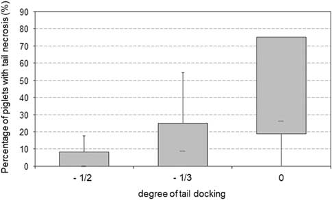
When the prevalence of alterations within litters was assessed, a negative correlation between docking length and the prevalence of tail lesions was noted. Litters with tails docked half showed the least occurrence of tail necrosis compared to litters with 1/3-docked or undocked tails. Percentage of piglets affected by tail necrosis increased from 8.3% within litters that were docked half to 14.8% of piglets with tip-clipped tails (1/3 docked) and 68% of undocked piglets. The differences between undocked piglets and those with docked tails were significant (P = 0.04 and P = 0.002 for tip docked and half docked piglets, respectively) (Figure 9).
Effect of degree of tail docking on percentage of piglets per litter affected by tail necrosis. Data were collected from four herds at which all individual docking methods were employed: 531 piglets in 48 litters docked at the half (−1/2) tail length; 337 piglets in 28 litters with tip docking (~one-third of the tail cut) (−1/3); 94 piglets in 10 litters without tail docking (0). Data show medians and boxplots (75%/25% percentiles) with whiskers (95%/5% percentiles). Significance was tested with a median test. The overall effect of degree of tail docking was significant at P <0.001). The difference between −1/2 tail and 0 (not docked) was significant at P = 0.002; the difference between −1/3 tail and 0 was statistically significant at P = 0.04. Exact values are given as Supplementary Table S1.

Effect of piglet age
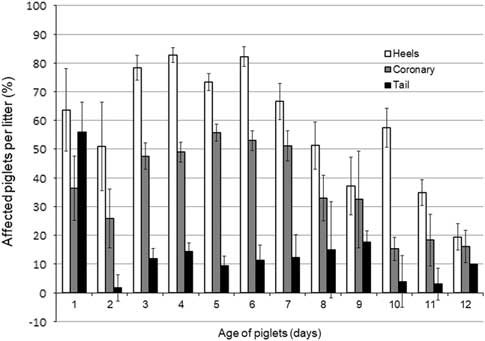
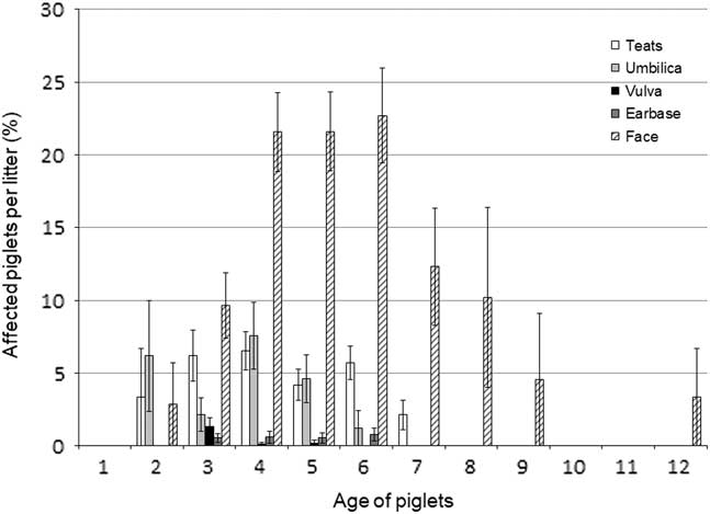
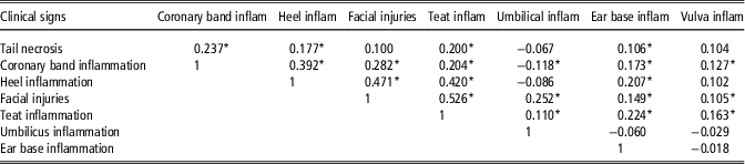
Although piglets in individual herds and litters were scored at different days of life, an overall trend analysis indicated that the clinical signs increased during the 1st week of life and then decreased again during the 2nd week (P <0.001) (Figures 10 and 11). Inflammation and necrosis of heels, coronary bands and tails were already visible at days 1 and 2 of life, while the other clinical signs (inflammation of vulva and ear base) started from days 2 or 3 onwards. Different clinical signs showed significant, mostly positive correlations with each other (Table 3).
Effect of the age of piglets on the percentage of animals per litter, affected by inflammation of the heels and coronary bands and inflammation/necrosis of the tail (mean ± standard error). The correlation of piglet age with inflammatory alterations was statistically significant (P <0.001). Exact values are given as Supplementary Table S2.

Effect of the piglets’ age on the percentage of piglets per litter, affected by inflammation/necrosis of teats, umbilicus, vulva and ear base and by inflammation/injuries of the face (mean ± standard error). The correlation of the piglet age was statistically significant (P <0.001), except for inflammation/necrosis of vulva and ear base. Exact values are given as Supplementary Table S3.

Spearman correlations between percentage of piglets in litters affected by the respective clinical signs (350 litters)

inflam = inflammation.
* P < 0.05.
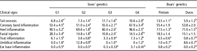
Table 4 shows the impact of the genetics of sows and boars on the assessed clinical signs. Besides the significant effects of sow and boar, there was also a significant herd effect for all assessed clinical signs (P <0.001). The genetics of the sow had a significant effect on inflammation and necrosis of the tail (even though all tails were docked), claw coronary bands, heels, teats, umbilica and ear base, as well as on the prevalence of facial injuries. In particular, litters of sows’ genetic G4 showed a higher overall susceptibility to developing inflammation and necrosis in some parts of the body of the piglets. Litters from the Pietrain boar line were more prone to develop inflammation and necrosis of the tail, heels, teats and ear base, but not to inflammation of the coronary bands.
Least square mean and standard error for the effects of sows’ and boars’ genetics on the percentage of piglets in litters, affected by clinical signs of inflammation (350 litters)

G1 to G4 = four different genetic lines of sows, representing typical production lines of various breeding companies.
Data represent least square means of the percentage of affected piglets per litter.
a,b,cBetween groups, means with different superscript letters differ significantly at P < 0.05.
Discussion
The present study aimed to get insight into the prevalence of clinically visible inflammatory processes in piglets in the first days of life. The scoring system included a visual scoring of inflammation and necrosis of the tail, ear base, claw coronary bands and heels, as well as umbilical area and vulva in piglets and is in line with current strategies in the assessment of animal-based welfare indicators (see European Network of Centres of excellence for Animal Welfare (ENCAW) report, 2016). Field observations and scientific studies have shown that discontinuing tail docking under current practical conditions can increase the prevalence of tail biting (Valros et al., Reference Valros, Ahlström, Rintala, Häkkinen and Saloniemi2004; Lahrmann et al., Reference Lahrmann, Busch, D’Eath, Forkman and Hansen2017). While various factors may induce tail biting (EFSA, 2007; D’ Eath et al., Reference D’ Eath, Arnott, Turner, Jensen, Lahrmann, Busch, Niemi, Lawrence and Sandøe2014), earlier studies focussed on tail-biting as a behavioural disorder attributable to the barren environment and housing conditions. However, even under extensive outdoor conditions as they are the common practice in Switzerland, a prevalence of tail biting between 14% and 20% was recorded in 2004 (Walker and Bilkei, Reference Walker and Bilkei2006). This suggests that three causalities have to be distinguished: tail biting as a behavioural disorder (also denoted primary biting; Taylor et al., Reference Taylor, Main, Mendl and Edwards2010), and secondary biting of pre-damaged tails, where the victim tolerates interactions with pen mates, due to a primary inflammatory process resulting in ischemic tail lesions. Moreover, tail necrosis without biting can be observed.
This third category of tail lesions occurs already in piglets at a very early stage of life and without any interference from other pigs (Penny et al., Reference Penny, Edwards and Mulley1971; Santi et al., Reference Santi, Gheller, Mores, Marques, Gonçalves, Gava, Zlotowski, Driemeier and Barcellos2008). In these cases, the authors described necrotic lesions at the base and tip of tails in new-born piglets. They assumed circular vascular constrictions and local ischemia as a possible pathogenesis and emphasized that this type of lesions can be clearly differentiated from tail biting. Penny et al. (Reference Penny, Edwards and Mulley1971) described that tail necrosis started as an erythema at the base of the tail of 4-day-old docked or undocked piglets, which agrees with the findings of the present study.
Inflammation and necrosis of the tail, claw coronary bands and inflamed heels have been detected in the current study involving piglets during the 1st day of life (2 litters with 23 piglets). This makes a major influence of external factors such as the housing conditions less likely and points towards a metabolic, endogenous stressor and/or systemic challenge from pro-inflammatory agents. Moreover, the obvious improvement in the 2nd week of life, especially in the signs of inflammation at the coronary bands and heels, supports this consideration. Any mechanical cause (such as poor floor conditions) would have led to a progression of the pathological alterations over time rather than the observed decrease in the severity of the lesions as recorded in the current study after day 6. The involvement of pathogens in the process cannot be completely excluded, because no further diagnostic investigations were conducted. However, none of the herds had specific health problems at the time of the investigation.
Taken together, these findings support the hypothesis that piglets regularly experience a generalized inflammatory reaction that can be described as SINS. According to Langbein et al. (Reference Langbein, Lechner, Schrade and Reiner2016), SINS, a syndrome that leads to inflammation and necrosis in piglets, has to be regarded as indicator of an impaired animal welfare.
The causal agents that induce this inflammatory syndrome, however, remain to be elucidated. One of the most common inducers of an inflammatory response is lipopolysaccharide (of Gram negative bacteria, particularly Escherichia coli). Lipopolysaccharide exposure may occur already during and shortly after parturition, and is followed by activation of toll-like-receptor 4 (TLR4)-receptors signalling and subsequent release of pro-inflammatory cytokines (for review see, Vallance et al., Reference Vallance, Zeuner, Williams, Widera and Vaiyapuri2017). The interplay between psychosocial stress factors, an inflammatory response and the vulnerability to infections in piglets and the links to animal welfare were shown recently by Tuchscherer et al. (Reference Tuchscherer, Puppe, Tuchscherer and Kanitz2018).
The current results, which included also the analysis of a possible correlation between the tail-docked and non-tail-docked piglets clearly indicated the higher prevalence of tail lesions in piglets with undocked tails. Although the cohort of undocked piglets was rather small in this study, these findings require further investigation, considering the demand (EU Directive 2008/120/EC) that routine tail docking of piglets should be phased-out. No additional blood or tissue samples were investigated to characterize the nature of the clinically observable lesions. However, there was no evidence of tail biting or other behavioural abnormalities in this age group. Whether or not these findings can be related to the prevalence of tail lesions during the fattening period remains to be elucidated. Individual tail lesion reports from the abattoir were not available for the scored piglets, but the mean prevalence reported from the selected farms reached 30% in fatteners.
In conclusion, the current data which are based on clinical investigations of individual suckling piglets show an unexpected high prevalence of inflammatory reactions in suckling piglets. The onset of the clinical signs during the 1st days of life, their combined appearance and the spontaneous improvement during the 2nd week of life, largely exclude commonly discussed causes such as physical injury induced by poor housing conditions and undesirable behaviour such as tail and ear biting. As the described clinical conditions clearly indicate an impairment of animal welfare, further studies should be devoted to identify the causative agents including (subclinical) infectious disease and anti-nutritional factors such as mycotoxins that are known to affect the intestinal integrity and the innate immune system in the early phase of life, thereby increasing the risk for an inflammatory syndrome.
Acknowledgements
The authors would like to express their gratitude to the pig farmers and their support during the scoring of the piglets at the individual farms.
Declaration of interest
The authors declare that they have no competing interests.
Ethics statement
In the current study piglets were scored by visual inspection, without applying any invasive intervention. The study was conducted with the consensus of the responsible authorities (Landesamt für Verbraucherschutz, Thuringia, Germany; AZ: 22.2684-04-07-001/16).
Software and data repository resources
None of the data were deposited in an official repository.
Supplementary materials
To view supplementary material for this article, please visit https://doi.org/10.1017/S1751731118003403