INTRODUCTION
Chagas' disease, caused by the haemoflagellate protozoan Trypanosoma cruzi (T. cruzi), is one of the few functional gastrointestinal disorders for which a causative agent has been identified. This disease affects 17–24 million people, ranging from between the United States-Mexico border thought to Argentina and Chile (Dias et al. Reference Dias, Silveira and Schofield2002). Although control programmes directed at decreasing transmission of T. cruzi to humans through the reduction of vector populations have been successful in some areas of Latin America, effective control measures are lacking in most regions of endemicity (Nowicki et al. Reference Nowicki, Chinchilla, Corado, Matsuoka, Selby, Steurer, Mone, Mendez and Aswad2006).
During the acute phase, the parasite has the ability to infect a wide variety of tissues including the viscera and central nervous system. In the chronic phase, lesions in the central, peripheral autonomic and enteric nervous systems lead to widely diverse disorders of the hollow muscular organs, including the heart, oesophagus and colon (Koberle, Reference Koberle1968). One of the most typical pathological features is the reduction in the number of ganglion cells in the enteric nervous system (ENS). Many investigators have attempted to explain the extensive neuronal loss associated with the digestive form of the Chagas' disease, mainly in the chagasic megacolon (Adad et al. Reference Adad, Cancado, Etchebehere, Teixeira, Gomes, Chapadeiro and Lopes2001). The participation of immune mechanisms in the destruction of neurons has received much attention (Lemos et al. Reference Lemos, Reis, Adad, Silva, Crema and Correa-Oliveira1998; Corbett et al. Reference Corbett, Ribeiro, Prianti, Habr-Gama, Okumura and Gama-Rodrigues2001; da Silveira et al. Reference da Silveira, Arantes, Vago, Lemos, Adad, Correa-Oliveira and D'Avila Reis2005).
Among inflammatory cells, macrophages, eosinophils and mast cells may be of particular importance, as they can produce a great number of substances involved in host defence and normal gut function, including cytokines and neurotrophins (Noga et al. Reference Noga, Hanf, Gorges, Thai Dinh, Groneberg, Suttorp and Kunkel2005; Kariyawasam and Robinson, Reference Kariyawasam and Robinson2006). These cells have physiological relevance in the intestinal functions (Straumann and Simon, Reference Straumann and Simon2004; Stepanyants and Chklovskii, Reference Stepanyants and Chklovskii2005) and may contribute to the development of chagasic megacolon (Vergara et al. Reference Vergara, Saavedra and Juanola2002; Pinheiro et al. Reference Pinheiro, Rua, Etchebehere, Cancado, Chica, Lopes and Adad2003; Cardoso et al. Reference Cardoso, Morato, Gomes, Rocha, Bonfim, Williams-Blangero, VandeBerg, Reis, Magalhaes and Correa-Oliveira2006). Many investigators have demonstrated that chagasic patients commonly exhibit an increased concentration of macrophages, eosinophils and mast-cells in the heart and intestine. However, the relationship between these inflammatory cells and the presence of fibrosis in the colon has yet to be determined (Milei et al. Reference Milei, Storino, Beigelman, Fernandez Alonso, Matturri and Rossi1991; Corbett et al. Reference Corbett, Ribeiro, Prianti, Habr-Gama, Okumura and Gama-Rodrigues2001; Cabral et al. Reference Cabral, Novak, Glocker and Castro-Viera2002; Pinheiro et al. Reference Pinheiro, Rua, Etchebehere, Cancado, Chica, Lopes and Adad2003; da Silveira et al. Reference da Silveira, Arantes, Vago, Lemos, Adad, Correa-Oliveira and D'Avila Reis2005; DosReis et al. Reference DosReis, Freire-de-Lima, Nunes and Lopes2005). Thus, the aim of this investigation was to characterize the presence of macrophages, eosinophils and mast cells in the colon of chagasic patients relative to unaffected individuals and to evaluate the correlation between the concentration of these and the occurrence of fibrosis in chagasic megacolon.
MATERIALS AND METHODS
Patients and samples
Samples of full thickness colon wall tissue were obtained from 8 chagasic patients with megacolon, 8 chagasic patients without megacolon and 10 non-infected individuals submitted to necropsy or surgical procedures at Faculdade de Medicina do Triângulo Mineiro (Uberaba, Minas Gerais, Brazil). Patients did not receive any parasite-specific treatment. Informed consent was obtained from the patient or family members prior to tissue procurement. This work was approved by UFMG Research Ethics Committee.
Serological tests indicative of Chagas' disease (complement fixation, haemagglutination, and immunofluorescence tests) were positive in all patients studied. All of the patients had left the endemic area more than 20 years before the tissue collection, and during that time, patients without megacolon did not present any symptom related to digestive disease. All patients originated from Uberaba, MG, Brazil, where the natural transmission of Chagas' disease was interrupted more than 20 years ago, and they had never received blood transfusions. The patients with and without megacolon had mean ages of 57±10 years, and 55±14 years respectively. The presence of megacolon was established based in clinical data reporting colon obstruction, and from radiological studies. Manometric studies of megacolon demonstrated decreased peristalsis and incomplete relaxation of the anal sphincter. These abnormalities precede clinical symptoms and dilatation seen by radiographic studies.
The control group was composed of non-infected individuals, as indicated by negative serology specific for Chagas' disease. Non-infected individuals were also from the state of Minas Gerais and had a mean age of 54±20 years.
Histology and peroxidase immunohistochemistry
Tissue samples were collected from the recto-sigmoid region. Each specimen was fixed in 4% neutral buffered formaldehyde solution and embedded in paraffin for immunohistochemistry studies. Sections of 7 μm were deparaffinized using xylene, and rehydrated through graded alcohols. Some sections were then stained for standard histology, using haematoxylin and eosin (H&E) (eosinophils), toluidine blue (mast cells) and Gomori trichrome (fibrosis), while others were prepared for immunohistochemistry. For immunohistochemistry, endogenous peroxidase was inhibited by incubation with 1% hydrogen peroxide and 30% absolute methanol for 30 min. The slides were then incubated with 2% normal swine serum (NSS) (Sigma, USA) in phosphate-buffered saline (PBS) for 15 min and subsequently with the anti-CD68 (DAKO, KP1, 1:100, USA) monoclonal antibody. Following this, the tissue sections were incubated with peroxidase-conjugated rabbit anti-mouse antibodies (DAKO) for 45 min, and peroxidase activity was detected by incubation with 3,3′-diaminobenzidine (Sigma) and hydrogen peroxide for 10 min. Slides were counterstained with Gill's haematoxylin (Sigma), dehydrated in graded alcohols, and mounted in synthetic mounting media. Negative control slides without primary antibody were performed for each case.
Cell quantification and morphometric studies
Enumeration of eosinophils and mast cells was performed in the enteric plexuses (submucosal and myenteric plexuses) and muscle layers, by counting 20 randomly-selected fields (total area of 1066 μm2) on a single slide per patient.
Morphometric studies of macrophages were performed by image analysis (Kontron KS300 v. 2.0), by measuring CD68-IR areas on 20 randomly-selected fields (total area of 1066 μm2) in each colon segment (submucosal plexus, inner muscular, myenteric plexus and outer muscular) on a single slide per patient.
Analysis of fibrosis was performed in 20 alternate fields in each muscle layer (total area of 1066 μm2 per muscle layer) on a single slide per patient. For the quantitative analysis of the area affected by fibrosis, images were obtained using a graphical measuring grid (Kontron KS 300) connected to an automatic image analysis system. All analyses were performed using the 40× objective lens.
Statistics
Statistical analysis among the different groups was performed by one-way ANOVA and Simple Linear Regression test. Differences were considered statistically significant at P<0·05.
RESULTS
Eosinophil analyses
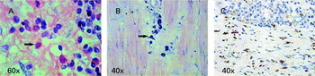
Eosinophils were observed in the neuronal plexuses and muscle layers in all analysed groups. In chagasic patients with megacolon, inflammation and myositis were observed in all examined cases. In this group, eosinophils were observed in the inflammatory foci in both nervous plexuses associated with damaged neuronal ganglia, as in the muscle layers (Fig. 1A). Chagasic patients without megacolon presented only discrete inflammation in 3 cases out of 8. Non-infected individuals did not show inflammatory foci or myositis in all analysed layers.
Characterization of eosinophils, mast cells and macrophages in the colon of chagasic patients with and without megacolon and non-infected individuals. (A) Haematoxylin-eosin section: eosinophils can be easily observed (arrow) in the colon associated with fibrosis and inflammatory mononuclear cells. (B) Toluidine blue section: chagasic patients with megacolon show clusters of mast cells associated with fibrosis foci (arrow). Others inflammatory cells were not seen near mast cell clusters. (C) Immunohistochemistry of macrophages CD68-IR: macrophages can be easily seen in the muscle layers and near inflammatory foci in the colon of chagasic patients with megacolon.

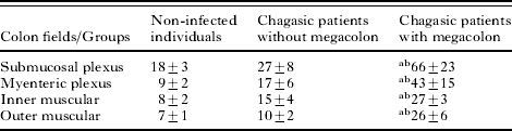
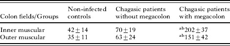
The number of eosinophils in all analysed layers was significantly greater in chagasic patients with megacolon than in the other 2 groups. When pairs of groups were analysed together, it was noted that the eosinophil count was greater in the chagasic patients with megacolon in comparison with the chagasic patients without megacolon and the non-infected individuals. There was no significant difference between the non-infected individuals and the chagasic patients without megacolon (Table 1).
Mean number and standard deviation of eosinophils in the enteric plexuses and muscle layers from chagasic patients' colon
(Quantification of eosinophils associated with the enteric plexuses and muscle layers from chagasic patients with and without megacolon (number of cells/mm2).)
a Statistically significant differences between this group and non-infected individuals.
b Statistically significant differences between this group and chagasic patients without megacolon (P<0·05). A total area of 1066 μm2 from each field for all patients was analysed.
Mast cell analyses
Chagasic patients with megacolon did not present any mast cells associated with enteric ganglia in nervous plexuses, although these cells were always associated with areas of fibrosis (Fig. 1B) and surrounding inflammatory foci. Moreover, mast cells were observed forming clusters and dispersed over all analysed layers. Analyses of mast cell distribution in chagasic patients without megacolon and non-infected individuals showed that, in contrast, cells were not observed associated with inflammatory foci and did not form clusters.
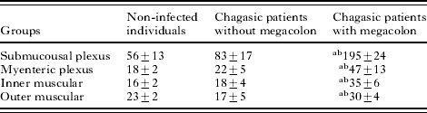
The mast cell count showed that chagasic patients with megacolon have a greater number of these cells relative to the other 2 groups investigated. Data analysis showed that the mast cell count was statistically higher in the chagasic patients with megacolon relative to the chagasic patients without megacolon and non-infected individuals. There was no significant difference between non-infected individuals and the chagasic patients without megacolon (Table 2).
Mean number and standard deviation of mast cells in the enteric plexuses and muscle layers from chagasic patients' colon
(Quantification of mast cells of enteric plexuses and muscle layers from chagasic patients with and without megacolon (number of cells/mm2).)
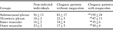
a Statistical difference between this group and non-infected individuals.
b Statistical differences between this group and chagasic patients without megacolon (P<0·05). A total area of 1066 μm2 from each field for all patients was analysed.
Macrophage CD68-IR area analyses
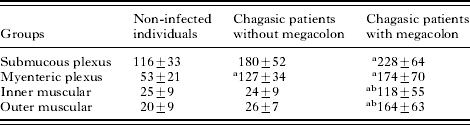
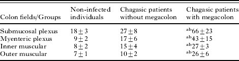
Quantification of the areas of sections occupied by macrophage CD68-IR area was performed on enteric plexuses (submucosal and myenteric plexus) and in the muscle layers of the colon. Chagasic patients with megacolon presented greater CD68-IR macrophage areas compared with the non-infected control group in all analysed fields (Fig. 1C), and, when compared with chagasic patients without megacolon, presented a higher area only in the muscle layers. Chagasic patients without megacolon showed a greater macrophage CD68-IR area than the non-infected control group only in myenteric plexus. No other statistically significant differences were observed (Table 3).
Mean number and standard deviation of macrophage CD68-IR area in the colon of chagasic patients
(Quantification of macrophage CD68-IR area in colon of chagasic patients with and without megacolon and non-infected individuals.)
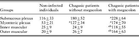
a Statistical differences between this group and non-infected individuals.
b Statistical differences between this group and chagasic patients without megacolon group (P<0·05). A total area of 1066 μm2 from each field for all patients was analysed. Reactive tissue area (μm2) per total tissue area (mm2).
Quantification of fibrosis area
Analyses of fibrosis areas in the muscle layers demonstrated that chagasic patients with megacolon presented a diffuse increase in endomysial and perimysial connective tissue and frequent fibrosis foci. Fibrous connective tissue was rare in the non-infected individuals group and it was represented only by endomysial or perimysial connective tissue. In chagasic patients without megacolon, the appearance of the connective tissue in the muscle layers was similar to that observed in non-infected individuals.
The fibrous connective tissue area relative to the total examined tissue area was greater in the chagasic patients with megacolon in relation to chagasic patients without megacolon and the non-infected individuals. There was no statistically significant difference between non-infected individuals and the chagasic patients without megacolon (Table 4).
Mean number and standard deviation of fibrotic area of muscle layers from chagasic patients' colon
(Quantification of proportional area occupied by fibrosis in colon of chagasic patients with and without megacolon and non-infected controls.)
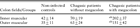
a Statistical differences between this group and non-infected individuals.
b Statistical differences between this group and chagasic patients without megacolon (P<0·05). A total area of 1066 μm2 from each field for all patients was analysed. Affected tissue area (μm2) per total tissue area.
Relation between inflammatory cells and fibrosis occurrence
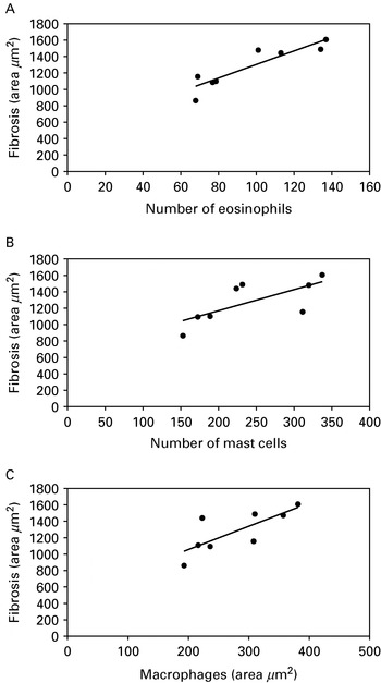
The statistical test Simple Linear Regression was used to evaluate the relation of the number of eosinophils, mast cells, macrophage area and fibrosis occurrence. Data analyses demonstrated that the presence of all studied inflammatory cells had a direct correlation with fibrosis occurrence (Fig. 2) and this was consistently observed in the chagasic group of patients with megacolon (P<0·05).
Relationship among the number of eosinophils (A), mast cells (B) macrophages area (C) and fibrosis occurrence in the colon of chagasic patients with megacolon. Data analyses demonstrated that the presence of all studied inflammatory cells have a direct relation with the fibrosis occurrence in these chagasic patients (P<0·05).

DISCUSSION
The main objective of this work was to quantify the presence of macrophages, eosinophils and mast cells in chagasic patients, with and without megacolon. We observed that chagasic patients with megacolon present increased numbers of eosinophils and mast cells, as well as a greater area occupied by CD68-IR macrophages, when compared with control individuals and even with chagasic patients without megacolon. These data are partly in agreement with the previous study by Pinheiro et al. (Reference Pinheiro, Rua, Etchebehere, Cancado, Chica, Lopes and Adad2003) which also described an increased concentration of mast cells in the colon of chagasic patients with megacolon.
We should consider that the increased concentration of these cells in the colon of chagasic patients with megacolon could be a consequence of the development of megacolon. Megacolon development is characterized by faecal accumulation in the intestinal lumen, which may compress the mucosa and cause ischaemia, with subsequent regressive cellular injuries and inflammation. The inflammatory process can reach the submucosa and, ultimately, the muscle layers and myenteric plexus (Tafuri et al. Reference Tafuri, Maria and Lopes1971). The accumulation of some inflammatory cells in patients with megacolon could be a consequence of this process, in which chronic inflammation induces cellular injury and necrosis. Studies carried out by Kobayashi et al. (Reference Kobayashi, Ohta, Inui, Yoshino and Nakazawa2002) and Noga et al. (Reference Noga, Englmann, Hanf, Grutzkau, Seybold and Kunkel2003) demonstrated that these cells are capable of producing, storing and secreting neurotrophins, essential for survival of neurons (Mendell et al. Reference Mendell, Albers and Davis1999; Sarkar et al. Reference Sarkar, Lakhtakia, Gill, Sharma, Mahapatra and Mehta2002; Micera et al. Reference Micera, Puxeddu, Aloe and Levi-Schaffer2003; Hashimoto et al. Reference Hashimoto, Nitta, Fukumitsu, Nomoto, Shen and Furukawa2005), and cytokines that participate in tissue regeneration (Hirschberg et al. Reference Hirschberg, Yoles, Belkin and Schwartz1994; Slawinska et al. Reference Slawinska, Majczynski and Djavadian2000; Kiefer et al. Reference Kiefer, Kieseier, Stoll and Hartung2001; Lakatos and Franklin, Reference Lakatos and Franklin2002; Smythies et al. Reference Smythies, Sellers, Clements, Mosteller-Barnum, Meng, Benjamin, Orenstein and Smith2005). In this context, we could speculate that the increased density of eosinophils, mast cells and macrophages in chagasic megacolon, can be perceived as an attempt of intestinal homeostasis reestablishment. The significant presence of those cells in the colon of non-infected individuals, implies their role in the maintenance of intestinal physiology, as has been well described in the literature (Levy et al. Reference Levy, Yamazaki, Van Keulen, Burgart, Sandborn, Phillips, Kephart, Gleich and Leiferman2001; Straumann and Simon, Reference Straumann and Simon2004).
We can also consider that eosinophils, mast cells and macrophages could be responsible for the parasite and/or host tissue damage. These cells when associated with the inflammatory process, can promote an increased tissue reaction by the secretion of cytokines such as IL-1, TNF-α and IL-6, which have the capacity to activate cytotoxic processes mediated by the cells of the immune system. Moreover, these cells by themselves can cause injury to parasites or host cells. Additionally, eosinophils and macrophages have cytotoxic capacity to produce, when activated, substances such as nitric oxide and free radicals (Geboes, Reference Geboes1994; Daryani et al. Reference Daryani, Hosseini and Dalimi2003).
The results of this study agree with quantitative studies of macrophages, eosinophils and mast cells in the human myocardium and oesophagus recognized previously (Almeida et al. Reference Almeida, Pereira and Tafuri1975; Laguens et al. Reference Laguens, Cabeza Meckert and Vigliano1999; Cabral et al. Reference Cabral, Novak, Glocker and Castro-Viera2002; da Silveira et al. Reference da Silveira, Arantes, Vago, Lemos, Adad, Correa-Oliveira and D'Avila Reis2005). Our data also agree with the description of mast cell counts in the circular musculature of the small and large intestines in chagasic patients (Pinheiro et al. Reference Pinheiro, Rua, Etchebehere, Cancado, Chica, Lopes and Adad2003). The data presented are in agreement with those of da Silveira et al. (Reference da Silveira, Arantes, Vago, Lemos, Adad, Correa-Oliveira and D'Avila Reis2005) that related augmented concentration of CD68-IR macrophages area in the oesophagus of chagasic patients with megaesophagus in relation with non-infected individuals. In addition, results obtained using an experimental model of Chagas' disease suggest the participation of eosinophils, mast cells and macrophages in the injury process of intrinsic nervous system components of the heart in rats infected with T. cruzi (Pinheiro et al. Reference Pinheiro, Beraldo, Junqueira Junior, Lopes and Chapadeiro1992; Melo and Machado, Reference Melo and Machado2001).
Morphometric analysis of areas occupied by fibrosis demonstrated that there was a significantly greater area of the colon muscle layers affected by fibrosis in chagasic patients with megacolon, relative to chagasic patients without megacolon and non-infected individuals. Going further, we investigated the relationship between the presence of analysed inflammatory cells and fibrosis in the colon from those patients. When we compared the Chagas' groups with and without megacolon, we observed a significant relationship between the mast cell and eosinophil count, CD68-IR macrophage area and the percentage area with fibrous connective tissue. These results agree with the observations of Tafuri et al. (Reference Tafuri, Maria and Lopes1971) and with the hypothesis that interstitial fibrosis is correlated with the increase in the number of eosinophils and mast cells present in pathologies that affect the gastrointestinal tract, as in Crohn's disease (Xu et al. Reference Xu, Rivkind, Pikarsky, Pappo, Bischoff and Levi-Schaffer2004). Furthermore, some studies had confirmed that macrophages are involved in fibrosis development as in the intestine, as well as in other organs (Bachem et al. Reference Bachem, Zhou, Zhou and Siech2006; Motomura et al. Reference Motomura, Khan, El-Sharkawy, Verma-Gandhu, Verdu, Gauldie and Collins2006).
These results infer that eosinophils, mast cells and macrophages are correlated with fibrosis occurrence in the muscle layers of the colon, and possibly with the megacolon development. Adad et al. (Reference Adad, Cancado, Etchebehere, Teixeira, Gomes, Chapadeiro and Lopes2001) showed that chagasic patients with megacolon have a significant reduction in the number of neurons in the colon as well as a considerable incidence of inflammatory process. These data agree with our hypothesis that these inflammatory cells are directly involved in the establishment of megacolon in chagasic patients. Our findings suggest that potential therapeutic agents could target these cells. It is possible that blocking components, that cause eosinophil, mast cell and macrophage activation or cause a temporarily inactivation of these cells may reduce the severity of Chagas' induced megacolon, thus avoiding fibrosis in the colon, or delaying megacolon development.