INTRODUCTION
Trypanosoma evansi is a flagellated monomorphic hemoparasite that infects a variety of domestic and wild animals, such as horses, camels, cows, dogs and some other economically important mammals (Silva et al. Reference Silva, Seidl, Ramirez and Dávila2002), but rarely parasitizes humans (Joshi et al. Reference Joshi, Shegokar, Powar, Herder, Katti, Salkar, Dani, Bhargava, Jannin and Truc2005; Chau et al. Reference Chau, Chau, Desquesnes, Herder, Lan, Campbell, Cuong, Yimming, Chalermwong, Jittapalalong, Franco, Tue, Rabaa, Carrique-Mas, Thanh, Thieu, Berto, Hoa, Hoang, Tu, Chuyen, Wills, Hien, Thwaites, Yacoub and Baker2016). This protozoan has a wide geographical distribution and it can be found in some countries of Africa, Europe, Asia, and Central and South America (Silva et al. Reference Silva, Seidl, Ramirez and Dávila2002). Trypanosoma evansi causes an important horse disease called ‘mal das cadeiras’ or ‘surra’, which is characterized by anaemia, motor incoordination, immunosuppression and severe neurological disorders (Rodrigues et al. Reference Rodrigues, Fighera, Souza, Schild, Soares, Milano and Barros2005).
The central nervous system (CNS) is protected by the blood–brain barrier (BBB), a physical barrier that defends the brain from microbial invasion. Recently, a study conducted by Ueno and Lodoen (Reference Ueno and Lodoen2015) reported that several parasites have evolved to breach the BBB, including T. evansi (Acosta et al. Reference Acosta, Rondón-Mercado, Avilán and Concepción2016). The mechanism by which trypanosomes penetrate the BBB remains unclear, but this invasion occurs where the BBB has been disrupted directly by the parasite or by the release of chemical mediators, such as pro-inflammatory cytokines (Masocha et al. Reference Masocha, Robertson, Rottenberg, Mhlanga, Sorokin and Kristensson2004). Usually clinical signs of neurological disorders are reported in experimentally and naturally infected animals (Da Silva et al. Reference Da Silva, Spanevello, Stefanello, Wolkmer, Costa, Zanette, Lopes, Santurio, Schetinger and Monteiro2010), as well as alterations linked to cognitive impairment in experimental models, such as memory impairment, depression and increased anxiety (Baldissera et al. Reference Baldissera, Rech, Da Silva, Nishihira, Ianiski, Gressler, Grando, Vaucher, Schwertz, Mendes and Monteiro2015, Reference Baldissera, Souza, Grando, Moreira, Schafer, Cossetin, da Silva, da Veiga, da Rocha, Stefani, da Silva and Monteiro2017). According to Baldissera et al. (Reference Baldissera, Rech, Da Silva, Nishihira, Ianiski, Gressler, Grando, Vaucher, Schwertz, Mendes and Monteiro2015), the capacity to cross the BBB is linked to these neurological disorders, which leads to necrotizing encephalitis (Rodrigues et al. Reference Rodrigues, Fighera, Souza, Schild and Barros2009).
The most common chemical compound used to treat infected animals is diminazene aceturate (D.A.), which provides the elimination of trypanosomes in the bloodstream few hours after its administration (Oliveira et al. Reference Oliveira, Rigo, Rosa, Gressler, Zimmermann, Ourique, Da Silva, Miletti, Beck and Monteiro2014). However, many cases of resistance and low efficacy are observed when this drug is used followed by parasitaemia relapses (Baker et al. Reference Baker, de Koning, Maser and Horn2013). This occurs when the parasite crosses the BBB, reaching the CNS, a well-known refuge area for T. evansi during the residual period of the drug that lasts approximately 21 days (Oliveira et al. Reference Oliveira, Rigo, Rosa, Gressler, Zimmermann, Ourique, Da Silva, Miletti, Beck and Monteiro2014). It is important to emphasize that D.A. does not cross the BBB, and consequently, the parasite may survive in the CNS even after D.A. treatment. In this sense, several studies have used nanotechnology as an attempt to improve the therapeutic efficacy of natural and synthetic trypanocidal drugs (Abamor and Allahverdiyev, Reference Abamor and Allahverdiyev2016; Want et al. Reference Want, Yadav and Afrin2016), such as nerolidol loaded in nanospheres (N-NS) against T. evansi (Baldissera et al. Reference Baldissera, Grando, Souza, Cossetin, Sagrillo, Nascimento, da Silva, Dalla Lana, Da Silva, Stefani and Monteiro2016a ).
Recently, a study conducted by Baldissera et al. (Reference Baldissera, Grando, Souza, Cossetin, Sagrillo, Nascimento, da Silva, Dalla Lana, Da Silva, Stefani and Monteiro2016a ) demonstrated that nanoencapsulation of nerolidol was able to improve its therapeutic efficacy against T. evansi. According to these authors, N-NS treatment showed 66% therapeutic effectiveness, while nerolidol on its free form (F-N) and D.A. treatment showed 0 and 33%, respectively. Also, Baldissera et al. (Reference Baldissera, Souza, Grando, Moreira, Schafer, Cossetin, da Silva, da Veiga, da Rocha, Stefani, da Silva and Monteiro2017) demonstrated that N-NS was able to prevent the cognitive deficit caused by T. evansi, a fact not observed after F-N treatment. Therefore, our hypothesis is that nanotechnology allows the passage of nerolidol through the BBB. Based on these evidences, the aim of this study was to investigate whether N-NS treatment is able to cross the BBB, to eliminate T. evansi from the CNS, and to cure infected animals.
MATERIALS AND METHODS
Nerolidol-loaded nanospheres
Preparation and characterization of N-NS: N-NS suspension was prepared according to the interfacial deposition of preformed polymers (Fessi et al. Reference Fessi, Puiseux, Devissaguet, Ammoury and Benita1989), recently described in details by Baldissera et al. (Reference Baldissera, Souza, Grando, Moreira, Schafer, Cossetin, da Silva, da Veiga, da Rocha, Stefani, da Silva and Monteiro2017). The components of the organic phase (eudragit S 100®, sorbitan monostearate, nerolidol and acetone) and aqueous phase (polysorbate 80 and milli-Q water) were placed in a beaker and kept under magnetic stirring in a water bath at 40 °C for 1 h, and the active principle was added to the organic phase at the end of the heating period. With the aid of a funnel, the organic phase was poured into the aqueous phase and stirred for 10 min. This mixture was concentrated to a final volume of 100 mL in a rotatory evaporator to eliminate the organic solvent, adjusting the final concentration of the active principle to 3 mg mL−1 from pure nerolidol (100%).
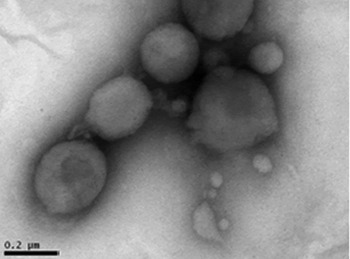
Morphological analysis was determined by transmission electron microscopy (TEM) (JEM 2010, Tokyo, Japan) at 200 kV. For this, the suspension was diluted (10×) in ultrapure water, placed on a specimen grid, and negatively stained with uranyl acetate (2% W/V). Moreover, the physical and chemical properties of N-NS, such as particle size, polydispersion index, zeta potential and pH, were performed according to the methodology recently described in details by Baldissera et al. (Reference Baldissera, Souza, Grando, Moreira, Schafer, Cossetin, da Silva, da Veiga, da Rocha, Stefani, da Silva and Monteiro2017). The particle size was 149·5 (±1·0 nm), and the polydispersion index was 0·117 (±0·0010), with a zeta potential of −12·8 (±1·1 mV), and pH 6·84 (±0·03). TEM analysis revealed spherical particles of similar sizes to those found by photon correlation spectroscopy technique (approximately 150–200 nm) (Fig. 1).
Transmission electron microscopy (TEM) image of nerolidol loaded in nanospheres (N-NS). Scale bar = 0·2 µ m.

Trypanosoma evansi isolate
Trypanosoma evansi was originally isolated from a dog naturally infected (Colpo et al. Reference Colpo, Monteiro, Stainki, Colpo and Henriques2005). During the first step, one rat (R1) was infected intraperitoneally with infected blood that was kept cryopreserved in liquid nitrogen. This procedure was performed to obtain a large amount of viable T. evansi for mice infection.
Animals and experimental design
Forty-eight (female) 60-day-old mice (outbred strain and heterogenic) weighing an average of 30 (±0·4) g were used in this study. Animals were maintained at 21–23 °C and 60% humidity with free access to water and food, under a 12:12 h light/dark cycle (7:00 AM to 7:00 PM). Animals were used according to the guidelines of the Committee on Care and Use of Experimental Resources (protocol number 2391230516) of the Universidade Federal de Santa Maria (UFSM). All efforts were made to minimize animal suffering and the number of animals.
The animals were divided into eight groups (A–H), with six animals each. Groups A, B, C and D were composed of uninfected animals, while animals in the groups E, F, G and H were inoculated intraperitoneally with 0·06 mL of blood obtained from one rat (R1), containing 2·0 × 105 trypanosomes. Groups A and E were used as negative control and positive control (saline treatment), respectively. Animals from groups B and F received F-N treatment, while animals of groups C and G received N-NS treatment, both at dose of 1·0 mL kg−1 (100% concentration) administered by oral gavage for 5 consecutive days before infection and 5 consecutive days after infection, according to the protocol described by Baldissera et al. (Reference Baldissera, Souza, Grando, Moreira, Schafer, Cossetin, da Silva, da Veiga, da Rocha, Stefani, da Silva and Monteiro2017). Animals from groups D and H received D.A. treatment 1 h post-infection (PI), according to manufacturer's recommendations (a single dose of 3·5 mg kg−1, intramuscularly). The animals were daily observed, and parasitaemia was daily monitored through blood smears. Each slide was prepared with fresh blood collected from the tail vein, stained by the panoptic method and visualized at a magnification of 1000×, according to the methodology described by Da Silva et al. (Reference Da Silva, Doyle and Monteiro2006).
Sample collection and tissue preparation
Five days PI, the animals were anaesthetized with isoflurane in an anaesthetic chamber, and blood samples were collected by cardiac puncture using tubes without anticoagulant. Serum was separated by centrifugation (3000 g for 15 min) in order to quantify seric concentrations of nerolidol and D.A. Brain samples were removed and separated into two parts: the left hemisphere was used to determine the concentration of nerolidol and D.A., while the right hemisphere was used to detect T. evansi by polymerase chain reaction (PCR). For nerolidol and D.A. quantification in the brain tissue, the left hemisphere was homogenized (1:3) in phosphate-buffered saline, and centrifuged (5000 g for 10 min at 4 °C). Acetone (1:1) was added to the supernatant for deproteinization and nerolidol extraction, followed by centrifugation (10 000 g for 10 min at 4 °C), as recommended by Trevisan et al. (Reference Trevisan, Rossato, Tonello, Hoffmeister, Klafke, Rosa, Pinheiro, Pinheiro, Boligon, Athayde and Ferreira2014). The supernatant was stored at −80 °C until utilization.
Nerolidol and D.A. concentrations in serum and brain tissue
Chemical, apparatus and general procedures
All chemicals used in this study were of analytical grade. Acetonitrile, methanol, acetic acid and ammonium acetate were purchased from Merck (Darmstadt, Germany). High-performance liquid chromatography (HPLC-DAD) was performed using a Shimadzu Prominence Auto Sampler (SIL-20A) HPLC system (Shimadzu, Kyoto, Japan), equipped with Shimadzu LC-20AT sharing pumps connected to a DGU 20A5 degasser with a CBM 20A integrator, SPD-M20A diode array detector and LC solution 1.22 SP1 software.
Quantification of seric and cerebral nerolidol levels by HPLC-DAD
Seric and cerebral samples were injected into reversed phase Phenomenex C18 column (250 × 4 mm) packed with 5 µ m diameter particles. The mobile phase consisted initially of 65% acetonitrile and 35% water for 18 min, followed by a gradient of 40–100% acetonitrile for 18–25 min and 100% acetonitrile for 25–40 min. The flow rate was 1·2 mL min−1 and the injection volume was 20 µL, as the method described by Gruber et al. (Reference Gruber, Kittipongpatana, Bloxton, Marderosian, Schaefer and Gibbs2004) with few modifications. The sample and mobile phase were filtered through a 0·45 µ m membrane filter (Millipore), and degassed by ultrasonic bath prior to use. Stock solutions of standard reference were prepared in the HPLC mobile phase at a concentration range of 0·050–0·250 mg mL−1. Quantifications were carried out by the integration of the peaks using the external standard method, at 210 nm with a variable wavelength UV-visible detector. All chromatography operations were carried out at room temperature and in triplicate. Calibration curve: Y = 13 089x + 8247·5 (r = 0·9997) (Y = sample absorbance; x = concentration; r = coefficient of determination).
Quantification of seric and cerebral D.A. levels by HPLC-DAD
Compounds were separated with a gradient reverse-phase system. A Phenomenex C8 column (4 × 150 mm) packed with 5 µ m diameter particles was used. The mobile phase consisted of acetonitrile/methanol/formic acid (10:88:2 v/v/v) at a flow rate of 0·7 mL min−1. The injection volume was 20 µL, and the procedure followed the method described by Baldissera et al. (Reference Baldissera, Gonçalves, Sagrillo, Grando, Ritter, Grotto, Brum, da Luz, Silveira, Fausto, Boligon, Vaucher, Stefani, da Silva, Souza and Monteiro2016b ). The sample and mobile phase were filtered through 0·45 µ m membrane filter (Millipore), and degassed by ultrasonic bath prior to use. Stock solutions of standard reference were prepared in the HPLC mobile phase at a concentration range of 0·050–0·500 mg mL−1. Quantifications were carried out by integration of peaks using the external standard method for D.A. at 254 nm. All chromatography operations were carried out at room temperature and in triplicate. Calibration curve: Y = 12 549x + 356·7 (r = 0·9980) (Y = sample absorbance; x = concentration; r = coefficient of determination).
Molecular diagnosis
In order to evaluate whether the treatments were able to eliminate T. evansi from the brain tissue, DNA extraction from the right hemisphere was performed using Trizol reagent (Thermo Fisher Scientific, Massachusetts, USA), according to manufacturer's recommendations.
Conventional PCR was performed using primers specific for T. evansi (fwd 5′ TGCAGACGACCTGACGCTACT 3′, rev 5′ CTCCTAGAAGCTTCGGTGTCCT3′) in a concentration of 0·25 µ m for each primer, and expected product of 227 pb. PCR mixture consisted of 2·0 U of homemade Taq DNA polymerase, 2 mm of MgCl2 and 200 µ m of each dNTP. Reactions were submitted to initial denaturation at 94 °C (7 min), followed by 30 cycles composed of three steps: 94 °C (1 min), 58 °C (1 min) and 72 °C (1 min) with final extension at 72 °C (5 min) according to Amaral et al. (Reference Amaral, Baldissera, Grando, Couto, Posser, Ramos, Sagrillo, Vaucher, Da Silva, Becker and Monteiro2016). Amplified products were analysed by 1·5% agarose gel electrophoresis at 80 V, stained with GelRed™ (Biotium, Hayward, California, USA) and photographed under UV light.
Statistical analysis
The data met the assumption for parametric testing according to Kolmogorov–Smirnov (normality) and Levene (variance homogeneity) tests. Therefore, statistical analysis was performed using three-way analysis of variance (ANOVA), followed by the Bonferroni post hoc analysis. Significance was set at P < 0·05. Results were expressed as average ± s.d.
RESULTS
Parasitaemia
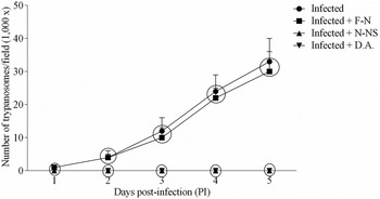
Figure 2 shows parasitaemia levels of infected untreated mice (the group E), and infected and F-N treated animals (the group F) with higher parasitaemia (an average of 33 ± 7 and 30 ± 5 trypanosomes/field – 1000×, respectively) than those infected and N-NS treated animals (the group G) and infected and D.A.-treated animals (the group H), both did not show parasites in the bloodstream (an average of 0 ± 0 trypanosomes/field – 1000×) on day 5 PI. None of the animals died during the experimental period.
Mice parasitaemia 5 days post-infection (PI) by Trypanosoma evansi. The results within a circle are not statistically different (Bonferroni's post hoc test with P > 0·05). Note: the group E (infected and untreated); the group F (infected and treated with nerolidol free); the group G (infected and treated with nerolidol loaded in nanospheres) and the group H (infected and treated with diminazene aceturate).

Seric quantification of nerolidol and D.A.
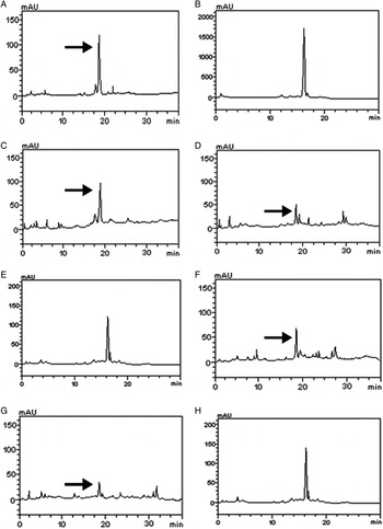
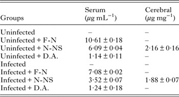
On day 5 PI, F-N-treated groups (B and F) showed an average of 10·61 (±0·18) and 7·08 (±0·02) µg mL−1 of F-N in sera samples, respectively. Regarding N-NS-treated animals (groups C and G), an average of 6·09 (±0·04) and 3·52 (±0·07) µg mL−1 of N-NS were found in sera samples, respectively. Moreover, animals treated with D.A. (groups D and H) showed an average of 1·14 (±0·11) and 1·24 (±0·18) µg mL−1 of D.A. in sera samples, respectively (Fig. 3). In summary, there was a reduction in the seric concentration of nerolidol in infected animals treated with F-N or N-NS compared with uninfected treated animals (Table 1).
Representative chromatograms of standards and mice sera: nerolidol standard (A), diminazene aceturate standard (B), uninfected and treated with nerolidol free (C), uninfected and treated with nerolidol loaded in nanospheres (D), uninfected and treated with diminazene aceturate (E), infected and treated with nerolidol free (F), infected and treated with nerolidol loaded in nanospheres (G), and infected and treated with diminazene aceturate (H). Arrow represents the peaks of nerolidol.

Mean and s.d. of nerolidol and diminazene aceturate (D.A.) levels in serum and cerebral tissue of uninfected and Trypanosoma evansi-infected mice treated with free nerolidol (F-N), nerolidol loaded in nanospheres (N-NS) and D.A.

Note: ‘–’ means that nerolidol or D.A. were not detected using high-performance liquid chromatography.
Cerebral quantification of nerolidol and D.A.
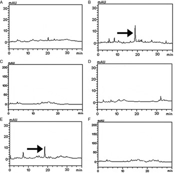
On day 5 PI, N-NS-treated animals (groups C and G) showed an average of 2·16 (±0·16) and 1·88 (±0·07) µg mg−1 in the brain tissue, respectively (Fig. 4). However, F-N and D.A. were not detected in this organ (Table 1). In summary, nanotechnology allowed the passage of nerolidol through the BBB, and consequently, the elimination of T. evansi from the CNS.
Representative chromatograms of brain tissue of mice: uninfected and treated with nerolidol free (A), uninfected and treated with nerolidol loaded in nanospheres (B), uninfected and treated with diminazene aceturate (C), infected and treated with nerolidol free (D), infected and treated with nerolidol loaded in nanospheres (E), and infected and treated with diminazene aceturate (F). Arrow represents the peaks of nerolidol.

Molecular diagnosis
On day 5 PI, PCR analyses was performed using samples from the brain and showed that all infected mice treated with N-NS were negatives for the presence of T. evansi, while all infected animals treated with F-N or D.A. were positives.
DISCUSSION
Although some studies have suggested that nanotechnology may enable natural and synthetic drugs to cross the BBB, eliminating an important refuge area for T. evansi like the brain, this is the first study proving this hypothesis. Our results confirm that D.A. does not cross the BBB at the therapeutic dose, as previously reported by Masocha et al. (Reference Masocha, Robertson, Rottenberg, Mhlanga, Sorokin and Kristensson2004). The most important finding of this study is that nanotechnology allows the passage of nerolidol through the BBB, which may eliminate T. evansi present in the CNS, since PCR analyses were negative in all infected mice treated with N-NS.
In this present study, nanoencapsulation of nerolidol allowed its passage through the BBB, possibiliting the elimination of T. evansi from the CNS. A fact not seen in D.A. treatments, which explains the inefficacy of D.A. (Masocha et al. Reference Masocha, Rottenberg and Kristensson2007; Vitouley et al. Reference Vitouley, Sidibe, Bengaly, Marcotty, Abbeele and Delespaux2012) since trypanosomes can hide in the brain. In this sense, formulations with trypanocidal action, such as N-NS (Baldissera et al. Reference Baldissera, Grando, Souza, Cossetin, Sagrillo, Nascimento, da Silva, Dalla Lana, Da Silva, Stefani and Monteiro2016a , Reference Baldissera, Gonçalves, Sagrillo, Grando, Ritter, Grotto, Brum, da Luz, Silveira, Fausto, Boligon, Vaucher, Stefani, da Silva, Souza and Monteiro b ), have the capacity to cross the BBB, and thus eliminate the parasite from the CNS, acting as an important resource to cure the disease. Recently, a study conducted by Oliveira et al. (Reference Oliveira, Rigo, Rosa, Gressler, Zimmermann, Ourique, Da Silva, Miletti, Beck and Monteiro2014) demonstrated that liposomes containing D.A. were able to improve its curative effectiveness compared with free D.A., and they concluded that this finding was related to the drug ability to cross the BBB. The mechanism of nerolidol absorption from oral via remains unknown, but study conducted by Krishnaiah et al. (Reference Krishnaiah, Al-Saidan, Chandrasekar and Satyanarayana2005) demonstrated the bioavailability of nerolidol by the transdermal route, possessing a maximum plasma concentration (C max) of 30·9 ng mL−1 and time to reach maximum concentration (T max) of 9·8 h. In order to investigate the mechanism of action involved, Garcia et al. (Reference Garcia-Garcia, Andrieux, Gil and Couvreur2005) demonstrated that nanoparticle size, type of the polymer and physical characteristics favour drug interaction and penetration into endothelial cells of the BBB. Petry et al. (Reference Petry, Bootz, Khalansky, Hekmatara, Muller, Uhl, Kreuter and Gelperina2007) demonstrated that alterations on the surface of nanospheres can enhance drug transportation through the BBB. In this sense, Zhang and Feng (Reference Zhang and Feng2006) demonstrated that polysorbate 80, the same polymer used in this study, is fundamental for cerebral vectorization. According to these authors, this could be explained by the fact that polysorbate 80 selectively promotes the adsorption of certain plasma proteins, such as apolipoprotein E, on the surface of the nanospheres. Thus, adsorbed apolipoprotein E nanospheres would be recognized as low-density lipoproteins, being transported by BBB endothelial cells through receptor-mediated endocytosis. Moreover, Xia et al. (Reference Xia, Liu, Lv, Zhang, Zhou and Zhao2017) demonstrated that nanoencapsulation of oleanolic acid, a lipophilic molecule (as nerolidol), was able to enhance the dissolution rate, intestinal absorption and bioavailability of oleanolic acid in the gastrointestinal tract, improving in 340% the bioavailability compared with free drug via oral administration. According to these authors, the use of nanoencapsulated compounds is considered a potential approach to enhance the oral absorption and bioavailability of lipophilic compounds in the gastrointestinal tract, as nerolidol. The action mechanism of these effects may be linked with the presence of lactoferrin, a globular protein that acts as a ligand that modifies the structures of nanomolecules and crosses the BBB via lactoferrin receptor-mediated transcytosis (Rempe et al. Reference Rempe, Cramer, Qiao and Galla2014). Furthermore, numerous studies have proven that lactoferrin is a promising target for the treatment of brain diseases (De Luca et al. Reference De Luca, Lai, Corrias, Caboni, Bimpisidis, Maccioni, Fadda and Di Chiari2015). Thus, the intact passage of nerolidol from BBB may be associated with interaction with lactoferrins.
Recently, several studies demonstrated that nanotechnology is considered an important approach to treat neurological disorders, such as neuroAIDS, brain tumours and Alzheimer's disease (Nair et al. Reference Nair, Jayant, Kaushik and Sagar2016; Pinto et al. Reference Pinto, Arce, Yameen and Vilos2017; Wen et al. Reference Wen, El-Salamouni, El-Refaie, Hazzah, Ali, Tosi, Farid, Blanco-Pietro, Billa and Hanafy2017). Recently, a study conducted by Baldissera et al. (Reference Baldissera, Souza, Grando, Moreira, Schafer, Cossetin, da Silva, da Veiga, da Rocha, Stefani, da Silva and Monteiro2017) demonstrated that nerolidol, when loaded in nanospheres, prevents cognitive deficits, such as memory impairment, depression and increased anxiety, while F-N was unable to prevent these alterations. According to Nair et al. (Reference Nair, Jayant, Kaushik and Sagar2016), the increased number of molecules available and of the surface of nanoparticles may result in higher drug binding/adsorption, which could be considered a factor involved in the neuroprotective effect of nerolidol's active principle associated with nanotechnology. Thus, we can infer that the passage of nerolidol through the BBB might be involved in the neuroprotective effects of N-NS.
Based on these evidences, it is possible to conclude that the passage of N-NS through the BBB allows the elimination of T. evansi from the cerebral tissue, an important factor involved in several cases of disease relapse. That BBB passage might also be involved in the neuroprotective effects of N-NS. In summary, nanotechnology can be considered an important approach to increase the therapeutic efficacy of several active principles, allowing the elimination of T. evansi from the CNS, a well-known refuge area for parasites and a reason for failure of many treatments.
ACKNOWLEDGEMENTS
The authors acknowledge support from Conselho Nacional de Desenvolvimento Científico e Tecnológico (CNPq). B. Baldisserotto, A. S. Da Silva and S. G. Monteiro received a CNPq research fellowship and M.D. Baldissera CAPES (Coordenação de Aperfeiçoamento de Pessoal de Nível Superior) PhD fellowships.
FINANCIAL SUPPORT
This study was supported by Coordenação de Aperfeiçoamento de Pessoal de Nível Superior (CAPES), Brazil.
CONFLICT OF INTEREST
None.