Introduction
Embryonic stem cells (ESCs), as the ideal seed cells, demonstrate both the capacity to self-renew and pluripotency, and have tremendous application potential in clinical therapy (Rideout et al., Reference Rideout, Hochedlinger, Kyba, Daley and Jaenisch2002; Passier and Mummery, Reference Passier and Mummery2003; Zwaka and Thomson, Reference Zwaka and Thomson2003). But there exist disadvantages such as ethical problems and material sourcing, which limit embryonic stem cell research (Malaren, Reference Malaren2001; Meng et al., Reference Meng, Shang and Ding2002).
De Coppi et al. (Reference De Coppi, Bartsch and Siddiqui2007) isolated a few stem cells from human amniotic fluid at the second trimester of gestation, and named them human amniotic fluid stem cells (hAFS cells). AFS cells expressed different specific markers compared to both ESCs and adult stem cells (ASCs) and could differentiate into cells of three germ layers. AFS cells pose no ethical problems, thereby providing a new route for stem cell research (Chiavegato et al., Reference Chiavegato, Bollini and Pozzobon2007). Since the study of AFS cells is in the early stages, there are still many factors that affect the primary culture of these cells. In this paper, we isolated AFS cells from human amniotic fluid during the second and third trimesters of gestation, and the factors influencing the primary culture were analysed.
Materials and methods
Collection of human amniotic fluid samples
Human amniotic fluid samples were collected and the parameters of each sample, including gestational age, the volume of fluid and the sample appearance (blood-cell-free, blood-cell, bloody or brown-coloured fluid), were carefully documented.
Isolation and culture of hAFS cells
Cells were isolated from human amniotic fluid after being centrifuged at 1000 rpm for 5 min, and cell pellets were washed three times with phosphate-buffered saline (PBS) containing 1% penicillin and streptomycin. Cells were plated with α-Minimal Essential Medium (α-MEM) (from Invitrogen Corp., Carlsbad, California, USA) supplemented with 10% fetal bovine serum (FBS) (from GIBCO/Invitrogen Corp.), 20% Chang medium (from Irvine Scientific, Santa Ana, California, USA), 1% glutamine (from GIBCO/Invitrogen Corp.), 1% penicillin and streptomycin under 5% CO2 at 37°C. Five to 12 days after the initiation of the culture, the medium was replaced with fresh medium, and the cell attachment time and numbers were recorded.
Immunocytochemistry
hAFS cells at the tenth and fifteenth passages were fixed with 4% paraformaldehyde at 4°C overnight. Cells were incubated in 3% hydrogen peroxidase for 30 min and then in blocking solution, consisting of 5% bovine serum albumin (BSA) in PBS for 20 min at room temperature. Cells were then incubated with several antibodies at 4°C overnight. The primary antibodies were anti-octamer-4 (anti-Oct-4) (from Chemicon International Inc., Temecula, California, USA), anti-human telomerase reverse transcriptase (anti-hTERT) (from Rockland Immunochemicals Inc., Gilbertsville, Pennsylvania, USA) and anti-stage-specific embryonic antigen (anti-SSEA)-1/3/4 (from Chemicon International Inc.) at 1:500 dilution; and CD29, 34, 44, 45, 73, 90, 105, 117 and 133 (from Abcam plc, Cambridge, Massachusetts, USA) at 1:200 dilution. Cells were treated with PBS as the negative control. After incubation with primary antibodies, slides were then incubated in biotinylated anti-goat, anti-mouse or anti-rabbit IgG for 20 min at room temperature. The slides were then incubated with streptavidin peroxidase for 20 min at room temperature. The results were visualized using an inverted microscope.
hAFS cell differentiation
For neuron differentiation, hAFS cells were initially treated with α-MEM containing 20% FBS, 1 mmol/l β-mercaptoethanol (from GIBCO/Invitrogen Corp.) and 5 ng/ml fibroblast growth factor 2 (FGF2), for 24 h. Then the medium was replaced with Dulbecco's modified Eagle's medium (DMEM) supplemented with 10 mmol/l β-mercaptoethanol. After culture for 5 h, Nestin expression was determined by the immunocytochemical method (1:200, antibody from Sigma-Aldrich Inc., St. Louis, Missouri, USA).
Cardiomyocytes were induced by culturing the cells in α-MEM supplemented with 10% FBS and 0.75% dimethylsulphoxide (DMSO) (from Sigma-Aldrich Inc.). Cardiomyocyte differentiation was assessed by staining with α-actin (from Sigma-Aldrich Inc.) (1:500).
Results
The biological characteristics
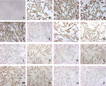
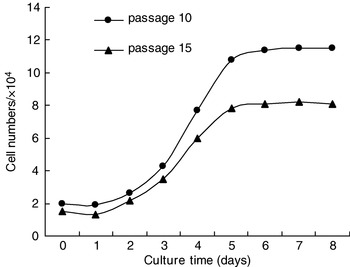
Human amniotic fluid samples at the second or third trimester of gestation were cultured and hAFS cells were isolated. The biological characteristics of hAFS cells were studied by immunocytochemistry. hAFS cells showing fibroblastoid-types (Fig. 1A) were obtained and positively expressed specific markers of embryonic stem cells, such as Oct-4, hTERT, SSEA-1, SSEA-4 and CD117, but not SSEA-3 (Fig. 1B–G). hAFS cells stained positively for mesenchymal stem cell markers, including CD29, CD44, CD73, CD90 and CD105 (Fig. 1H, J, L, M and N), but negatively for haematopoietic stem cells and haematopoietic lineage markers, including CD34, CD45 and CD133 (Fig. 1I, K and O). hAFS cells at different passages were in a logarithmic growth phase after 2 days' culture and the doubling times of the cell populations were 34 h (10 passages) and 39 h (15 passages), respectively (Fig. 2). The isolated hAFS cells expressed specific markers and had strong proliferation ability.
Characteristics of human amniotic fluid stem (hAFS) cells. (A) Morphology of hAFS cells (passage 8, P8); (B) Oct-4 positive (passage 10, P10); (C) hTERT positive (P10); (D) SSEA-1 positive (P10); (E) SSEA-4 positive (P10); (F) SSEA-3 negative (P10); (G) CD117 positive (passage 15, P15); (H) CD29 positive (P15); (I) CD34 negative (P15); (J) CD44 positive (P15); (K) CD45 negative (P15); (L) CD73 positive (P15); (M) CD90 positive (P15); (N) CD105 positive (P15); (O) CD133 negative (P15); (P) negative control. (See online for a colour version of this figure.)

Growth curve of human amniotic fluid stem cells.

hAFS cell differentiation
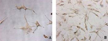
The differentiated neural cells were smaller, with cytoplasmic contractions, cone-shaped or triangular, and were positive for Nestin (Fig. 3A). In cardiomyocyte differentiation, the differentiated cells were rod shaped, smaller than fibroblast cells, α-actin positive, and were similar to cardiomyocytes (Fig. 3B). The results showed that the isolated hAFS cells can differentiate into neural cells and cardiomyocytes.
Differentiation of amniotic fluid stem cells into neural cells (A) and cardiomyocytes (B). (See online for a colour version of this figure.)

Influencing factors
The cell attachment time (4.7±0.6 days) in the second trimester of gestation was significantly different from that in the third trimester of gestation (6±0.5 days) (P<0.05), suggesting that cells collected from the fluid during the third trimester of gestation need a longer time for attachment.
We also found that blood contamination can significantly affect the time for cell attachment. The attachment time of the brown-coloured fluid (10.8±0.3 days) was significantly different from the blood-cell-free group (6±0.5 days) and the blood-cell group (6.3±0.6 days) (P<0.05) (Fig. 4).
Effect of the blood contamination level on the cell attachment time (days). (1) Blood-cell-free fluid group; (2) blood-cell group; (3) bloody group; (4) brown-coloured fluid group.

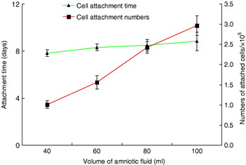
The effects of the volume of amniotic fluid were also investigated regarding numbers and time of cell attachment. The volume of amniotic fluid did not affect cell attachment time, but affected the cell attachment numbers (P<0.05) (Fig. 5).
Effect of the volume of the amniotic fluid on the cell attachment time and the cell attachment number.

Discussion
Human amniotic fluid samples at the second or third trimester of gestation were cultured. hAFS cells showing fibroblastoid types were obtained and maintained proliferative potential after expansion to several populations. hAFS cells positively expressed specific markers of embryonic stem cells, such as Oct-4, hTERT, SSEA-1, SSEA-4 and CD117, but not SSEA-3. hAFS cells stained positively for mesenchymal stem cell markers, including CD29, CD44, CD73, CD90 and CD105, but negatively for haematopoietic stem cells and haematopoietic lineage markers, including CD34, CD45 and CD133. The characteristics of hAFS cells separated in this paper suggested that hAFS cells represented an intermediate stage between pluripotent embryonic stem cells and lineage-restricted adult stem cells (De Coppi et al., Reference De Coppi, Bartsch and Siddiqui2007), which is consistent with other reports (Tsai et al., Reference Tsai, Hwang, Tsai, Cheng, Lee and Chang2006; Kim et al., Reference Kim, Lee and Kim2007). hAFS cells could be induced to differentiate into neural cells which were positive for Nestin, and cardiomyocytes which were positive for α-actin, which suggested that hAFS cells may offer an alternative approach for cell and tissue engineering.
In this study, the factors that affected the growth of hAFS cell primary culture were investigated. The results showed that gestational age had no influence on cell attachment numbers, explained by a decrease in the number of viable cells and only a fraction of these cells forming clones, although the total number of cells increased with gestational age (Prusa and Hengstschlager, Reference Prusa and Hengstschlager2002). On the other hand, gestational age influenced cell attachment time significantly. Cells collected from the fluid during the third trimester of gestation needed a longer time for attachment. The possible reason may be that there were many cells in the fluid of the third trimester of gestation that did not have the capacity to attach, which inhibited the growth of hAFS cells.
Most of the amniotic fluid samples collected during the third trimester of gestation were contaminated with blood. Blood contamination significantly affected the time of cell attachment but did not affect cell attachment numbers. Seguin and Palmer (Reference Seguin and Palmer1983) suggested from their results that bloody amniotic fluid inhibited cell growth, and a grossly bloody specimen could inhibit growth by 30% (Chang and Jones, Reference Chang and Jones1991). Our results showed that bloody amniotic fluid delayed cell growth, but the cell attachment numbers were minimally affected.
The effects of the volume of amniotic fluid on cell attachment numbers and time were also investigated. The volume of amniotic fluid did not affect total cell attachment time, but affected the cell attachment numbers to only a minimal extent (P<0.05). This was associated with cell plating density (Sikkema-Raddatz et al., Reference Sikkema-Raddatz, van Echten, van der Vlag, Buys and te Meerman2002). When cells were seeded with a high density, the capacity of cells to attach was inhibited and cell attachment numbers decreased.
The present studies systematically examine factors affecting the primary culture of hAFS cells and provide useful data for AFS cell research. hAFS cells described in this paper had characteristics of embryonic stem cells and could differentiate into neural cells and cardiomyocytes. hAFS cells could be used as seed cells for cell and tissue engineering.
Acknowledgements
This work was supported by grants from the Outstanding Talents Fund of Northwest A&F University. We would like to thank Dr Rick Rochester and his organization, International College of China Agricultural University, for proofreading the English manuscript of this paper.