




No CrossRef data available.
Article contents
Resistance against migrating Ascaris suum larvae in pigs immunized with infective eggs or adult worm antigens
Published online by Cambridge University Press: 12 July 2001
Abstract
Resistance to Ascaris suum infections was investigated in 8- and 15-week-old Iberian pigs. Groups of 3 or 5 pigs were immunized weekly for 6 weeks with antigens of adult A. suum: a 97 kDa body wall (BW) fraction, a 42 kDa fraction of pseudocoelomic fluid (PF) or a 14 kDa PF-fraction; or were inoculated with increasing doses of infective eggs (500–20000), with or without abbreviation by pyrantel pamoate. All immunized pigs and unimmunized control pigs, were challenged with 10000 infective eggs 7 days after the last immunization. The number of liver lesions and lung larvae was substantially lower in the older pigs than in the younger ones 7 days after challenge, but the resistance in immunized pigs of both age groups was similar in comparison to the challenge controls of the same age. The highest degree of resistance against lung larvae was observed in pigs immunized with A. suum eggs (97–99%). The pigs immunized with the 14 kDa and 42 kDa PF-fractions were also well protected (67–93%), while no protection was produced by the 97 kDa BW fraction (0–49%). The reduction of white spots following immunization was less evident, with a maximum of 82% reduction in egg-inoculated young pigs.
Keywords
Information
- Type
- Research Article
- Information
- Copyright
- © 2001 Cambridge University Press
INTRODUCTION
The nematode Ascaris suum is generally regarded as highly immunogenic in pigs (reviewed by Urban, 1986) and the characteristic age relationship, with high prevalences in young growing pigs and low prevalences in the adult stock, has been attributed to a gradual acquirement of resistance (reviewed by Nansen & Roepstorff, 1999). While inoculation with a single (Helwigh & Nansen, 1999) or a few immunizing doses (Jungersen et al. 1999) of infective eggs only causes a moderate reduction in the number of migrating larvae of a subsequent challenge infection, chronic exposure of pigs has been shown to cause strong resistance at the intestinal level, precluding hepatic migration of the majority of larvae (Urban, Alizadeh & Romanowski, 1988). A similar reduced migration has been found in fatteners and sows from heavily infected herds (Eriksen et al. 1992a).
Attempts to vaccinate against A. suum have focused on immunization with eggs and larvae (Crandall & Arean, 1965), UV-attenuated eggs (Urban & Tromba, 1982), and drug-abbreviated infections (Stewart et al. 1985; Stankiewicz, Jeska & Froe, 1990; Stankiewicz, Jonas & Froe, 1992), but without much success. Furthermore, these procedures include the risk that some of the larvae survive and develop to patent infections. Immunization with non-infective material, therefore, seems more attractive. Some attempts have used protective antigens, focusing on excretory–secretory (E–S) antigens of larvae. Stromberg (1979) found that the allergenic E–S antigen of L3/4 is a 67 kDa molecule which, given intraperitoneally with adjuvant to guinea-pigs, produced a strong IgE response and very little IgG1 activity was seen; intramuscular administration induced only low titres of both immunoglobulins. Kennedy & Qureshi (1986) found that components of both L2 and L3/4 E–S are heterogeneous, ranging from 14 to 410 kDa, stimulating an antibody response which was superior to that of pseudocelomic fluid (PF) in non-natural hosts (rabbits, rats and mice). However, each host recognized a different subset of antigens. Significantly, a major 14 kDa E–S antigen, homologous to the major constituent of PF, was not recognized by mice. In pigs, parenteral immunization with L2/3 E–S antigens stimulated a moderate protection (Urban & Romanowski, 1985) while significant protection has been obtained with adult crude antigens (Lukes, 1992) and cuticle components of larval and adult worms in pigs (Hill et al. 1994).
Iberian pigs are an old traditional race principally bred on acorn pastures in the West of Spain, mainly in the Extremadura region, and usually slaughtered at 16 months of age and 150 kg of body weight. This expensive management is profitable only due to the high quality of Iberian pork products. However, the economy is threatened by a high prevalence of A. suum of 37% in Iberian finisher pigs (Pérez-Martín et al. 1991), stressing the need for prevention and control of A. suum infection.
In the present study, 3 partly purified antigenic fractions of adult A. suum were used to investigate the acquisition of resistance against larval migration in Iberian pigs, 8 and 15 weeks of age.
MATERIALS AND METHODS
Experimental animals
Thirty-five, 15-week-old Iberian pigs (25.9±4.5 kg), obtained from an intensively managed swine herd but non parasite-free, and 21 young Iberian pigs (8 weeks old, 11.4±1.5 kg), born in A. suum-free pens at the Veterinary Faculty of the University of Extremadura, were randomly allocated into 7 groups of 5 older pigs and 3 young pigs, in order to compare the influence of age and parasite infections in the experimental design. The absence of patient A. suum infections was recorded by individual faecal examinations before and at weekly intervals during the experiment. Due to logistic limitations, the experiment was conducted separately in time for the 2 age groups. The groups of pigs were maintained in separate units, cleaned thoroughly, that had not been used for pigs for at least 3 years.
Infective eggs
A. suum eggs were removed from the uteri of adult females collected at the local abattoir. They were embryonated for 2 months in 100 ml of 0.1 M NaOH at 20 °C at a maximum concentration of 20000 eggs/ml, and aerated by daily agitation. The infectivity of the eggs was tested in an 8-week-old Iberian pig, in which 26% of the inoculated eggs were recovered as migrating larvae in the lungs 7 days post-inoculation (pi).
Parasite antigens
Adult worms were collected from naturally infected pigs at local abattoirs, washed in sterile PBS and dissected to obtain the pseudocoelomic fluid (PF) and the body wall (BW) after the digestive and reproductive systems had been removed. A BW extract was prepared by maceration and ultrasonic disruption at 55 W in cycles of 2 sec for 5 min in a Vibra-cellTM VC300 (Sonics & Materials Inc, CT, USA). Sonication was performed 3 times on ice-water and continuous temperature control. PF and soluble BW extracts were centrifuged (2500 g for 30 min), filter-sterilized through a 0.28 μm filter, and stored at −80 °C until use. SDS–PAGE of the PF and BW soluble extracts was carried out in 12% (w/v) polyacrylamide slab gels under reducing conditions, according to the method of Laemmli (1970). Two strips of about 2 cm long were cut out of the right and left side of each gel, respectively, containing the lane of molecular weight marker (M-4038 SigmaMarkerTM, Sigma) and some part of the Ascaris extract line, and stained using Coomassie blue. The central parts of the gels were reversibly stained using a Zinc-stain kit (161-0440, Bio–Rad). Two thick bands of approximately 42 kDa and 14 kDa from the PF gels and a double band of approximately 97 kDa from the BW gels were carefully removed from the Zinc-stained gels and cut into pieces of 1–2 mm (Fig. 1). The 97 kDa and 42 kDa fractions were selected among most clearly recognized antigens by pigs inoculated with infective A. suum eggs and conventional pigs in Western blot analysis (unpublished observations). The 14 kDa fraction was selected because it contains a major component of the PF and well-known allergen (ABA-1), although this fraction was poorly recognized by pig sera in Western blot analysis. Each minced band was placed in 4 electroelution sample chambers (MicrotrapTM) containing dialysis membranes of 3.5 kDa molecular weight exclusion with 12 mM Tris–glycine (pH 8.5) and 0.1% SDS and electroeluted at 400 V, 40 mA and 12 W limits in a Little Blue TankTM (Model 1750, Isco) with the same buffer for 12 h. In order to avoid protein precipitation in the dialysis membranes, the eluted protein was collected every 2 h and the sample chambers refilled with fresh Tris–glycine buffer. The concentration of the eluted protein was estimated using a commercial Bradford dye binding assay (Coomassie® Plus Protein Assay, Pierce) and stored at −80 °C until use.

Fig. 1. Coomassie blue-stained 10% (A) and 16% (B) SDS–polyacrylamide gels of body wall extracts (BW) and pseudocelomic fluid (PF) of adult worms of Ascaris suum and purified fractions used for immunization of Groups 5, 6 and 7. Lane m, molecular weight markers; lane 1, BW extracts; lane 2, 97 kDa BW fraction; lane 3, 42 kDa PF fraction; lane 4, 14 kDa PF fraction; lane 5, PF.
Experimental design
The experimental protocol is outlined in Table 1. The pigs of Group 1 remained uninoculated throughout the experiment (49 days) to monitor any uncontrolled A. suum infections in the pigs. The pigs of Groups 2–7 were all challenge inoculated with 10000 infective eggs on day 42 of the experiment. The inoculations were performed by means of a stomach intubation. Pigs were killed using a captive bolt pistol, bled and eviscerated 7 days after challenge. Group 2 pigs were challenged to record the numbers of lesions in the liver and the numbers of migrating larvae in the lungs of naïve pigs. The aim of Group 3 was to estimate the level of protection acquired after 6 weekly inoculations with increasing numbers of infective eggs. Group 4 was identical to Group 3 except that all immunizing infections were abbreviated by adding pyrantel pamoate in the feed (96 mg/kg) from 2 days before until 2 days after each pre-challenge inoculation. In Group 5, each pig was injected subcutaneously with 1 μg of the 14 kDa fraction of PF per kg body weight, emulsified in 500 μl of Freund's incomplete adjuvant (Sigma ImmunoChemical, F-5506) on the same days as the pigs of both Group 3 and 4 were inoculated. The pigs in Groups 6 and 7 were immunized with the 42 kDa fraction of PF and the 97 kDa fraction of BW, respectively, using a protocol identical to that of Group 5.
Table 1. Experimental design (The symbols represent necropsy ([bull ]), challenge inoculation with 10000 infective Ascaris suum eggs (○), immunization with infective eggs ([utri ], numbers of eggs given), days of treatment with pyrantel pamoate (x) and days of immunization with the 14 kDa PF ([lozf ]), the 42 kDa PF ([squf ]) or the 97 kDa BW (□) fractions from adult A. suum.)

Post-mortem procedures
At necropsy, the lungs of the pigs were cut into tissue blocks and slightly homogenized in a blender to obtain pieces of about 5 mm in diameter and digested in pepsin–HCl as described for the isolation of Trichinella larvae from muscle samples (Serrano et al. 1992). Samples of 100 g liver tissue were digested using the same method. Gross hepatic lesions were recorded by counting white spots on the liver surface, differentiating between lymphonodular spots (LNS), mesh-like white spots (MWS), unusually large and raised white spots, and opalescences of the liver capsule or ‘foggy' spots. These liver lesions have been described histologically by Roneus (1966) and others. Briefly, LNS are pearly small nodules of 1–3 mm originating as a lymphofollicular hyperplasia in the form of several lymphoid follicles surrounded by a thin connective tissue. The LNS are visibly different from MWS which appear as diffuse increases of interlobular septa of variable size, sometimes showing a compact whitish centre, arising from an eosinophilic interstitial hepatitis accompanied by fibroblastic proliferation and intralobular necrosis. Large white spots caused a strong focal fibrosis and atrophy of lobules, that disappeared completely in the centre. Foggy spots are, presumably, almost completely healed MWS where a pale area in the liver surface can still be differentiated, generally showing smaller lobules than the surrounding parenchyma.
The contents of the small intestine, the large intestine and the caecum were sieved and examined separately to record the presence of macroscopic intestinal worms originating from the pre-challenge doses or from uncontrolled infections.
Calculations and statistical analyses
The degree of protection in the immunized pigs was calculated as the reduction in numbers of lung larvae and liver white spots in comparison to the post-mortem results of the Group 2 pigs. Correlations between the number of white spots and the number of larvae in the lungs were estimated by Pearson's linear correlation coefficient. Differences between groups were analysed by analysis of variance (ANOVA) and post hoc comparisons of pairs of groups, using the Ryan–Einot–Gabriel–Welsch multiple range test (Day & Quinn, 1989) with a significance level of P = 0.05. All calculations were performed using the SPSS 9.0 software package.
RESULTS
Young pigs, in general, had many more liver lesions than older pigs at a significant level (Fig. 2). However, the confluence of adjacent white spots and the diffuse appearance of intralobular connective tissue made it difficult to count the exact number of MWS and foggy spots. Overall, young pigs had numerous MWS (Fig. 2A), which were in general intense, small and flat, with no generalized liver fibrosis. However, in Group 5, many of the MWS had slightly raised and nodular centres, often with a haemorrhagic lesion of about 1 mm. The MWS of older pigs (Fig. 2B) were clearly different from that of young pigs, as there were relatively few lesions, that were all of uneven size and intensity. A small number of large (>6–10 mm), hard and raised white spots, usually with a defined centre, were observed in old pigs only and especially in those of Groups 4, 5, and 6 (Fig. 2D), but there were no significant differences between groups. Pigs in Groups 3 and 4 had several LNS of 1–2 mm (Fig. 2E,F), as well as ‘foggy' spots of about 5–10 mm (Fig. 2G,H). Both types were most numerous in young pigs (P<0.001). There was a significant positive correlation between LNS and foggy white spots (r = 0.69, P<0.01) in Groups 3 and 4.

Fig. 2. Numbers (mean±S.D.) of mesh-like white spots (A, B), large white spots (C, D), lymphonodular white spots (E, F), and foggy white spots (G, H) in the livers of 8-week-old (A, C, E, G) and 15-week-old (B, D, F, H) pigs 7 days post-challenge with 10000 infective Ascaris suum eggs in relation to immunizing procedure (see Table 1). All differences between groups were significant except between those labelled with identical characters (a, b, c, d, e, f).
The reduction in number of liver lesions and migrating lung larvae due to the immunization protocols are shown in Table 2. The egg-inoculated pigs of Groups 3 and 4 showed the highest reduction in spots (53–82%). The 14 kDa PF immunized pigs of Group 5 had also showed significantly reduced numbers of white spots (absence of significant differences between two groups are represented as shared characters in Fig. 2), while this was not the case for the 42 kDa PF and the 97 kDa BW immunized pigs (Groups 6 and 7). The number of larvae recovered in 8-week-old pigs of Group 2 (x¯ = 3180) was markedly higher than in older animals of this group (x¯ = 66.4). However, age had overall only a small effect on the reduction of percentages with respect to the challenge control group. Groups 3 and 4 showed also the highest reductions for lung larvae (97–99%). These groups did not show significant differences compared to the negative control. In 15-week-old pigs this was also applicable for Groups 5 and 6, whereas Group 7 was very similar to the challenge group. In young pigs the number of larvae was increased successively in Groups 5, 6 and 7, which also showed significant differences compared to the challenge group.

The number of lung larvae obtained by pepsin digestion was positively associated with the total number of liver lesions with treatment (Groups 2–7) and age (P<0.01). However, numbers were slightly lower for Groups 3 (r = 0.80) and 4 (r = 0.83) than in the remaining challenged groups (r = 0.93–0.99). This association was found in all challenge infected experimental groups mainly due to the MWS that was the only type of liver lesion with a significant association with lung larvae (r = 0.99, P<0.01). The ratio between lung larvae and white spots was highest in young pigs and ranged from 3.87 in younger pigs of Group 2 to 0.04 in the older pigs of Group 4. Except for 2 larvae found in one 15-week-old pig of Group 7, no larvae were recovered in the liver samples.
No pigs excreted A. suum eggs during the experiment. However, a total of 5 adult A. suum females (18–34cm) was found in 3 pigs in Group 1 at necropsy. Thus this group had significantly more large worms than the remaining experimental groups, in which only 1 female was found in 1 pig of Group 5. All the 15-week-old pigs had a low excretion of strongylid eggs (79% positive samples), and similarly all pigs, but 1, excreted eggs of Trichuris suis (37% positive samples), usually <100 eggs/gram of both species. Only very low numbers of adult Oesophagostomum sp. And T. suis were found at necropsy in 15-week-old pigs. Neither helminth eggs nor adult worms were detected in the 8-week-old pigs.
DISCUSSION
Pigs inoculated weekly with A. suum eggs were almost completely resistant to larval migration to the lungs (Groups 3 and 4) at day 7 post-challenge, while the level of protection obtained by immunization with antigenic A. suum fractions was variable.
The effect of the various protocols for immunizing pigs against A. suum appeared largely independent of age and parasitological parameters, although the actual numbers of white spots and lung larvae were generally lower in older pigs. Liver white spots and especially the number of lung larvae were most markedly reduced in the pigs immunized with infective eggs, irrespective of simultaneous anthelmintic treatment. Pigs immunized with the 14 kDa PF fraction also had reduced numbers of white spots and lung larvae although less significantly (37–53% and 88–93%, respectively). To a lesser extent this was also observed for the 42 kDa PF fraction, while immunization with the 97 kDa BW fraction did not result in significant protection.
In the challenge control group, the recovery of lung larvae was 31.8% in young pigs, showing that the eggs were highly infective, while recovery was only 0.7% in older pigs. In spite of the absence of Ascaris eggs, the finding of a few adult A. suum in these pigs indicates that these animals had been moderately exposed to infection before the start of the experiment, as well as by the higher ELISA reactivity against A. suum L2/L3 E–S antigens at day 0 than in the younger pigs (unpublished observations) and the presence of Oesophagostomum and Trichuris which are, in general, more sensitive to transmission-limiting factors than A. suum (Roepstorff & Jorsal, 1989; Nansen & Roepstorff, 1999). It seems unlikely that the presence of a few large worms was deleterious to the responses of the pigs to the immunization protocols. Urban et al. (1988) found that adult worm burdens in presumably helminth-free pigs were unable to stimulate any protection to a challenge, and correspondingly Jungersen et al. (1999) showed that the presence of adult A. suum had no significant effect on acquired resistance to migrating larvae in the lungs. Heavy O. dentatum infections slightly increase the antibody response against A. suum L2/L3 E–S antigens but are unable to reduce larval migration (Helwigh et al. 1999). Although a gradual reduction in the percentage of successfully migrating A. suum larvae with increasing age of helminth-naive pigs has been reported (Eriksen et al. 1992a), it is more likely that the lower numbers of white spots and lung larvae in older pigs were caused by a moderate level of acquired homologous resistance, which can be induced experimentally by high single doses of eggs (Helwigh et al. 1999), or a moderate trickle dosing regime for 3 weeks (Jungersen et al. 1999), but not by a very low single dose (Roepstorff, unpublished).
The absence of liver lesions in old pigs of Group 1 does not provide any information on pre-experimental exposure, as mesh-like white spots gradually fade to become foggy and disappear within a few weeks (Roneus, 1966; Roepstorff et al. 1997). Although inoculation with infective eggs or hatched L3 may stimulate more severe liver lesions after a challenge, the rapid healing process in general means that white spots are evidence of recent infections (Eriksen, 1982, Eriksen et al. 1980). In contrast, few large/raised white spots and lymphonodular white spots may remain for 5–11 weeks (Roneus, 1966; Buchwalder et al. 1984) and are common in natural infections (Roneus, 1966; Bindseil, 1967). In our study, LNS were a characteristic of pigs immunized with infective eggs (Groups 3 and 4) and were most probably due to the immunizing infections, since LNS do not appear until day 10 pi (Schwartz & Alicata, 1963; Roepstorff et al. 1997) and were related to the presence of foggy spots. The absence of LNS in older pigs of Groups 5, 6, 7 and all older pigs except 1 in Group 2, indicates that these pigs were only lightly exposed to infective eggs of A. suum prior to the experiment, while the presence of LNS and foggy spots in pyrantel-treated pigs (Group 4) shows that larvae migrated to the liver despite anthelmintic treatment.
Urban et al. (1988) suggested the existence of a pre-hepatic immunity causing reduced white spot formation. However, the low relative number of lung larvae in comparison to the number of milk spots in older pigs suggests that the increase in resistance was more pronounced at the lung stage, which fits with the finding of a higher frequency of large white spots in older pigs, probably reflecting the trapping of larvae in the liver tissue. In spite of the overall higher pre-hepatic resistance of older pigs, differences between experimental groups remained basically unchanged in both ages, suggesting that parenteral immunization by means of antigens from adult worms may be useful to reinforce the resistance even when pigs are somewhat exposed to natural infections.
Stankiewicz et al. (1992) and Stankiewicz & Froe (1995) suggested that an immunosuppressive effect of larval stages can be reduced by repeated pyrantel treatments during immunizations, producing a higher protection level than unabbreviated infections. Stankiewicz et al. (1990) obtained a 99% protection with this strategy, although the pre-challenge infections produced relatively high numbers of milk spots. The infections of the Group 4 pigs were correspondingly abbreviated, but it is noteworthy that almost the same protection level was obtained in the non-treated animals of Group 3. Similarly, Hill et al. (1994) obtained 98% protection in pigs maintained for 6 weeks on a contaminated dirt lot. Therefore, the continuous pyrantel dosing regime may not increase resistance against migrating larvae, but instead aid long-term A. suum control.
Targeting immunity to adult worm antigens may be a practical goal, considering the epidemiological importance of even a few adults in pigs with high resistance against larval migration (Eriksen et al. 1992b). Ascaris cuticle collagen induces antibody, eosinophil and mast cell responses which usually must be prevented by the cuticlin layer (Archer et al. 1985). The double band around 97 kDa BW in our study probably consisted of the 90 and 99 kDa polypeptides of collagen which together with a 65 kDa fraction comprises 95% of the cuticular collagen (Winkfein et al. 1985). Haswell-Elkins et al. (1989) found a unique negative association between the infection level in humans and the recognition of the 99 kDa band in L3/L4 E–S antigens, suggesting a protective role of the response to this antigen. However, antigen shedding of migrating larvae may be a general strategy of nematodes to direct the immune response away from the larvae surface (Smith, 1991). The present immunization against the 97 kDa BW band produced an antibody recognition of L2/L3 antigens (unpublished observations) without affecting the larval migration and may even enhance the formation of white liver spots, suggesting that this antigen may act as a misleading target for these stages or induces an inappropriate type of response. This might be relevant for significance of results where animals are immunized with complex antigenic mixtures.
Oral immunization with PF may cause very intense liver lesions after challenge without protection against larval migration (Urban et al. 1988; Rhodes et al. 1986). However, PF has numerous antigenic fractions, including one recognized by pigs immunized with egg antigens (Rhodes & Staudinger, 1983) and screening for protective adult antigens has not been systematic. Using a Western blot analysis, we have observed that a marked 42 kDa PF fraction is usually recognized by A. suum-infected pigs (data not shown), probably corresponding to the strong recognition of similar bands in extracts of L2/L3 and L3/L4 (Jungersen et al. 1999). A major component of this fraction can be the erythrocrouin. Darawshe, Tsafadyah & Daniel (1987) found that this protein of A. suum showed an apparent molecular weight of 41–45 kDa in SDS–PAGE. However, the present results indicate that the 42 kDa PF fraction seems to sensitize the pigs rather than cause a protective response.
The 14 kDa fraction contains the most abundant protein of the PF of adult A. suum and it has received particular attention as an allergen, named Allergen A, Asc-1 or ABA-1 (McReynolds, Kennedy & Selkirk, 1993). ABA-1 is also found in migrating larvae (Kennedy & Qureshi, 1986). However, ABA-1 is not commonly recognized by humans infected with A. lumbricoides or by some mouse strains (Haswell-Elkins et al. 1989; Kennedy, 1991). Christie, Fraser & Kennedy (1992) showed that ABA-1 induces a strong IgG response, but not an IgE response, after adjuvant-assisted immunization, for that the presentation of the antigen to the immune system seems crucial. In the present study, the significant reduction in lung larvae and liver lesions in pigs immunized with the 14 kDa PF fraction supports the hypothesis that the ABA-1 may be useful for the induction of protective immunity against larval migration, although further studies using a recombinant ABA-1 allergen of Ascaris, as used by McSarry et al. (1999) for diagnostic purposes, are needed to clarify the role of this polyprotein in the host response.
The authors would like to acknowledge Dr Lis Eriksen, Dr Jaap Boes and Ph.D. student Helena Mejer for their useful suggestions concerning the manuscript.
References
ARCHER, G., COULITS, N., JINDRA, J. & ROBSON, J. (1985). Eosinophilia, mast cell hyperplasia and antibody production in rats following an intraperitoneal injection of Ascaris cuticle including in-vitro studies of immune eosinophil granule lysis. Pathology 17, 101–107.CrossRefGoogle Scholar
BINDSEIL, E. (1967). Larvae of ascarids as the cause of liver lesions in pigs. Nordisk Veterinœr-Medicin 19, 209–235.Google Scholar
BUCHWALDER, R., HIEPE, TH., RIBBECK, R. & RUMMEL, CH. (1984). Investigations into the course of experimental Ascaris suum invasions in pigs under field conditions. Helminthology 21, 221–229.Google Scholar
CHRISTIE, J. F., FRASER, E. M. & KENNEDY, M. W. (1992). Comparison between the MHC-restricted antibody repertoire to Ascaris antigens in adjuvant-assisted immunization or infection. Parasite Immunology 14, 59–73.CrossRefGoogle Scholar
CRANDALL, C. A. & AREAN, V. M. (1965). The protective effect of viable and nonviable Ascaris suum larvae and egg preparations in mice. American Journal of Tropical Medicine and Hygiene 14, 765–769.CrossRefGoogle Scholar
DARAWSHE, S., TSAFADYAH, Y. & DANIEL, E. (1987). Quaternary structure of erythrocruorin from the nematode Ascaris suum. Evidence for unsaturated haem-binding sites. The Biochemical Journal 242, 689–694.Google Scholar
DAY, R. W. & QUINN, G. P. (1989). Comparisons of treatments after analysis of variance in ecology. Ecological Monographs 59, 433–463.CrossRefGoogle Scholar
ERIKSEN, L. (1982). Experimentally induced resistance to Ascaris suum in pigs. Nordisk Veterinœr-Medicin 34, 177–187.Google Scholar
ERIKSEN, L., ANDERSEN, S., NIELSEN, K., PEDERSEN, A. & NIELSEN, J. (1980). Experimental Ascaris suum infection in pigs. Serological response, eosinophilia in peripheral blood, occurrence of white spots in the liver and worm recovery from the intestine. Nordisk Veterinœr-Medicin 32, 233–242.Google Scholar
ERIKSEN, L., LIND, P., NANSEN, P., ROEPSTORFF, A. & URBAN, J. (1992a). Resistance to Ascaris suum in parasite naïve and naturally exposed growers, finishers and sows. Veterinary Parasitology 41, 137–149.Google Scholar
ERIKSEN, L., NANSEN, P., ROEPSTORFF, A., LIND, P. & NILSSON, O. (1992b). Response to repeated inoculations with Ascaris suum eggs in pigs during the fattening period. I. Studies on worm population kinetics. Parasitology Research 78, 241–246.Google Scholar
HASWELL-ELKINS, M. R., KENNEDY, M. W., MAIZELS, R. M., ELKINS, D. B. & ANDERSON, R. M. (1989). The antibody recognition profiles of humans naturally infected with Ascaris lumbricoides. Parasite Immunology 11, 615–627.CrossRefGoogle Scholar
HELWIGH, A. B., CHRISTENSEN, C. M., ROEPSTORFF, A. & NANSEN, P. (1999). Concurrent Ascaris suum and Oesophagostomum dentatum infections in pigs. Veterinary Parasitology 82, 221–234.CrossRefGoogle Scholar
HELWIGH, A. B. & NANSEN, P. (1999). Establishment of Ascaris suum in pigs: development of immunity following a single primary infection. Acta Veterinaria Scandinavica 40, 121–132.Google Scholar
HILL, D. E., FETTERER, R. H., ROMANOWSKI, R. D. & URBAN, J. F. (1994). The effect of immunization of pigs with Ascaris suum cuticle components on the development of resistance to parenteral migration during a challenge infection. Veterinary Immunology and Immunopathology 42, 161–169.CrossRefGoogle Scholar
JUNGERSEN, G., ERIKSEN, L., ROEPSTORFF, A., LIND, P., MEEUSEN, E. N. T. & NANSEN, P. (1999). Experimental Ascaris suum infections in the pig: protective memory response after three immunizations and effect of intestinal worm population. Parasite Immunology 21, 619–630.CrossRefGoogle Scholar
KENNEDY, M. W. (1991). The antibody repertories in nematode infections. In Parasitic Nematodes-Antigens, Membranes and Genes (ed. KENNEDY, M. W.), pp. 219–236. Burgess Science Press, Basingstoke, UK.
KENNEDY, M. W. & QURESHI, F. (1986). Stage-specific secreted antigens of the parasitic larval stages of the nematode Ascaris. Immunology 58, 515–522.Google Scholar
LAEMMLI, U. K. (1970). Cleavage of structural proteins during the assembly of the head of bacteriophage T4. Nature, London 277, 680–685.CrossRefGoogle Scholar
PÉREZ-MARTÍN SERRANO, F., REINA, D., BREÑA, M. & NAVARRETE, I. (1991). Efecto de la climatología sobre la parasitofauna del cerdo ibérico de montanera en el sur de Extremadura (España). In I Congreso Internacional de las Asociaciones Suboccidental – Europeas de Parasitologia (ed. MAS-COMA, S. ESTEBAN, J. G. BARGUES, M. D. VALERO, M. A. & GALÁN-PUCHADES, M. T.), pp. 297. J. Aguilar Press, Valencia.
LUKES, S. (1992). Ascaris suum – vaccination of mice with liposome encapsulated antigen. Veterinary Parasitology 43, 105–113.CrossRefGoogle Scholar
MCSHARRY, C., XIA, Y., HOLLAND, C. V. & KENNEDY, M. W. (1999). Natural immunity to Ascaris lumbricoides associated with immunoglobulin E antibody to ABA-1 allergen and inflammation indicators in children. Infection and Immunity 67, 484–489.Google Scholar
MCREYNOLDS, L. A., KENNEDY, M. W. & SELKIRK, M. E. (1993). The polyprotein allergens of nematodes. Parasitology Today 9, 403–406.CrossRefGoogle Scholar
NANSEN, P. & ROEPSTORFF, A. (1999). Parasitic helminths of the pig: factors influencing transmission and infection levels. International Journal for Parasitology 29, 877–891.CrossRefGoogle Scholar
RHODES, M. B., KERALIS, M. B., STAUDINGER, L. A. & BAKER, P. K. (1986). Immunity of swine to Ascaris suum. Veterinary Parasitology 22, 87–94.CrossRefGoogle Scholar
RHODES, M. B. & STAUDINGER, L. A. (1983). Antigens in perienteric fluid of Ascaris suum as detected through antibodies in pigs orally inoculated with fully embryonated eggs. Veterinary Parasitology 12, 179–186.CrossRefGoogle Scholar
ROEPSTORFF, A., ERIKSEN, L., SLOTVED, H.-C. & NANSEN, P. (1997). Experimental Ascaris suum infection in the pig: worm population kinetics following single inoculations with three doses of infective eggs. Parasitology 115, 443–452.CrossRefGoogle Scholar
ROEPSTORFF, A. & JORSAL, S. E. (1989). Prevalence of helminth infections in swine in Denmark. Veterinary Parasitology 33, 231–239.CrossRefGoogle Scholar
RONEUS, O. (1966). Studies on the aetiology and pathogenesis of white spots in the liver of pigs. Acta Veterinaria Scandinavica 7 (Suppl. 16), 1–112.Google Scholar
SCHWARTZ, B. & ALICATA, J. E. (1963). Ascaris larvae as a cause of liver and lung lesions in swine. Journal of Parasitology 19, 17–24.Google Scholar
SERRANO, F., PÉREZ MARTÍN, E., REINA, D. & NAVARRETE, I. (1992). Trichinella strain, pig race and other parasitic infections as factors in the reliability of ELISA for the detection of swine trichinellosis. Parasitology 105, 111–115.CrossRefGoogle Scholar
SMITH, H. V. (1991). Immune evasion and immunopathology in Toxocara canis infection. In Parasitic Nematodes-Antigens, Membranes and Genes (ed. KENNEDY, M. W.), pp. 116–139. Burgess Science Press, Basingstoke, UK.
STANKIEWICZ, M. & FROE, D. L. (1995). Unabbreviated Ascaris suum immunizing infections of piglets lead to immunosuppression and increased numbers of intestinal parasites. Acta Parasitologica 40, 152–155.Google Scholar
STANKIEWICZ, M., JESKA, E. L. & FROE, D. L. (1990). Acquired resistance to migrating larvae of Ascaris suum in young pigs by repeated drug-abbreviated infections. Journal of Parasitology 76, 383–388.CrossRefGoogle Scholar
STANKIEWICZ, M., JONAS, W. & FROE, D. L. (1992). Patent infections of Ascaris suum in pigs: effect of previous exposure to multiple, high doses of eggs and various treatment regimes. International Journal for Parasitology 22, 597–601.CrossRefGoogle Scholar
STEWART, T., SOUTHERN, L., GIBSON, R. & SIMMONS, L. (1985). Immunization of pigs against Ascaris suum by sequential experimental infections terminated with fenbendazole during larval migration. Veterinary Parasitology 17, 319–326.CrossRefGoogle Scholar
STROMBERG, B. E. (1979). IgE and IgG1 antibody production by soluble products of Ascaris suum in the guinea-pig. Immunology 38, 489–495.Google Scholar
URBAN, J. F. (1986). The epidemiology and control of swine parasites. Veterinary Clinics of North America – Food Animal Practice 2, 765–778.CrossRefGoogle Scholar
URBAN, J. F., ALIZADEH, H. & ROMANOWSKI, R. D. (1988). Ascaris suum: Development of intestinal immunity to infective second-stage larvae in swine. Experimental Parasitology 66, 66–77.CrossRefGoogle Scholar
URBAN, J. F. & ROMANOWSKI, R. D. (1985). Ascaris suum: Protective immunity in pigs immunized with products from eggs and larvae. Experimental Parasitology 60, 245–254.CrossRefGoogle Scholar
URBAN, J. F. & TROMBA, F. G. (1982). Development of immune responsiveness to Ascaris suum antigens in pigs vaccinated with ultraviolet-attenuated eggs. Veterinary Immunology and Immunopathology 3, 399–409.CrossRefGoogle Scholar
WINKFEIN, R. J., PASTERNAK, J., MUDRY, T. & MARTIN, L. H. (1985). Ascaris lumbricoides: characterization of the collagenous components of the adult cuticle. Experimental Parasitology 59, 197–203.CrossRefGoogle Scholar
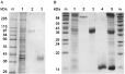
Fig. 1. Coomassie blue-stained 10% (A) and 16% (B) SDS–polyacrylamide gels of body wall extracts (BW) and pseudocelomic fluid (PF) of adult worms of Ascaris suum and purified fractions used for immunization of Groups 5, 6 and 7. Lane m, molecular weight markers; lane 1, BW extracts; lane 2, 97 kDa BW fraction; lane 3, 42 kDa PF fraction; lane 4, 14 kDa PF fraction; lane 5, PF.
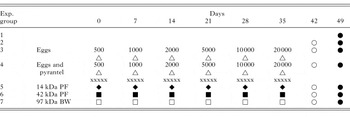
Table 1. Experimental design
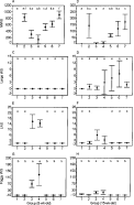
Fig. 2. Numbers (mean±S.D.) of mesh-like white spots (A, B), large white spots (C, D), lymphonodular white spots (E, F), and foggy white spots (G, H) in the livers of 8-week-old (A, C, E, G) and 15-week-old (B, D, F, H) pigs 7 days post-challenge with 10000 infective Ascaris suum eggs in relation to immunizing procedure (see Table 1). All differences between groups were significant except between those labelled with identical characters (a, b, c, d, e, f).
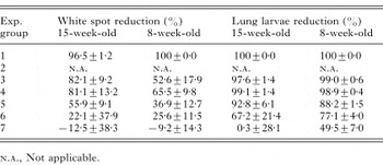
Table 2. Percentage reduction (mean and S.D.) in the total numbers of liver white spots and migrating Ascaris suum larvae in the lungs of immunized pigs in relation to the numbers in the challenge control pigs of Groups 2
 You have
Access
- 15
- Cited by