INTRODUCTION
The brain receives more sensory information than it can fully process. Attention is the process by which the brain selects those stimuli, or features thereof, that will be further processed. This down-selection system allows for successful interaction with the environment (Heilman & Valenstein, Reference Heilman and Valenstein1979; Heilman et al., Reference Heilman, Valenstein and Watson2012, Reference Heilman, Watson, Bower and Valenstein1983; Vecera & Rizzo, Reference Vecera and Rizzo2003). With sustained attention to particular stimuli, they lose their novelty and a concomitant reduction in the allocation of attention to said stimuli occurs, a phenomenon called habituation or temporal adaptation (Mennemeier et al., Reference Mennemeier, Chatterjee, Watson, Wertman, Carter and Heilman1994). The current study is designed to assess the influence of directed focal attention and prolonged viewing on bisection of both horizontal and vertical lines.
Individuals who suffer from brain injury such as stroke, tumor, trauma, or even neurodegenerative diseases can experience disturbances in their ability to attend to a certain portion of space. This inattention frequently manifests as “unilateral neglect” or “hemispatial neglect.” Patients with this disorder appear to be unaware or to have reduced awareness of stimuli in one half of egocentric (body-centered) space or, less frequently, allocentric (other- or object-centered) space. For example, individuals with a right-sided stroke often present with a left-sided neglect, manifested by a the failure to report, respond, or orient to stimuli presented on their contralesional, egocentric side of space (Heilman et al., Reference Heilman, Valenstein and Watson2012). With allocentric hemispatial neglect, patients are unaware of the one side of objects independent of the position of this object in relation to their body (Marsh & Hillis, Reference Marsh and Hillis2008). The presence of egocentric spatial neglect or inattention is often detected clinically using a line bisection task. When performing this test, patients with the neglect syndrome may be inattentive to the portion of the line contralateral to their lesioned hemisphere or hyper-attentive to the portion of stimuli ipsilateral to their injured hemisphere, resulting in misplacement of the attempted bisection toward the ipsilesional side of space (Heilman & Valenstein, Reference Heilman and Valenstein1979; Kerkhoff, Reference Kerkhoff2001). However, when healthy (i.e., neurologically normal) adult participants attempt to bisect horizontal lines, they tend to deviate toward the left of their true center, a phenomenon termed pseudoneglect (Bowers & Heilman, Reference Bowers and Heilman1980; Jewell & McCourt, Reference Jewell and McCourt2000; Shelton et al., Reference Shelton, Bowers and Heilman1990). There are reports, however, that some individuals exhibit rightward biases or a mixed pseudoneglect including variation in both the direction of deviation as well as the magnitude of deviation. Further, there can be an effect of handedness as well as older age, but not gender (Jewell & McCourt, Reference Jewell and McCourt2000; Manning et al., Reference Manning, Halligan and Marshall1990; Scarisbrick et al., Reference Scarisbrick, Tweedy and Kuslansky1987; Thomas et al., Reference Thomas, Aniulis and Nicholls2016). In addition to the horizontal biases, healthy individuals often have a vertical visuospatial attentional bias, with an upward pseudoneglect, although vertical neglect after brain injury has received less attention than horizontal (Heber et al., Reference Heber, Siebertz, Wolter, Kuhlen and Fimm2010; James, Reference James1890; McCourt & Olafson, Reference McCourt and Olafson1997; Scarisbrick et al., Reference Scarisbrick, Tweedy and Kuslansky1987; Suavansri et al., Reference Suavansri, Falchook, Williamson and Heilman2012).
The exact mechanisms of horizontal pseudoneglect are not fully understood; however, it has been postulated that this deviation could be related to right hemispheric dominance in the mediation of spatial attention and especially global attention (Lee et al., Reference Lee, Kim, Kang, Park, Kim, Adair and Na2004; Zago et al., Reference Zago, Petit, Jobard, Hay, Mazoyer, Tzourio-Mazoyer and Mellet2017). This asymmetric hemispheric activation could result in asymmetric allocation of spatial attention, resulting in a portion of the line (i.e., the left portion of the line) being perceived as having a larger magnitude. Vertical attentional asymmetries might be related to the relative activation of the ventral attentional network. Studies of patients with ventral temporal and dorsal inferior parietal lobe lesions suggest that the ventral system primarily allocates attention upward and the dorsal system downward (Rapcsak et al., Reference Rapcsak, Cimino and Heilman1988; Shelton et al., Reference Shelton, Bowers and Heilman1990). Studies have also revealed that the ventral stream mediates allocentric (object centered) attention and the dorsal system mediates egocentric attention. Since the line bisection test is an allocentric task, activation of the ventral stream may induce upward deviation (Medina et al., Reference Medina, Kannan, Pawlak, Kleinman, Newhart, Davis and Hillis2009).
Sustained Attention and Troxler Fading
With temporally extended attentional focus, the stimuli that are not the focus of attention lose their novelty, and the attentional resources allocated to it are reduced. For example, with sustained fixation on a central stimulus, peripheral stimuli will eventually fade from awareness after approximately 17 s in neurologically normal individuals, a phenomenon known as Troxler fading (Bonneh et al., Reference Bonneh, Donner, Cooperman, Heeger and Sagi2014; Mennemeier et al., Reference Mennemeier, Chatterjee, Watson, Wertman, Carter and Heilman1994; Troxler, Reference Troxler1804). For this effect to occur, the stimuli must remain stationary on the retina, allowing for both retinal and cortical adaptation (Bonneh et al., Reference Bonneh, Donner, Cooperman, Heeger and Sagi2014; Poletti & Rucci, Reference Poletti and Rucci2010). In addition to attentional habituation, Troxler fading and other types of visual fading may be the result of a decrease in visual microsaccades resulting in sensory adaptation (Bonneh et al., Reference Bonneh, Donner, Sagi, Fried, Cooperman, Heeger and Arieli2010; Poletti & Rucci, Reference Poletti and Rucci2010). Individuals with focal parietal lobe lesions typically exhibit significantly shorter fading times (Mennemeier et al., Reference Mennemeier, Chatterjee, Watson, Wertman, Carter and Heilman1994). Consistent with this, transcranial magnetic stimulation induced interruption of parietal lobe activity results in decreased awareness of a stimulus (Kanai et al., Reference Kanai, Muggleton and Walsh2008). The results of these studies suggest that the parietal lobe plays an important role in sustained attention to peripheral stimuli. In contrast, Troxler fading is absent in individuals with focal frontal lobe lesions, suggesting that the frontal lobe plays a critical role for habituation of attention to peripheral stimuli that are no longer novel (Mennemeier et al., Reference Mennemeier, Chatterjee, Watson, Wertman, Carter and Heilman1994).
We posited that directed allocation of focal attention to a portion of the line during the performance of line bisections would result in a bias toward the attended portion of the line. Further, we predicted that when focal attention is directed toward a portion of the line and the participants increase their viewing time of this line before attempting line bisection, a greater deviation toward the attended portion of the line would be produced (based on Troxler fading of the unattended portion of the line). Alternatively, increased viewing time could result in sensory habituation to the attended portion of the line and a reduction of the focal deviation.
METHODS
Participants
Thirty-six healthy, right-handed individuals, 19 women, between the ages of 18-31 (mean age = 23.25 years, SD = 2.66) were recruited for this study. The mean number of years of education was 17.58 (SD = 1.61). Of these participants, 27 were right eye dominant, 6 were left eye dominant, and 3 had mixed eye dominance as assessed by variations of both the Porta and Miles tests for eye dominance. For the Porta variant, participants were asked to extend their thumb to cover a viewed distant fixed object, and then alternate closing each eye while keeping the other eye open. The dominant eye was determined to be the one that, while open, kept the object covered by their thumb. For the Miles variant, the participants were asked to extend their arms in front of them and bring both hands together to make a small opening between their hands. They were then asked to view the same object through the opening in their hands, and slowly bring their hands with the opening toward their face. The dominant eye was determined to be the one that kept the object in focus through the opening in their hands. Handedness was assessed using the Benton Handedness Questionnaire. All participants signed an informed consent that was approved by the University of Florida Institutional Review Board in compliance with the Helsinki Declaration.
Apparatus
A white foam board measuring 20 in. x 30 in. (508 mm x 762 mm) was attached to the wall in a vertical orientation. The white board was mounted on the wall such that the center of the board was at the participants’ eye level at a distance of 14 in. (356 mm) from the bridge of their nose. The participants’ midsagittal plane was aligned with the center of the board. Pin-point sized holes were used to mark the correct location to place the stimulus for each trial. The stimuli used for this study were black lines measuring 240 mm long and 2 mm thick centered on a white sheet of ANSI A (US letter) size paper (8.5 in. x 11 in. or 215.9 mm x 274.9 mm). Each stimulus was presented separately, that is, one line per piece of paper for each trial. Both vertically and horizontally oriented lines were used in the study.
Experimental Procedure
For each trial, either a horizontal or vertical line, 240 mm in length, was placed on the board in front of the participants and they were verbally instructed to focus on either the whole line or a specified end of the line and then to bisect the line either immediately or after a 20-s delay indicated by an auditory (verbal) cue. For vertical lines, the participants were asked to focus either on the whole line, the top or the bottom end of the line. For horizontal lines, they were asked to focus either on the whole line, the left or the right end of the line. There were 30 immediate and 30 delay trials for horizontal lines (10 left-end focus immediate bisection, 10 left-end focus delayed bisection; 10 whole line focus immediate bisection, 10 whole line focus delayed bisection; 10 right-end focus immediate bisection, 10 right-end focus delayed bisection) as well as similar top, bottom, and whole line focus for vertical lines, for a total of 120 trials per subject. The participants were asked to bisect the line quickly after they were instructed to, and no feedback was given regarding their performance during execution of the task. The order that each attentional task was given was counterbalanced and randomized. All vertical lines bisection tests were given sequentially together and all horizontal line bisection tests were also given sequentially together within each task, and between participants the order in which the vertical versus horizontal lines were presented was counterbalanced. The immediate and delay trials (including which portion of the line to focus on) were given in a pre-determined, randomized order.
Analyses
All line measurements were made using Mitutoyo CD-S8”CT calipers (Kanagawa, Japan). Deviations toward the top of vertical lines or the left of horizontal lines were assigned a positive (+) value, whereas deviations toward the bottom of a vertical line or the right of a horizontal line were given negative values (-). Bisections were measured from the left of the horizontal lines and from the top of the vertical lines to the point where the line was marked. Statistical analyses were completed using R 3.1.3 using lmerTest (linear mixed effects models), applying the Satterthwaite’s method for degree of freedom calculation, and phia (post hoc interaction analysis) for evaluation of the effect of attentional focus on the size of the delay effect. A mixed effects model with attentional focus (whole line, left, right; whole line, top, bottom), the delay condition (20 s delayed or immediate bisection), an interaction between the attentional focus and delay condition, and a random effect of intercept and interaction slope by subject ID were used for analysis of each of the two experiments. A second set of mixed effects models with the same structure as was used for each of the primary analyses was used in which the bisection values were multiplied by -1 for the left and top attentional focus conditions to evaluate the magnitude of pairwise interaction between attentional focus and delay, with the p values of these post hoc interaction analyses being Holm corrected to reduce the risk of type 1 error but with less risk of type 2 error than Bonferroni correction.
RESULTS
Horizontal Line Bisections
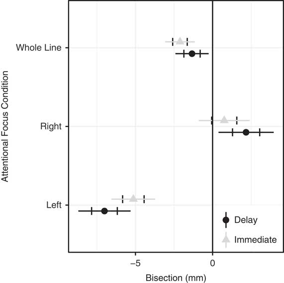
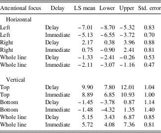
We found that there was a main effect of attentional focus position (left, right, whole line) {F[3, 35.09] = 29.00, p<0.0001}, no main effect of delay {F[1, 35.06] = 0.32, p = 0.578}, and an interaction effect between delay and side of the attentional focus {F[2, 43.72] = 11.74, p<0.0001} when participants performed horizontal line bisections (see Figure 1 and Table 1 for least square means and confidence intervals). The effect of directing attention to a certain portion of the line (left or right end) or the whole line, whether the line was bisected immediately or after a delay, was 6.1 mm {95%CI: [5.1-7.0], Cohen’s d = 1.39} leftward with a left-sided focus, 1.5 mm {95%CI: [0.5-2.4], Cohen’s d = 0.30} rightward with a right-sided focus, and 1.7 mm {95%CI: [0.8-2.7], Cohen’s d = 0.60} leftward with a whole line focus. Thus, when they were asked to view the left end of the line, there was deviation toward the left, away from the true center. When they were asked to view the right end of the line, there was a deviation toward the right end of the line, again away from the true center; and with attention directed to the whole line, there was a slight leftward bias, consistent with pseudoneglect.
Horizontal line bisection results shown as least square means and 95% confidence intervals. Standard errors are indicated with an overlaid error bar. True center is indicated with a vertical line at 0.

LS means (estimated marginal means), 95% confidence interval, and standard error estimates

When the participants were asked to fixate on a certain portion of the line for a delay, there was no significant main effect between the amount of deviation in comparison to the trials where they were asked to bisect immediately; however, this is because of the cancelling effects of the interaction between the attentional focus and the delay. Increased viewing time to the left {t[35.0] = 3.84, p = 0.0005} or right {t[36.4] = 3.47, p = 0.0013} side of the line, however, did significantly enhance their deviation toward the end of the line to which they were attending or toward the true center with whole line attentional focus {t[68.6] = 2.75, p = 0.0076}, with the effect of delay with left focus being 1.9 mm {95%CI: [1.2-2.6], Cohen’s d = 0.64} leftward, the effect of delay with right focus being 1.4 mm {95%CI: [0.7-2.1], Cohen’s d = 0.58} rightward, and 0.8 mm {95%CI: [0.1-1.5], Cohen’s d = 0.46} toward the true center (rightward). To evaluate the potential effect of attentional focus on the magnitude of the delay-induced shift in bisection, we repeated the analysis with the bisection of left-directed attention multiplied by -1. With this analysis, we found an effect of attentional focus (p<0.0001) and delay (p<0.0001), but no interaction (p = 0.146). Post hoc analysis of the interaction found no pairwise effects for delay (Left-Right χ 2 = 0.789, p = 0.394; Left-Whole Line χ 2 = 3.946, p = 0.141; Right-Whole Line χ 2 = 1.664, p = 0.394, Holm adjusted p values), thus we found no effect of attentional focus on the effect size of the delay, although there may be a trend toward such an effect.
Vertical Line Bisections
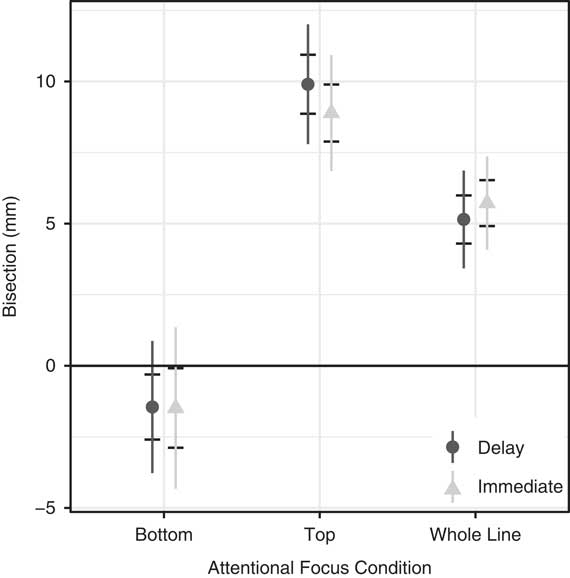
We found that there was a main effect for the position of the attentional focus (top, bottom, whole line) {F[3, 35.0] = 30.41, p<0.0001}, no main effect of delay {F[1, 35.11] = 0.23, p = 0.633}, and an interaction effect between delay and side of the attentional focus {F[2, 48.61] = 4.54, p = 0.0156} when participants performed vertical line bisections (see Figure 2 and Table 1 for least square means and confidence intervals). The effect of directing attention to a certain portion of the line (top or bottom) or the whole line, whether the line was bisected immediately or after a delay, was 9.4 mm {95%CI: [7.4-11.4]} upward for top focus, 1.5 mm {95%CI: [-4-1]} downward with bottom focus, and 5.4 mm {95%CI: [3.8-7.0]} upward for whole line focus. Increased viewing time to the top {t[36] = 2.03, p = 0.05}, but not bottom {t[35.9] = 0.05, p = 0.95} or whole line {t[38.5] = 1.29, p = 0.2}, resulted in an enhanced deviation in the direction of focused attention. Thus, when the participants were asked to view the top end of the line, there was deviation toward the top, away from the true center. When they were asked to view the bottom end of the line, there was a deviation toward the bottom end of the line, again away from the true center; and with attention directed to the whole line, an upward bias was observed, consistent with prior reports.
Vertical line bisection results shown as least square means and 95% confidence intervals. Standard errors are indicated with an overlaid error bar. True center is indicated with a vertical line at 0.

When the participants were asked to fixate on a certain portion of the line, with a delay in their attempted bisection, there was no significant main effect between the amount of deviation in comparison to the trials where they were asked to bisect immediately. Increased viewing time to the top, but not the bottom end or the whole line, significantly enhanced their deviation toward the top of the line, with the effect of delay with top focus being 1.01 mm {95%CI: [0.0-2.0], Cohen’s d = 0.34} upward, there was no significant effect of delay with bottom focus being 0.03 mm {95%CI: [-1.14-1.07], Cohen’s d = 0.01}, and 0.57 mm {95%CI: [-0.33-1.47], Cohen’s d = 0.22} toward the true center (downward). To evaluate the potential effect of attentional focus on the magnitude of the delay-induced shift in bisection with upward focus, we repeated the analysis with the bisection of upward-directed attention multiplied by -1. With this analysis, we found an effect of attentional focus (p<0.0001) but not delay (p = 0.0855) or an interaction (p = 0.0288). Post hoc analysis of the interaction found no pairwise effects for delay (Bottom-Top χ 2 = 2.247, p = 0.402; Bottom-Whole Line χ 2 = 0.993, p = 0.638; Top-Whole Line χ 2 = 0.322, p = 0.638, Holm-adjusted p values). Thus, we found no effect of attentional focus on the effect size of the delay, although there was a significant effect of delay with top-directed attention. This finding may be due to the lack of power to detect this interaction magnitude effect.
DISCUSSION
The primary results of this study demonstrate that directed allocation of vertical or horizontal focused attention prior to attempted line bisection increases deviations toward attended spatial locations in healthy participants. Further, leftward attentional focus and upward attentional focus, respectively, resulted in the largest deviations. Attentional delay amplified deviation toward the attentional focus. In horizontal space, the effect of attentional delay was essentially symmetrical. In vertical space, the deviation was strongest with upward focus, with no effect of delay when focusing attention downward.
Additionally, when the participants were asked to focus on either the top of a vertical line or the left of a horizontal line, the distance of their deviation away from the true center was larger than they were asked to focus on the bottom or the right of a vertical or horizontal line, respectively. Healthy adults often have “pseudoneglect” with a leftward and upward attentional bias when performing horizontal and vertical line bisection tasks. The leftward bias, that is, pseudoneglect, on the line bisection test has been attributed to right hemispheric dominance in mediating horizontal spatial attention (Heilman & Van Den Abell, Reference Heilman and Van Den Abell1980; McCourt & Jewell, Reference McCourt and Jewell1999). The upward bias with vertical line bisections is thought to be related to activation of the ventral attentional stream that mediates allocentric, or object-centered, as well as upward attention (Falchook et al., Reference Falchook, Mody, Srivastava, Williamson and Heilman2013). Therefore, focusing attention on the left or top of a line might have been more likely to activate the right parietal and ventral attentional networks than focusing attention to the right or downward.
Prior work has suggested that, by directing the participants to willfully attend to a specific portion of the line, a “top-down” attentional network including the intraparietal cortex and superior frontal cortex is probably engaged (Corbetta & Shulman, Reference Corbetta and Shulman2002; Katsuki & Constantinidis, Reference Katsuki and Constantinidis2014). Additionally, “bottom-up” attention may have played a role in the allocation of attention toward the other portions of the line. “Bottom-up” attention relies on distinctiveness and novelty of the stimulus and appears to be primarily mediated by the right hemisphere’s temporoparietal cortex and inferior frontal cortex (Corbetta & Shulman, Reference Corbetta and Shulman2002; Ibos et al., Reference Ibos, Duhamel and Ben Hamed2013).
Although not as frequently studied, patients with brain injuries such as stroke often also reveal vertical neglect in addition to the commonly reported contralesional horizontal neglect. As with left-sided hemispatial neglect, lower vertical neglect is more common than upper vertical neglect. Therefore, with engagement of the “top-down” attentional network, there should have been greater activation of the more dorsal attentional system than the ventral system. In addition, with activation of this dorsal network, we would have expected that with both immediate and prolonged viewing of vertical lines, there would have been greater downward deviation when viewing the bottom of the lines than upward deviation when viewing the top of the lines. However, our finding of the opposite results suggests that a different network might be mediating allocentric upward attention.
This upward bias in the vertical line bisection test may be induced by activation of the allocentric (object-centered) ventral visual system (Falchook et al., Reference Falchook, Mody, Srivastava, Williamson and Heilman2013). This hypothesis and prediction are opposite to that proposed by Corbetta and Shulman (Reference Corbetta and Shulman2002). Whereas the brain mechanism accounting for this top-bottom asymmetry is not entirely known, a patient with bilateral damage to the ventral temporal and occipital lobes had neglect of the upper altitudinal space (Shelton et al., Reference Shelton, Bowers and Heilman1990). This patient also revealed an apperceptive agnosia with an impaired ability to mediate focal attention. This patient’s focal inattention is in contrast to patients parietal lesions who may present with simultanagnosia, an inability to allocate global attention (Bálint, Reference Bálint1909).
Patients with a parietal lobe lesion have been reported to more often have left egocentric neglect (body-centered spatial neglect) and those with temporal lobe lesions to have allocentric neglect (object-centered neglect) (Medina et al., Reference Medina, Kannan, Pawlak, Kleinman, Newhart, Davis and Hillis2009). Thus, focusing on a segment of a line might be more like object-oriented attention and with focused attention the ventral object centered network may be more activated than the dorsal network. This asymmetry of ventral versus dorsal cerebral activation might help to account for the greater upper than lower bias seen with the vertical line bisections.
To further learn whether this object-centered ventral stream or the “top down” dorsal stream might mediate focal attention, prior studies had normal participants perform a vertical line quadrisection task (Falchook et al., Reference Falchook, Mody, Srivastava, Williamson and Heilman2013). This quadrisection task requires more focal attention than does the line bisection test. That study found that the mean upward bias (deviation error) for the quadrisection task was significantly greater than the mean error for the line bisection test and that upper quadrisection induced a greater upward bias than lower quadrisection induced a lower bias. These results are consistent with the hypothesis that activation of the ventral stream by a task that requires allocentric as well as upward focal attention can induce an upward vertical bias that is relatively greater than the downward bias when focal attention is allocated to the lower portion of the line.
Habituation to the portion of the line to which the participants directed their attention did not seem to occur because over time when performing the line bisection, the participants did not deviate away from the targeted portion, but instead toward the target. This implies that novelty of the stimulus is not necessarily the most significant factor in terms of the allocation of spatial attention. Perhaps sustained attention, “top-down,” or goal directed attention supersedes the mechanisms underlying habituation to a non-novel stimulus.
Alteration in line bisection performance during the delayed bisection test could be caused by increased allocation of attention to one location or decreased attention to the opposite location. For example, when a person fixates on a particular area or object for several seconds, a fixed stimulus that is located away from the fixation point will fade away (Troxler fading) (Troxler, Reference Troxler1804). Although it is not known whether Troxler fading occurs to the unattended portion of the line when participants sustain their attentional focus to one end of a line, inattention to an object might make that object appear smaller. Allocation of attention to an object or a portion of an object can also make that object appear larger (Anton-Erxleben et al., Reference Anton-Erxleben, Henrich and Treue2007).
Although the mechanism accounting for the deviation toward the end of the line with sustained attention is not known and needs to be further investigated, when the participants in this study were asked to sustain their attention to the entire line, their pseudoneglect bias decreased. Taken together, the findings of reduction in pseudoneglect with sustained attention to the entire line and increased bias with sustained attention to the ends of the line suggest that these results are due to a reduction in attention to unattended portions of the stimuli. Attending to the whole line might have allowed the peripheral portions of the line to fade. With fading of the ends of the line, they have been perceived as shorter, and it has been shown that the magnitude of pseudoneglect reduces with shorter lines (Nicholls et al., Reference Nicholls, Beckman and Churches2016).
With a delay in vertical line bisection, the increased viewing time to the top, but not the bottom end, significantly enhanced these participants’ deviation toward the top of the line. With a delay of bisection during a unilateral focus of attention to the end of a horizontal line, there was also a greater deviation toward the end of the line to which the participants focused their attention. The underlying mechanism for the top-bottom asymmetry in the delayed vertical line bisection test, but no left-right asymmetry with the horizontal line stimuli is unknown.
This study has limitations. Our neuropsychological hypotheses were based on prior literature and were not directly measured with an imaging technology such as functional magnetic resonance imaging. Further, an independent test of Troxler fading rate in this sample was not performed. Because Troxler fading can be altered with eye movements, future research should incorporate eye tracking. Further, eye tracking would be useful to verify compliance with directed attention and to address potential individual differences in the ability to sustain attentional focus. Moreover, it may be important to learn whether different lengths of delay might produce different results.
Neurologic patients with hemispatial neglect are inattentive or even unaware of stimuli in the hemispace contralateral to their lesion (Heilman et al., Reference Heilman, Valenstein and Watson2012); however, many patients with this disorder recover their ability to perform normally on tests such as line bisection. Despite their improved performance on this test, most of these patients remain disabled. Many of the daily tasks we perform, whether they be activities of daily living or instrumental activities require sustained attention. It has been demonstrated that Troxler fading is often more rapid on the side contralateral to a hemispheric lesion in the temporoparietal region (Mennemeier et al., Reference Mennemeier, Chatterjee, Watson, Wertman, Carter and Heilman1994). Perhaps patients with neglect who have recovered and performed well on tests, such as line bisection, may still have rapid fading of contralesional stimuli and thus allocate less attention to these contralesional stimuli. Being inattentive might lead to a greater disability. Therefore, in patients with frontal or posterior temporoparietal hemispheric lesions, it may be valuable to evaluate the attentional impacts of prolonged viewing during attentional tasks, particularly in conjunction with modifications of current clinical tests (McIntosh et al., Reference McIntosh, Ietswaart and Milner2017). Such modifications to clinical assessments may offer more clinically sensitive tests for the detection of elements of the neglect syndrome.
ACKNOWLEDGMENTS
This material is based upon work supported in part by the US Department of Veterans Affairs, Veterans Health Administration, Office of Research and Development Grant I01CX000744, and Brain Rehabilitation Research Center (BRRC) Center Grant B6793C. The views expressed in this article are those of the authors and do not necessarily reflect the position or policy of the Department of Veterans Affairs or the United States government. The authors declare no conflicts of interest.