





Introduction
Acanthamoeba species are ubiquitous free-living amoebae widely distributed in soil, water and air (Visvesvara et al., Reference Visvesvara, Moura and Schuster2007; Mungroo et al., Reference Mungroo, Khan, Maciver and Siddiqui2022; Rayamajhee et al., Reference Rayamajhee, Willcox, Henriquez, Petsoglou, Subedi and Carnt2022). While most human exposures are asymptomatic, certain species can cause severe opportunistic infections, including granulomatous amoebic encephalitis (GAE) (Visvesvara et al., Reference Visvesvara, Moura and Schuster2007; Seas and Legua, Reference Seas and Legua2022). Acanthamoeba encephalitis is rare but associated with high mortality, largely due to delayed diagnosis and limited therapeutic options (Cope et al., Reference Cope, Ali and Visvesvara2020; Damhorst et al., Reference Damhorst, Watts, Hernandez-romieu, Mel, Palmore, Ali, Neill, Kalapila and Cope2022). The disease most commonly affects individuals with impaired immune function, although cases in immunocompetent hosts have also been reported (Haston et al., Reference Haston, O’laughlin, Matteson, Roy, Qvarnstrom, Ali and Cope2023). In contrast to primary amoebic meningoencephalitis caused by Naegleria fowleri, which is acute and rapidly progressive, Acanthamoeba encephalitis typically follows a subacute or chronic course (Visvesvara et al., Reference Visvesvara, Moura and Schuster2007; Mungroo et al., Reference Mungroo, Khan, Maciver and Siddiqui2022). Disease progression reflects prolonged persistence of the organism within neural tissue and a sustained host immune response rather than overwhelming cytolytic injury (Siddiqui and Khan, Reference Siddiqui and Khan2012). Nonetheless, comparative neuropathological analysis across species, including humans, naturally infected animals and experimental models, provides a framework for identifying conserved mechanisms of tissue invasion, inflammation and vascular involvement, and for assessing the translational relevance of experimental models. Here, we discuss pathological findings from human cases, naturally infected animals and experimental studies to define the core pathological features of Acanthamoeba granulomatous encephalitis.
Pathogenesis and routes of central nervous system invasion
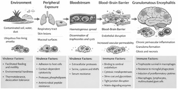
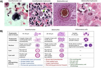
Infection with Acanthamoeba typically begins at extracranial sites, most commonly the respiratory tract or skin (Kofman and Guarner, Reference Kofman and Guarner2022). Environmental exposure occurs through inhalation of cysts or trophozoites, contact with contaminated water or entry through skin lesions (Bahrami et al., Reference Bahrami, Zarei, Henriquez and Nzelu2025). Following peripheral colonization, amoebae disseminate haematogenously and interact with vascular endothelium to gain access to the central nervous system (Figure 1) (Khan, Reference Khan2007; Siddiqui and Khan, Reference Siddiqui and Khan2012). Crossing the blood-brain barrier is associated with endothelial disruption, degradation of tight junction proteins and increased vascular permeability (Khan, Reference Khan2007; Siddiqui and Khan, Reference Siddiqui and Khan2012). Acanthamoeba produces a range of proteases and other virulence factors that contribute to endothelial injury and facilitate paracellular migration (Siddiqui and Khan, Reference Siddiqui and Khan2012). Once within the brain, the organism exhibits a predilection for perivascular and parenchymal niches (Martinez, Reference Martinez1991; Visvesvara et al., Reference Visvesvara, Moura and Schuster2007). Both trophozoites and cysts are present in neural tissue, indicating ongoing differentiation and long-term persistence (Figure 2). Unlike Naegleria infection, where rapid trophozoite proliferation dominates pathology, Acanthamoeba encephalitis reflects a dynamic balance between parasite survival and host immune containment (Mungroo et al., Reference Mungroo, Khan, Maciver and Siddiqui2022).
Pathogenic sequence and host-pathogen interactions in granulomatous amoebic encephalitis caused by Acanthamoeba spp.
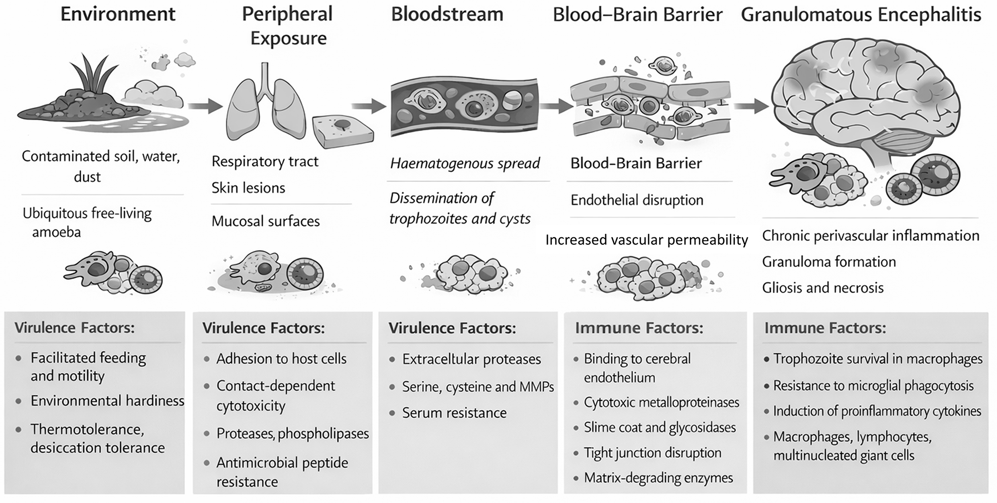
Figure 1 Long description
The diagram illustrates the progression of granulomatous amoebic encephalitis through five stages: Environment, Peripheral Exposure, Bloodstream, Blood-Brain Barrier and Granulomatous Encephalitis. Starting from the left, the Environment stage depicts contaminated soil, water and dust, highlighting ubiquitous free-living amoebae. Virulence factors include facilitated feeding, environmental hardness, thermotolerance and desiccation tolerance. The Peripheral Exposure stage shows the respiratory tract and skin lesions, with mucosal surfaces as entry points. Virulence factors here are adhesion to host cells, contact-dependent cytotoxicity, proteases, phospholipases and antimicrobial peptide resistance. The Bloodstream stage illustrates haematogenous spread and dissemination of trophozoites and cysts. Virulence factors include extracellular proteases, serine, cysteine and MMPs and serum resistance. The Blood-Brain Barrier stage shows endothelial disruption and increased vascular permeability. Immune factors involve binding to cerebral endothelium, cytokine neurotoxicity, slime coat and glycosides, tight junction disruption and matrix-degrading enzymes. Finally, the Granulomatous Encephalitis stage depicts chronic perivascular inflammation, granuloma formation and gliosis and necrosis. Immune factors include trophozoite survival in macrophages, resistance to microglial phagocytosis, induction of inflammatory cytokines and the presence of macrophages, lymphocytes and multinucleated giant cells. Arrows indicate the progression from one stage to the next.
Histopathological features of Acanthamoeba spp. and Balamuthia mandrillaris infections.
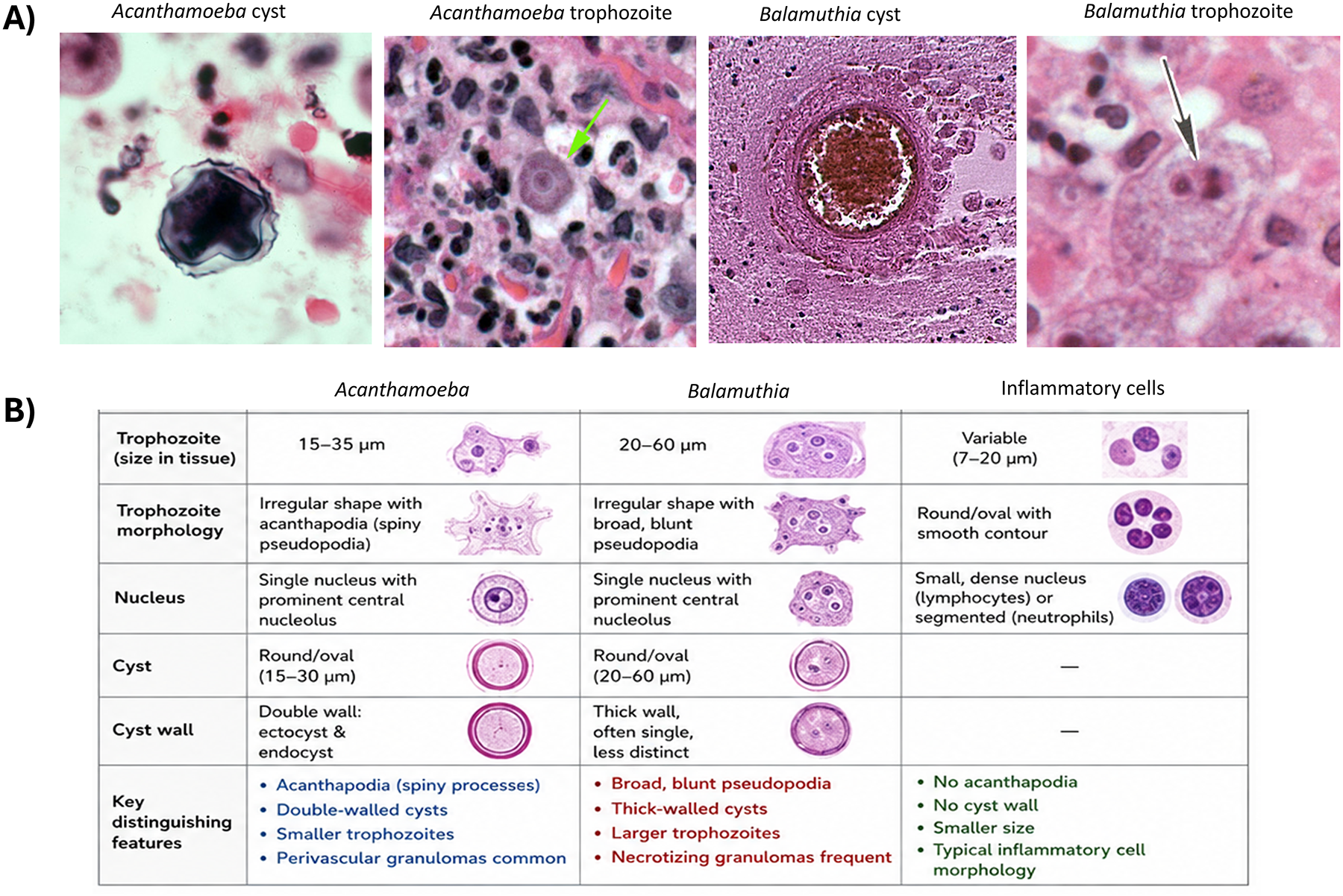
Figure 2 Long description
Consists of two parts. A) Four micrographs showing: Acanthamoeba cyst, Acanthamoeba trophozoite, Balamuthia cyst and Balamuthia trophozoite. B) A table comparing Acanthamoeba, Balamuthia and inflammatory cells. Columns include: Trophozoite morphology, nucleus, cyst, cyst wall and key distinguishing features. Acanthamoeba shows irregular shape with pseudopodial tapering, single nucleus and double-walled cysts. Balamuthia has broad pseudopodes, large walled cysts and necrotizing granulomas. Inflammatory cells are round with dense nuclei. Key features are highlighted in different colors for each organism.
Pathology in humans and animals
Human disease
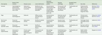
In humans, Acanthamoeba granulomatous encephalitis presents with multifocal lesions distributed throughout the cerebral hemispheres, basal ganglia, brainstem and cerebellum (Martinez, Reference Martinez1991; Mungroo et al., Reference Mungroo, Khan, Maciver and Siddiqui2022). Gross pathological findings include cerebral oedema, focal haemorrhage and areas of necrosis. Lesions are often poorly demarcated and may mimic neoplastic, demyelinating or vasculitic processes on neuroimaging (Damhorst et al., Reference Damhorst, Watts, Hernandez-romieu, Mel, Palmore, Ali, Neill, Kalapila and Cope2022; Mungroo et al., Reference Mungroo, Khan, Maciver and Siddiqui2022). Microscopically, the defining feature is granulomatous inflammation composed of macrophages, lymphocytes, plasma cells and multinucleated giant cells (Martinez, Reference Martinez1991; Visvesvara et al., Reference Visvesvara, Moura and Schuster2007). Perivascular cuffing is prominent, reflecting the angiotropic nature of the infection (Martinez, Reference Martinez1991; Khan, Reference Khan2007). Necrosis is typically focal rather than diffuse and may be accompanied by gliosis and microhaemorrhage. Amoebae are detected within granulomas, perivascular spaces and necrotic foci, with both trophozoites and cysts present (Martinez, Reference Martinez1991; Visvesvara et al., Reference Visvesvara, Moura and Schuster2007; Mungroo et al., Reference Mungroo, Khan, Maciver and Siddiqui2022). Organisms may be sparse and difficult to identify without specialized staining or molecular techniques, contributing to frequent diagnostic delay. The principal neuropathological features of Acanthamoeba encephalitis observed across species are summarized in Table 1.
Animal infections and experimental models
Naturally occurring Acanthamoeba encephalitis has been documented in a variety of animals, including dogs, non-human primates and livestock species (Dubey et al., Reference Dubey, Benson, Blakeley, Booton and Visvesvara2005; Visvesvara et al., Reference Visvesvara, Moura and Schuster2007; Kent et al., Reference Kent, Platt, Rech, Eagleson, Howerth, Shoff, Fuerst, Booton, Visvesvara and Schatzberg2011; Gaide et al., Reference Gaide, Pélandakis, Robveille, Albaric, Jouvion, Souchon, Risler and Abadie2015; Bahrami et al., Reference Bahrami, Zarei, Henriquez and Nzelu2025; Barrantes Murillo et al., Reference Barrantes Murillo, Berrocal, Ali and Uzal2025). Clinical and pathological features closely resemble human disease, with progressive neurological dysfunction and multifocal granulomatous lesions centred on blood vessels (Dubey et al., Reference Dubey, Benson, Blakeley, Booton and Visvesvara2005; Kent et al., Reference Kent, Platt, Rech, Eagleson, Howerth, Shoff, Fuerst, Booton, Visvesvara and Schatzberg2011). Post-mortem examination reveals chronic meningoencephalitis with perivascular inflammation, focal necrosis and persistent amoebae (Kent et al., Reference Kent, Platt, Rech, Eagleson, Howerth, Shoff, Fuerst, Booton, Visvesvara and Schatzberg2011; Barrantes Murillo et al., Reference Barrantes Murillo, Berrocal, Ali and Uzal2025). Experimental models, primarily in rodents but also including early studies in non-human primates, reproduce key aspects of human pathology, particularly when immunosuppression is employed (Wong et al., Reference Wong, Karr and Balamuth1975; Siddiqui and Khan, Reference Siddiqui and Khan2012; Borges et al., Reference Borges, De Macedo, Da Silva, Ataíde, Mendes, Passos, De Moraes, Herculano, Oliveira, Bahia and Dolabella2024). Following intranasal or intravenous inoculation, animals develop haematogenous dissemination and angiocentric brain lesions (Borges et al., Reference Borges, De Macedo, Da Silva, Ataíde, Mendes, Passos, De Moraes, Herculano, Oliveira, Bahia and Dolabella2024). Granulomatous inflammation, perivascular amoebae and cyst formation are consistently observed (Siddiqui and Khan, Reference Siddiqui and Khan2012; Borges et al., Reference Borges, De Macedo, Da Silva, Ataíde, Mendes, Passos, De Moraes, Herculano, Oliveira, Bahia and Dolabella2024). Disease progression is slower than in Naegleria models and reflects chronic infection rather than acute lethality. These models have been instrumental in elucidating host–pathogen interactions and evaluating therapeutic strategies, although they may not fully capture the heterogeneity observed in human disease.
Reports of Acanthamoeba encephalitis in diverse mammalian hosts further support the conserved nature of the neuropathological process. Naturally occurring infections have been described in domestic animals such as dogs, wildlife species including sloths and non-human primates, where lesions frequently involve necrotizing or granulomatous meningoencephalitis associated with amoebic trophozoites and cysts within neural tissue (Dubey et al., Reference Dubey, Benson, Blakeley, Booton and Visvesvara2005; Kent et al., Reference Kent, Platt, Rech, Eagleson, Howerth, Shoff, Fuerst, Booton, Visvesvara and Schatzberg2011; Gaide et al., Reference Gaide, Pélandakis, Robveille, Albaric, Jouvion, Souchon, Risler and Abadie2015; Barrantes Murillo et al., Reference Barrantes Murillo, Berrocal, Ali and Uzal2025). Although host species vary in physiology and immune responses, the fundamental pattern of angiocentric inflammation, tissue necrosis and persistent amoebic stages appears broadly conserved. These observations reinforce the concept that Acanthamoeba neuropathogenesis follows similar pathological trajectories across mammalian hosts and support the translational relevance of experimental models.
Cross-species neuropathological features of granulomatous amoebic encephalitis caused by Acanthamoeba spp
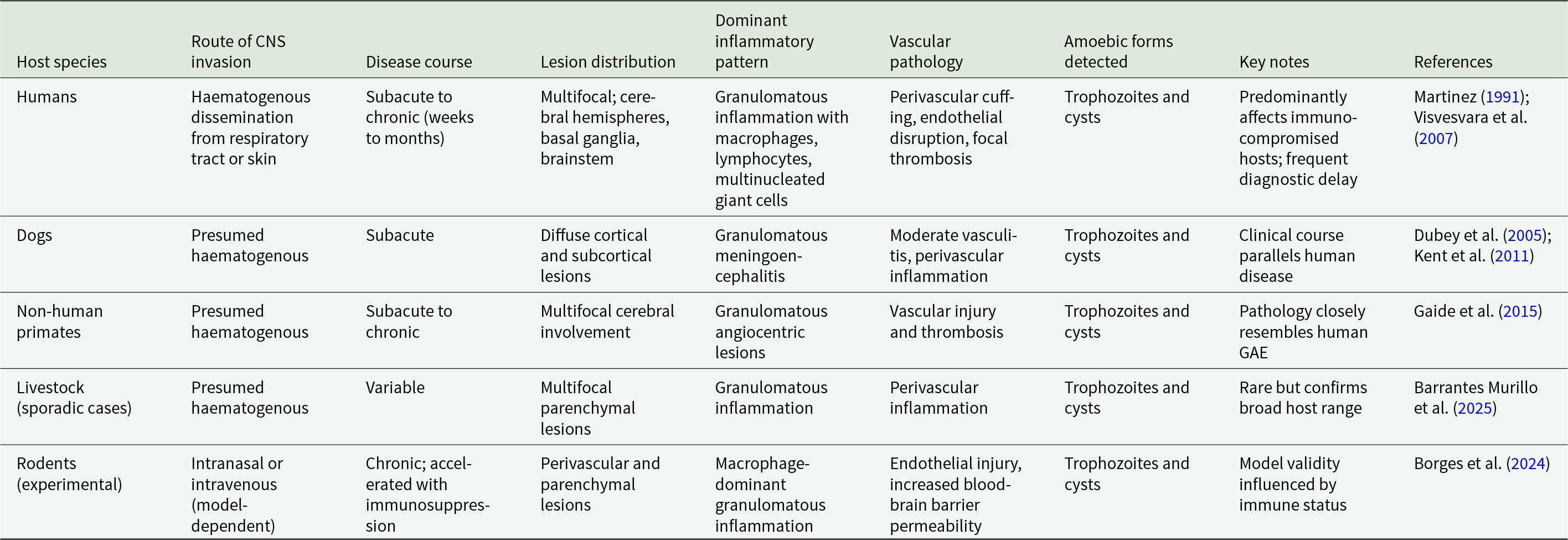
Table 1 Long description
Columns compare host species by route of CNS invasion, disease course, lesion distribution, dominant inflammatory pattern, vascular pathology, amoebic forms detected, key notes, and references. Humans are listed with haematogenous dissemination from respiratory tract or skin, a subacute to chronic course over weeks to months, multifocal lesions in cerebral hemispheres, basal ganglia, and brainstem, granulomatous inflammation with macrophages, lymphocytes, and multinucleated giant cells, and vascular findings of perivascular cuffing, endothelial disruption, and focal thrombosis; trophozoites and cysts are detected, with notes of predominant immunocompromised hosts and frequent diagnostic delay, and references Martinez, 1991 and Visvesvara et al., 2007. Dogs show presumed haematogenous invasion, subacute course, diffuse cortical and subcortical lesions, granulomatous meningoencephalitis, moderate vasculitis with perivascular inflammation, trophozoites and cysts, and a note that the clinical course parallels human disease, with references Dubey et al., 2005 and Kent et al., 2011. Non human primates list presumed haematogenous invasion, subacute to chronic course, multifocal cerebral involvement, granulomatous angiocentric lesions, vascular injury and thrombosis, trophozoites and cysts, and a note that pathology closely resembles human GAE, with reference Gaide et al., 2015. Livestock, described as sporadic cases, list presumed haematogenous invasion, variable course, multifocal parenchymal lesions, granulomatous inflammation, perivascular inflammation, trophozoites and cysts, and a note that cases are rare but confirm broad host range, with reference Barrantes Murillo et al., 2025. Rodents in experimental models list intranasal or intravenous invasion depending on model, chronic course that is accelerated with immunosuppression, perivascular and parenchymal lesions, macrophage dominant granulomatous inflammation, endothelial injury with increased BBB permeability, trophozoites and cysts, and a note that model validity is influenced by immune status, with reference Borges et al., 2024.
Cross-species comparison
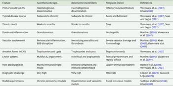
Comparative analysis across species reveals a conserved pathological signature of Acanthamoeba encephalitis characterized by haematogenous dissemination, perivascular localization, granulomatous inflammation and chronic tissue injury. In humans, companion animals and experimental models, lesions consistently cluster around blood vessels and are associated with sustained inflammatory responses (Visvesvara et al., Reference Visvesvara, Moura and Schuster2007; Kent et al., Reference Kent, Platt, Rech, Eagleson, Howerth, Shoff, Fuerst, Booton, Visvesvara and Schatzberg2011; Borges et al., Reference Borges, De Macedo, Da Silva, Ataíde, Mendes, Passos, De Moraes, Herculano, Oliveira, Bahia and Dolabella2024). The presence of both trophozoites and cysts within neural tissue is a universal finding and underscores the organism’s capacity for long-term persistence. Despite this conservation, interspecies differences influence disease kinetics and severity. Immunocompromised hosts exhibit more extensive dissemination and reduced granuloma organization, whereas immunocompetent hosts may contain infection more effectively but still sustain progressive neurological damage. Lesion distribution varies with host anatomy and immune status, but the fundamental mechanisms of vascular invasion and granulomatous inflammation remain consistent across species. Key pathological distinctions between Acanthamoeba, Balamuthia and N. fowleri are outlined in Table 2.
Comparative pathological distinctions in CNS infection due to major free-living amoebae
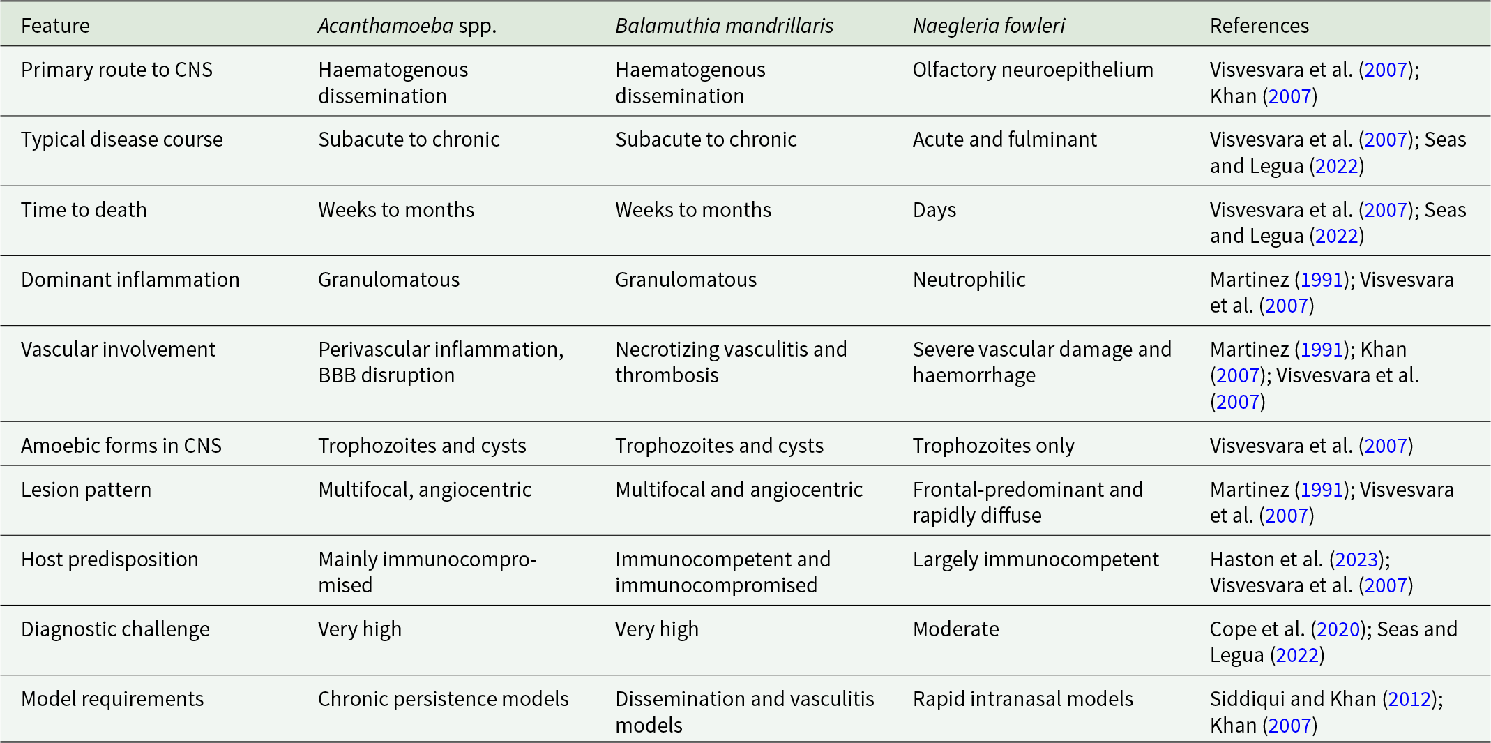
Table 2 Long description
Features compared across Acanthamoeba spp., Balamuthia mandrillaris, and Naegleria fowleri include primary route to the central nervous system, disease course, time to death, inflammation, vascular involvement, amoebic forms, lesion pattern, host predisposition, diagnostic challenge, and model requirements. For primary route to the central nervous system, Acanthamoeba spp. and Balamuthia mandrillaris list haematogenous dissemination, while Naegleria fowleri lists olfactory neuroepithelium. Typical disease course is subacute to chronic for Acanthamoeba spp. and Balamuthia mandrillaris, and acute and fulminant for Naegleria fowleri. Time to death is weeks to months for Acanthamoeba spp. and Balamuthia mandrillaris, and days for Naegleria fowleri. Dominant inflammation is granulomatous for Acanthamoeba spp. and Balamuthia mandrillaris, and neutrophilic for Naegleria fowleri. Vascular involvement lists perivascular inflammation and blood brain barrier disruption for Acanthamoeba spp., necrotising vasculitis and thrombosis for Balamuthia mandrillaris, and severe vascular damage and haemorrhage for Naegleria fowleri. Amoebic forms in the central nervous system are trophozoites and cysts for Acanthamoeba spp. and Balamuthia mandrillaris, and trophozoites only for Naegleria fowleri. Lesion pattern is multifocal and angiocentric for Acanthamoeba spp. and Balamuthia mandrillaris, and frontal predominant and rapidly diffuse for Naegleria fowleri. Host predisposition is mainly immunocompromised for Acanthamoeba spp., immunocompetent and immunocompromised for Balamuthia mandrillaris, and largely immunocompetent for Naegleria fowleri. Diagnostic challenge is very high for Acanthamoeba spp. and Balamuthia mandrillaris, and moderate for Naegleria fowleri. Model requirements list chronic persistence models for Acanthamoeba spp., dissemination and vasculitis models for Balamuthia mandrillaris, and rapid intranasal models for Naegleria fowleri.
Environmental niche and host susceptibility
Acanthamoeba species are among the most ubiquitous protozoa in the environment, yet invasive central nervous system infection is exceedingly rare. This discrepancy highlights the importance of host susceptibility in disease development. Immunosuppression, including HIV infection, organ transplantation and corticosteroid therapy, is a major risk factor (Damhorst et al., Reference Damhorst, Watts, Hernandez-romieu, Mel, Palmore, Ali, Neill, Kalapila and Cope2022; Kofman and Guarner, Reference Kofman and Guarner2022; Haston et al., Reference Haston, O’laughlin, Matteson, Roy, Qvarnstrom, Ali and Cope2023), although cases in immunocompetent individuals demonstrate that host factors alone do not fully explain disease occurrence. The ability of Acanthamoeba to form cysts that resist environmental stress and immune clearance likely contributes to both environmental persistence and chronic infection.
The conserved neuropathological features of Acanthamoeba encephalitis across species validate the use of animal models for mechanistic and translational research. Chronic perivascular inflammation, cyst persistence and focal necrosis identify key processes that distinguish Acanthamoeba infection from the acute pathology of Naegleria and the angiocentric vasculitis of Balamuthia. These differences have important implications for diagnosis and therapy. Future studies should focus on elucidating mechanisms of blood–brain barrier traversal, immune evasion and cyst resilience within neural tissue. Improved experimental models that capture chronic disease dynamics and immune heterogeneity are needed to evaluate therapeutic strategies. Early recognition of infection and targeted intervention remain critical challenges in improving outcomes for this devastating disease.
In conclusion, comparative analysis demonstrates that Acanthamoeba species produce a conserved pattern of granulomatous encephalitis across mammalian hosts. Regardless of species, infection is characterized by haematogenous dissemination, perivascular localization, chronic inflammation and persistent tissue injury mediated by both trophozoites and cysts. While disease kinetics and clinical presentation vary, the core pathological mechanisms remain consistent. These findings reinforce the translational relevance of animal models and provide a framework for advancing diagnosis, treatment and prevention of Acanthamoeba granulomatous encephalitis.
Acknowledgements
R.S., N.A.K. and S.K.M. are supported by the Air Force Office of Scientific Research (AFOSR), USA. An AI-based tool (GPT-5.5) was used to improve the manuscript. All scientific content, analysis and interpretation were performed by the authors, who take full responsibility for the final manuscript.
Author contributions
R.S. and N.A.K. conceptualized the study amid discussions with D.L. and S.K.M. R.S. and N.A.K. carried out the study and prepared the first draft of the manuscript. D.L. and S.K.M. added to the manuscript. All authors approved the final manuscript.
Financial support
This research received no specific grant from any funding agency, commercial or not-for-profit sectors.
Competing interests
The authors have stated explicitly that there are no conflicts of interest in connection with this article.
Ethical standards
Not applicable.

 Open access

