Introduction
In vitro development of mammalian embryos is compromised by sub-optimal culture conditions because embryos are sensitive to the medium components and environmental conditions in which they develop (Rizos et al., Reference Rizos, Lonergan, Boland, Arroyo-Garcia, Pintado, de la Fuente and Gutierrez-Adan2002a, Fleming et al., Reference Fleming, Kwong, Porter, Ursell, Fesenko, Wilkins, Miller, Watkins and Eckert2004, Pupera et al., 2009). There are several different media and systems available for in vitro embryo culture, including simple optimised salt solutions such as synthetic oviductal fluid (SOF) or potassium simplex optimisation medium (KSOM), and tissue culture medium such as TCM-199 or Ham's F10 (Lane & Gardner, Reference Lane and Gardner2007). The media can be used in a non-renewal, renewal or sequential media system (Biggers & Summers, Reference Biggers and Summers2008). While the main purpose of renewal is to replace degraded essential nutrients and avoid accumulation of toxic substances such as ammonium (Gardner et al., Reference Gardner, Lane, Spitzer and Batt1994; Trounson et al., Reference Trounson, Pushett, Maclellan, Lewis and Gardner1994; Vatja et al., Reference Vatja, Rienzi, Cobo and Yovich2010), sequential media systems are designed to supply the dynamic physiology of the preimplantation embryo, so that a change in medium formulation is used dependent upon the stage of development (Gardner and Lane Reference Gardner and Lane1998).
Rabbits are used as experimental models in embryology, developmental biology and genetic engineering due to their reproductive characteristics (Yang and Foote, Reference Yang and Foote1987). Rabbit zygotes have been successfully developed to blastocysts using different media formulations, ranging from simple salt solutions to complex defined media (Adams, Reference Adams1960, Reference Adams1970; Carney & Foote Reference Carney and Foote1991; Hohn et al., Reference Hohn, Mootz and Denker1992; Li et al., Reference Li, Foote and Simkin1993; Li & Foote Reference Li and Foote1993; Escribá et al., Reference Escribá, Silvestre, Saeed and García-Ximénez2001; Sultana et al., Reference Sultana, Hatori, Shimozawa, Ebisawa and Sankai2009). However, these systems do not mimic the uterine environment and in vitro developed embryos differ from their in vivo counterparts. Specifically, in vitro cultured rabbit embryos have fewer cells, smaller diameters (Adams Reference Adams1970) and morphological signs of degeneration that appear principally in mitochondria, lysosomes and cytoplasm (Hegele-Hartung et al., Reference Hegele-Hartung, Fischer and Beier1988). Additionally, as a result of in vitro conditions, it has been reported that rabbit embryos undergo a retarded development and do not forming mucin coat, which reduces pregnancy rates after embryo transfer (Seidel et al., Reference Seidel, Bowen and Kane1976; Jin et al., Reference Jin, Kim, Im and Choi2000).
Moreover, the detrimental effects of culture under sub-optimal conditions has been found to induce changes in the mRNA abundance, such that in vitro and in vivo developed preimplantation embryos show different gene expression patterns (Niemann & Wrenzycki Reference Niemann and Wrenzycki2000; Lonergan et al., Reference Lonergan, Rizos, Kanka, Nemcova, Mbaye, Kingston, Wade, Duffy and Boland2003; Corcorán et al., Reference Corcorán, Fair, Park, Rizos, Patel, Smith, Coussens, Ireland, Boland, Evans and Lonergan2006). Fukuda et al. (Reference Fukuda, Katarigi and Morita2010) have recently showed how gene expression in early mouse embryos is related to culture medium components and oxygen concentration. In this way, the study of quantitative genes appears to be a valuable tool to estimate embryo development and implantation competence, and this approach has been used to access blastocyst quality development (Lonergan et al., Reference Lonergan, Rizos, Kanka, Nemcova, Mbaye, Kingston, Wade, Duffy and Boland2003; Gutiérrez-Adán et al., Reference Gutiérrez-Adán, Rizos, Fair, Moreira, Pintado, de la Fuente, Boland and Lonergan2004, Balasubramanian et al., Reference Balasubramanian, Son, Mohana Kumar, Ock, Yoo, Im, Choe and Rho2007; Wrenzycki et al., Reference Wrenzycki, Herrmann and Niemann2007; Rizos et al., Reference Rizos, Clemente, Bermejo-Alvarez, de la Fuente, Lonergan and Guitiérrez-Adán2008; Purpera et al., Reference Purpera, Giraldo, Ballard, Hylan, Godke and Bondioli2009).
The current study aims to examine the effect of renewal TCM-199 and Ham's F10 during in vitro development of pronuclear embryos to hatched blastocyst, and analyse the influence on the mRNA expression of a set of genes that are related to embryo development and implantation.
Materials and methods
Unless stated otherwise, all chemicals in this study were purchased from Sigma-Aldrich Química S.A.
Animals
Rabbit does from New Zealand White lines from the ICTA (Instituto de Ciencia y Tecnología Animal) at the Polytechnic University of Valencia (Spain) were used to obtain embryos for this study.
Embryo production and collection
Receptive does were artificially inseminated with pooled sperm from fertile males and pronuclear embryos were recovered at 16 h post-insemination (hpi) by perfusion of each oviduct with 5 ml of pre-warmed Dulbecco's phosphate-buffered saline (DPBS) supplemented with 0.1% bovine serum albumin (BSA). To obtained blastocysts for controls, uterine horns were flushed at 4 and 6 days' post-insemination with 20 ml of DPBS supplemented with 1% BSA.
Culture of zygotes to the blastocyst stage
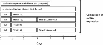
Pronuclear embryos divided into two equal groups; embryos in one group were cultured, in TCM-199 and embryos from the second group were cultured in Ham's F10 medium, both media were supplemented with 0.1% of BSA. After 48 h, a proportion of the embryos was transferred to a new medium, and the others remained in the initial culture system until the fifth day (experimental groups are shown at Fig. 1). Culture was performed in 500 μl of medium layered under paraffin oil at 38.5 °C in 5% CO2 in air and saturated humidity.
Experimental design. In vivo produced embryos (IVP) were recovered at pronuclear stage and cultured in vitro until the hatching blastocyst stage (6 days old embryos) in two different media (Ham's F10 or TCM-199). After 48 h, one group of embryos was transferred to new medium (Ham's F10 renewal or TCM-199 renewal), and the rest remained in the initial medium. Finally, mRNA expression of all in vitro developed groups was compared with two control groups consisting of 4- and 6-day in vivo developed embryos.

Evaluation of in vitro developmental ability
In vitro developed embryos were examined after 5 days of culture and the percentages of hatching blastocysts (cell mass extruding through zona pellucida) were noted.
Evaluation of differential mRNA expression
RNA extraction and reverse transcription
For in vitro cultured embryos, RNA was isolated from five embryo pools of each in vitro condition using a MicroRNA Isolation Kit (Stratagene, Agilent Technologies) according to the manufacturer's instructions, with minor modifications (Hartshorn et al. Reference Hartshorn, Rice, Wangh and Bustin2004). For in vivo cultured embryos, RNA was extracted from eight embryo pools from each stage (4th and 6th) using Trizol reagent (Invitrogen S.A).
RNA was isolated and was reverse transcribed into cDNA using SuperScript III Reverse Transcriptase (Invitrogen S.A) in a total volume of 20 μl. The first reaction mixture consisted of 200 ng of random primers, 9 μl of RNA, 1 μl of 10 mM dNTP mix (10 mM of each dNTP) and 13 μl of water. This reaction was incubated for 5 min at 65 °C and then 4 μl of First-Strand Buffer, 1 μl DTT, 1 μl water and 1 μl SuperScript III RT (reverse transcriptase) (200 U/μl) was added to each tube. The RT reaction was carried out at 25 °C for 5 min and 50 °C for 60 min, followed by an inactivation step at 75 °C for 15 min.
SYBR green assay (qRT-PCR)
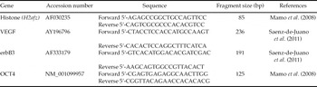
The mRNA expression was analysed by real-time polymerase chain reaction (PCR) in an Applied Biosystems 7500 reader (Applied Biosystems). Each PCR was performed from 5 μl diluted cDNA template, 200 nM each of forward and reverse primers (Table 1) and 10 μl of PowerSYBR Green PCR Master Mix in a final volume of 20 μl. The PCR protocol included an initial step of 50°C (2 min), followed by 95°C (10 min) and 40 cycles of 95°C (15 s) and 60°C (60 s). After real-time PCR, a melting curve analysis was performed by slowly increasing the temperature from 65°C to 95°C, with a continuous registration of changes in fluorescence emission intensity. The products from the qRT-PCR were confirmed by ethidium bromide-stained 2% agarose gel electrophoresis in 1× TBE (Tris–borate–EDTA) buffer. The fragments were visualised on a UV-transilluminator and the image was analysed quantitatively with GENETOOLS software (Syngene, IZASA, Spain). The relative level expression of each target gene was determined using the 2−ΔCt method (Pfaffl Reference Pfaffl2001), employing histone (H2afz) as the housekeeping gene.
Primers used for real-time PCR

erbB3, epidermal growth factor receptor 3; OCT4, transcription factor Oct4; VEGF, vascular endothelial growth factor.
Statistical analysis
Differences in the rate that embryos reached the hatched blastocyst stage in two commercial media (TCM-199 and Ham's F10) and in two different culture systems (renewal or non-renewal single medium) were analysed by chi-squared analysis. Differences in mRNA expression between different groups were analysed after data normalisation test (asymmetric coefficients and curtosis) by one-way ANOVA (analysis of variance), using the General Linear Models (GLM) procedure of Statgraphics Plus 5.1. Differences in p-values <0.05 were considered significant.
Results
Effect of media composition and culture system on embryo developmental ability
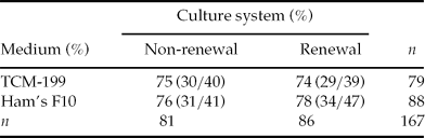
A total of 167 pronuclear embryos were cultured in two commercial media (TCM-199 and Ham's F10) and two culture systems (renewal and non-renewal), and the rates of hatched blastocyst developed are described in Table 2. There was no significant difference in number of blastocyst hatching between groups (Table 2).
Percentage of rabbit embryos that developed to hatching blastocyst stage in two different culture systems (renewal or non-renewal) and two commercial media (Ham's F10 and TCM-199)
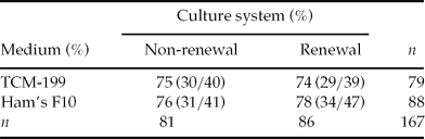
n, number of embryos.
Effect of medium composition and culture system on mRNA expression
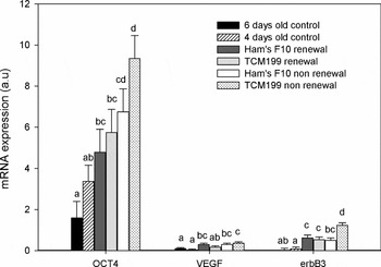
The relative abundance of gene transcripts of OCT 4, VEGF and erbB3 in in vitro cultured rabbit embryos in TCM-199 and Ham's F10 with renewal or non-renewal media at 48 h of culture are shown in Fig. 2. The type of culture system (renewal or non-renewal) and the choice of medium effected on the expression of the selected genes. Overall expression of OCT4 mRNA in in vitro cultured embryos was higher than in vivo 6-day-old blastocysts. However, when the medium was renewed, OCT4 mRNA abundance was similar to that found with 4-day-old embryos that had been developed in in vivo conditions. In the case of VEGF, similar mRNA expression was observed between embryos cultured in renewal TCM-199 medium and in vivo developed blastocysts, whereas for erbB3 only embryos cultured in non-renewal Ham's F10 showed similar mRNA expression to 6-day-old blastocysts produced in in vivo conditions. For all genes, the highest transcript abundance was observed in non-renewal TCM-199 medium.
Relative abundance of OCT4, VEGF (vascular endothelial growth factor) and erbB3 (epidermal growth factor receptor 3) mRNA expression for in vitro rabbit embryos cultured during 5 days in different commercial media formulation (TCM-199 and Ham's F10) and different culture systems (medium and non-renewal) compared with 4- and 6-day-old in vivo rabbit embryos (Controls). Relative abundance values are expressed in arbitrary units (a.u.), showing the mean value ± SEM. Different letters represent significant differences.

Discussion
Early developmental events are regulated by maternal RNAs and proteins synthesised during oogenesis (Gandolfi & Gandolfi, Reference Gandolfi and Gandolfi2001). The embryonic nuclei become transcriptionally active when maternal-embryonic transition occurs, and the length of this period depends on the species, and is at the 8-cell stage in rabbit (Telford et al., Reference Telford, Watson and Schultz1990; Gandolfi & Gandolfi, Reference Gandolfi and Gandolfi2001). A recent microarray study has demonstrated that the rabbit embryo transcriptome is dynamic, and has an over-expression of molecular processes involved in RNA and DNA metabolism at the embryonic genome activation period (Léandri et al., Reference Léandri, Archilla, Bui, Peynot, Liu, Cabau, Chastellier, Renard and Duranthon2009). At the 48 h time point of in vitro culture this essential event takes place in the nuclei of rabbit embryo, therefore the environmental conditions of the medium in which it develops play a crucial role. As stated before, the renewal of medium in a two-step embryo culture protocol may remove any damaged product from the used medium, such as ammonium, pyrrolidone-5-carboxylic acid or the breakdown products of glutamine, and thus re-supply beneficial compounds that have been depleted in the initial culture (Gardner, Reference Gardner1994). However, no significant differences have been reported in the blastocyst proportions, hatching rates and cell numbers in the inner cell mass and trophectoderm, which suggested that the renewal of medium is not necessary to embryo development (Fukui et al., Reference Fukui, Lee and Araki1996; Biggers et al. Reference Biggers, McGinnis and Lawitts2005). In accordance with this study, the rates of rabbit blastocysts that hatch at 5 days of in vitro culture were similar between renewal and non-renewal systems in both media tested (Ham's F10 and TCM-199). The media chosen for this study differ in their amino acid concentration and in the presence of three amino acids: l-asparagine · H2O, l-cysteine · 2H2O and hydroxy-l-proline. Energy is mainly supported by glucose concentration, similar for both culture media (1 and 1.1 g/l for TCM-199 and Ham's F10, respectively) and pyruvic acid (sodium), which is only present in Ham's F10 formulation. Karja et al. (Reference Karja, Kikuchi, Fahrudin, Ozawa, Somfai, Ohnuma, Noguchi, Kaneko and Nagai2006) reported that pyruvate and lactate appear to be important energy substrates for both early embryonic development in vitro and improvement in the number of cells in the blastocysts.
It is known that the cost of adaptation to sub-optimal media formulations could alter the maintenance of cellular physiology, the gene expression or the pregnancy potential, and many of these alterations might not be evident until later in development (Lane & Gardner, Reference Lane and Gardner2007). Therefore, this finding supports the idea that it is necessary to combine traditional measures of development capacity with new tools as mRNA expression analysis to determine and describe in more detail the real consequences of a given culture environment (Purpera et al., Reference Purpera, Giraldo, Ballard, Hylan, Godke and Bondioli2009). Although no significant difference in hatching blastocyst rates was found, our results show changes in mRNA expression levels, specifically in OCT4, VEGF and erbB3 genes. The findings of this work are in agreement with other previous studies that report that the post-fertilisation culture environment mainly interferes with the mRNA expression and embryo quality, but have no major effect on the yield of blastocyst produced (Rizos et al., Reference Rizos, Ward, Duffy, Boland and Lonergan2002b; Lonergan et al., Reference Lonergan, Rizos, Kanka, Nemcova, Mbaye, Kingston, Wade, Duffy and Boland2003; Gutiérrez-Adán et al., Reference Gutiérrez-Adán, Rizos, Fair, Moreira, Pintado, de la Fuente, Boland and Lonergan2004).
The set of genes evaluated have previously been related to regulation of transcription, pluripotency maintenance, angiogenesis, proliferation, differentiation and implantation (Carson et al., Reference Carson, Bagchi, Dey, Enders, Fazleabas, Lessy and Yoshinaga2000; Lee & DeMayo, Reference Lee and DeMayo2004; Artini et al., Reference Artini, Valentino, Montleone, Simi, Parisen-Toldin, Cristello, Cela and Genazzani2008; Medvedev et al., Reference Medvedev, Shevchenko, Mazurok and Zakian2008; Saenz-de-Juano et al., Reference Saenz-de-Juano, Peñaranda, Marco-Jiménez, Llobat and Vicente2010). The findings obtained show an important divergence in mRNA expression between embryos developed under in vivo and in vitro environments. It is known that transcription factor OCT4, regarded as a key regulator of the pluripotency maintenance system, is essential to preimplantation embryo development (Boiani & Schöler Reference Boiani and Schöler2005, Medvedev et al., Reference Medvedev, Shevchenko, Mazurok and Zakian2008), and its mRNA expression was detected in rabbit embryos in previous studies (Mamo et al., Reference Mamo, Gal, Polgar and Dinnyes2008; Kobolak et al., Reference Kobolak, Kiss, Polgar, Mamo, Gaillard, Tancos, Bock, Baji, Tar, Pirity and Dinnyes2009; Saenz-de-Juano et al., Reference Saenz-de-Juano, Peñaranda, Marco-Jiménez, Llobat and Vicente2010). In the present work, the results show that mRNA expression in in vitro developed embryos is higher than their in vivo chronological counterparts (6 days old). These results are agreement with other works performed in mouse (Li et al., Reference Li, Kato and Tsunoda2005) and bovine (Purpera et al., Reference Purpera, Giraldo, Ballard, Hylan, Godke and Bondioli2009), in which an up-regulation of OCT4 expression appeared as a consequence of the in vitro culture conditions. However, similar mRNA abundance was observed between hatching blastocysts cultured with renewal media and in vivo 4-day-old embryos. It is well known that development of rabbit blastocyst is lower in in vitro than in vivo conditions because the culture system cannot mimic the nutritional role of uterine environment (Hegele-Hartung et al., Reference Hegele-Hartung, Fischer and Beier1988; Hohn et al., Reference Hohn, Mootz and Denker1992; Jin et al., Reference Jin, Kim, Im and Choi2000). Previously, Saadelhin et al. (2010) suggested that the presence of glucose and other additives in renewal culture medium causes harmonic gene expression for OCT4.
Transcript abundance of VEGF and erbB3 showed marked variation, depending on the type of development (in vivo or in vitro) and media and culture system employed. The main function of erbB family during early embryo development is to activate specific intracellular signalling pathways to control mRNA expression of differential genes that are involved cell proliferation, differentiation and prevent apoptosis (Collins et al. Reference Collins, Perkins, Rodriguez-Tarduchy, Nieto and Lopez-Rivas1994). VEGF has also been associated with the novo angiogenesis process (Lee and DeMayo, Reference Lee and DeMayo2004), and its expression and function has been regarded as ensuring a suitable vasculogenesis during implantation and early placentation (Torry et al., Reference Torry, Leavenworth, Chang, Maheshwari, Groesch, Ball and Torry2007). Fleming et al. (Reference Fleming, Kwong, Porter, Ursell, Fesenko, Wilkins, Miller, Watkins and Eckert2004) reported that the embryo environment affects the maintenance of genomic imprinting, which leads to alterations in gene expression. Furthermore, a comparison between five commercial media found a differential loss of imprinting dependent upon the media and culture system employed (Market-Velker et al., Reference Market-Velker, Fernandes and Mann2010). Therefore, as gene expression in preimplantation embryos of VEGF and erbB3 is regulated by several epigenetic mechanisms (Kim et al., Reference Kim, Hwang, Zhou, Shin, Noh, Song, Kim, Lee and Kim2010; Hoque et al., Reference Hoque, Brait, Rosenbaum, Poeta, Pal, Begum, Dasgupta, Carvalho, Ahrendt, Westra and Sidransky2010), a variable and altered mRNA expression might be a consequence of altered epigenetic modifications due to in vitro conditions.
In conclusion, it was observed that different culture media and culture systems affect mRNA expression of different genes without changes in embryo development to hatched blastocyst stage. Despite the enormous efforts carried out in recent years on rabbit embryo culture systems, with in vitro conditions still far from mimetic the maternal reproductive tract environment and embryo developmental competence remain compromised by media composition. As the study of specific transcript abundance in rabbit blastocyst provides a more detailed description of which alterations occur at mRNA expression level, further studies should be carried out to determine which components present in media culture modify gene expression patterns, in order to optimise in vitro culture conditions and reduce current limitations of long-term culture rabbit preimplantation embryo.
Acknowledgements
This work was supported by the Spanish Research Projects (CICYT AGL2008–03274) and the Generalitat Valenciana research program (Prometeo 2009/125). The authors thank Neil Macowan Language Services for revising the English version of the manuscript.