INTRODUCTION
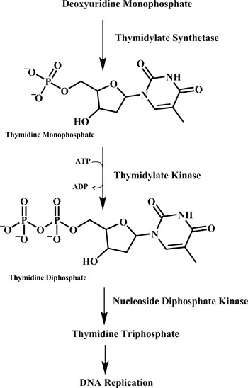
Human malaria is a worldwide problem and more than one-third of the world's population (about 2 billion people) live in malaria-endemic areas, and 1 billion people are estimated to be carriers of the causative parasite, Plasmodium spp., at any one time (Guerin et al. Reference Guerin, Olliaro, Nosten, Druilhe, Laxminarayan, Binka, Kilama, Ford and White2002). About 500 million people become infected annually, resulting in more than 2 million deaths (Breman et al. Reference Breman, Egan and Keusch2001). Moreover, the increasing problem of multidrug-resistant strains necessitates the discovery of new and highly efficacious drugs. The rational step in drug development involves the characterization of a molecular target unique for survival of the parasite. Thymidylate kinase (TMK; ATP: TMP phosphotransferase; dTMP kinase, deoxythymidine monophosphate kinase, EC 2.7.4.9) belongs to the nucleoside monophosphate kinase superfamily and is critical for thymidine triphosphate synthesis (Fig. 1). It catalyses the reversible phosphorylation of dTMP to its diphosphate form, deoxythymidine diphosphate, in the presence of the divalent cation Mg2+ with ATP as the preferred phosphate donor (Li de la Sierra et al. Reference Li de la Sierra, Munier-Lehmann, Gilles, Bârzu and Delarue2001). TMKs have medical and therapeutic importance for the activation of nucleoside analogue pro-drugs used as antiviral and anticancer drugs (Ostermann et al. Reference Ostermann, Segura-Peña, Meier, Veit, Monnerjahn, Konrad and Lavie2003).
Malarial parasites are completely dependent on de novo pyrimidine biosynthesis and cannot salvage pyrimidines. As the parasite cannot manipulate pre-formed pyrimidine nucleotide precursors, it depends on its own enzymes for the complete synthesis of dUMP which is utilized by thymidylate synthetase to give TMP. Thymidylate kinase is the last specific enzyme in the synthesis of thymidine triphosphate (TTP) which is important for DNA replication. TMKs catalyse the phosphorylation of thymidine monophosphate (TMP) using ATP as a preferred phosphate donor to form thymidine diphosphate (TDP).

Recently, we have reported that Plasmodium falciparum thymidylate kinase (gene ID PFL2465c) can tolerate a range of substrates, which distinguishes it from other TMKs (Kandeel and Kitade, Reference Kandeel and Kitade2008). Compared with the human enzyme, recombinant PfTMK shows a broader spectrum of substrate specificity. The enzyme not only phosphorylates deoxythymidine monophosphate (dTMP) and deoxyuridine monophosphate (dUMP) but can also tolerate the bulkier purines. PfTMK can efficiently utilize deoxyguanosine monophosphate (dGMP), whereas it can utilize guanosine monophosphate (GMP) and deoxyinosine-5′-monophosphate (dIMP) with a lower catalytic efficiency. In our previous work, we demonstrated that P. falciparum guanylate kinase efficiently uses GMP (Kandeel et al. Reference Kandeel, Nakanishi, Ando, El-Shazly, Yosef, Ueno and Kitade2008); however, the catalytic efficiency for dGMP was much lower than known guanylate kinases. Thus, PfTMK is not a dual function thymidylate/guanylate kinase enzyme; however, the differences in catalytic properties for dGMP kinase activity provide an interesting contribution to the knowledge of nucleic acid metabolism in P. falciparum. Viral thymidine kinases (TKs) have broader substrate specificity than the corresponding human enzyme; therefore, they become important targets for chemotherapy (De Winter and Herdewijn, Reference De Winter and Herdewijn1996; El Omari et al. Reference El Omari, Liekens, Bird, Balzarini and Stammers2006). The functional determinants within PfTMK that elucidate substrate recognition and specificity remain unknown. In the present study, we aim to clarify the dual purine and pyrimidine nucleoside monophosphate (NMP) kinase activity of PfTMK; such dual activity was an attractive finding in previous enzymology and drug design studies. In human TMK, the cavity in which a base binds is very tight to accommodate purine bases, and thus reduces specificity to pyrimidines. Thus, the parasite enzyme can be targeted by the development of highly selective purine-based inhibitors.
Based on molecular modelling, we hypothesized that few amino acid replacements may contribute to the difference in the catalytic properties of PfTMK. We sought to characterize the functional properties of PfTMK-based mutational, kinetic, inhibitory and isothermal analyses. We used isothermal titration calorimetry (ITC) to investigate the binding affinities which can give insights into either productive or non-productive binding of ligands. In contrast, the other conventional assay methods, such as spectrophotometric assays or high performance liquid chromatography separations, depend mainly on the detection of products. Thus, combining the ITC results with those of other conventional methods will validate the catalytic properties of the mutant enzymes. Our results highlight the differences in substrate binding, which should enable the development of selective inhibitors.
MATERIALS AND METHODS
Materials
Nucleosides, nucleotides, nucleoside derivatives and phosphoenolpyruvate were obtained from Sigma (MO, USA). Escherichia coli JM109, Sephacryl S200HR were obtained from GE Healthcare (NJ, USA). AccuPrime Pfx polymerase was obtained from Invitrogen (CA, USA). Talon metal affinity resin was obtained from Clontech (Palo Alto, CA). The vector pQE-30 was obtained from Qiagen (CA, USA). Lactate dehydrogenase and pyruvate kinase were obtained from Wako Pure Chemicals (Osaka, Japan). Protein concentrations were determined by using a protein assay reagent (Bio-Rad, USA) with bovine serum albumin as the standard.
Site-directed mutagenesis and production of mutant enzymes
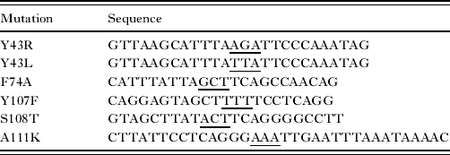
Site-directed mutagenesis was performed to produce mutant enzymes containing a single amino-acid substitution. The target mutations are shown in Table 1. The rationale for constructing Y43R and Y43L enzymes was based on comparative structure-function studies of TMKs from other sources. The equivalent residues Arg36 of Staphylococcus aureus TMK (Kotaka et al. Reference Kotaka, Dhaliwal, Ren, Nichols, Angell, Lockyer, Hawkins and Stammers2006) and Arg39 of Escherichia coli TMK (Lavie et al. Reference Lavie, Ostermann, Brundiers, Goody, Reinstein, Konrad and Schlichting1998) directly interact with O1 of the dTMP phosphate group. The bulky aromatic Phe74, which is important for stacking interaction with pyrimidine ring, is replaced by the small-sized alanine. Consequently, this mutation will help in identifying the proposed position for dGMP. S108T, Y107F and A111K mutants are conservative and non-conservative substitutions of the PfTMK residues based on the human TMK structure. The small alanine residue is mutated to the bulkier lysine to yield the A111k mutant.
Oligonucleotide primers used for point mutations
(Base changes from wild-type PfTMK are underlined.)
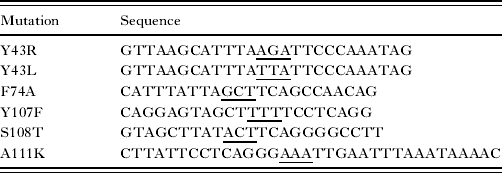
The single-tube ‘megaprimer’ PCR method was used to make the point mutations, in which 2 sequential PCR reactions were carried out (Ke and Madison, Reference Ke and Madison1997). In brief, the first PCR reaction included the mutational primer (Table 1) and the low Tm megaprimer (GATTCTCACCAATAAAAAAC). In the second PCR reaction the high Tm megaprimer was added (GGCGTATCA CGAGGCCCTTTCGTCTTC ACCTCG) and the full-length mutants were obtained after carrying out the annealing and extension steps at 68°C. The product of PCR was digested with BamHI and HindIII and subcloned into pQE-30 vector pre-digested with the same enzymes. The wild type and mutations were confirmed by sequencing all the plasmids. Expression of the wild type and mutant PfTMK was carried out in JM109 E. coli cells. The recombinant E. coli cells were grown in LB medium containing 50 μg/ml ampicillin at 37°C to the mid-log phase (D 600=0·6). Induction of expression was carried out by the addition of IPTG (isopropyl β-D-1-thiogalactopyranoside) to a final concentration of 1 mm and cell growth was continued at 37°C for 4 h. Cells were harvested by centrifugation at 5000 g for 15 min and then stored at −20°C until use. To obtain the cell extract, cells were lysed in an extraction buffer (25 mm Tris-HCl buffer, pH 7·2, containing 150 mm NaCl) and disintegrated by sonication for 40 s (3 cycles with 3 min intervals). The lysate was centrifuged at 16 000 g for 15 min at 4°C, and the precipitate discarded.
Affinity and preparative size exclusion chromatography
The hexahistidine-tagged PfTMK was purified from the soluble cell extract by using talon metal affinity resin. The wild type and mutants were purified by the batch purification method. The eluted fractions were further purified by removal of potential low molecular weight contaminants using a Sephacryl S200HR column.
Enzyme kinetics
The TMP kinase activity of wild-type and mutant PfTMK was measured spectrophotometrically using an enzyme coupling assay (Blondin et al. Reference Blondin, Serina, Wiesmüller, Gilles and Bârzu1994). In the spectrophotometric assay, the standard assay of 1 ml contained the following: 50 mm Tris-HCl, pH 7·2, 0·5 mm phosphoenolpyruvate, 40 mm KCl, 2 mm MgCl2, 0·15 mm NADH, 2 U pyruvate kinase, 2 U lactate dehydrogenase, and various concentrations of NMP or ATP. The reaction was initiated by the addition of the recombinant enzyme. The decrease in absorbance at 335 nm was monitored at 30°C. One unit of enzyme activity is defined as the amount of enzyme catalysing the production of 1 μmol nucleoside diphosphate/min under the above conditions.
Inhibitor kinetics
For inhibitor studies, the enzyme was incubated with TMP in the presence or absence of an inhibitor and the progress curve was monitored by spectrophotometric assay as well as by HPLC. The data were analysed by non-linear regression analysis. HPLC assay was used to characterize PfTMK activity and to avoid the disadvantages of the coupled enzyme assay (Manallack et al. Reference Manallack, Pitt, Herdewijn, Balzarini, De Clercq, Sanderson, Sohi, Wien, Munier-Lehmann, Haouz and Delarue2002; Haouz et al. Reference Haouz, Vanheusden, Munier-Lehmann, Froeyen, Herdewijn, Van Calenbergh and Delarue2003). The assay for inhibitors is complicated by sensitivity of the coupling enzymes. The reaction was carried out in a final volume of 1 ml of a solution of 50 mm Tris-HCl, pH 7·2, 40 mm KCl, 2 mm MgCl2, 0·5 mm dithiothreitol and variable concentrations of ATP or TMP. The buffer used for elution was 50 mm sodium phosphate, pH 6·5, 2·5% (v/v) ethanol, 40 mm terabutylammonium bromide. The concentration of separated nucleotides was monitored at 260 nm using a Mightysil C18, 5 μm column (Kanto Chemicals Co.).
Isothermal titration microcalorimetry
Binding of nucleotides was investigated by isothermal titration calorimetry (MicroCal Inc., USA). The reference cell was filled with water. The PfTMK protein was dialysed extensively overnight in 25 mm sodium phosphate buffer, pH 7, containing 2·5 mm MgCl2. Since Mg2+ was found to participate in the bonding network of the binding pocket in many kinases, Mg2+ was added to the dialysis buffer to insure proper binding of substrates. The protein solution, at a concentration of 20–30 μm, was loaded into a 1·4 ml sample cell and titrated with the ligand (at concentrations of 0·1–1 mm) in the 250 μl injection syringe. The substrate solution was made in the final dialysis buffer to minimize artifacts due to subtle differences in buffer composition. The protein was diluted to the appropriate concentration with dialysis buffer immediately before titration. ITC experiments were performed at 30°C with a stirring speed of 350 rpm and 300 s duration between each 10 μl injection. Control experiments were performed by injection of the nucleotide into the dialysis buffer to determine the heat of dilution. The apparent heat change after each injection was determined by integration and corrected for heat of dilution of the nucleotide. The data were fitted to a single-binding site model or two set sites model by a non-linear regression analysis to yield the thermodynamic parameters K a, association constant; ΔH, enthalpy of binding; and n, stoichiometry of binding. The affinity of the nucleotides to protein is given as the dissociation constant (K d=1/K a). The free energy of binding (ΔG) was calculated from the equations: ΔG=RT ln K a and ΔG=ΔH−TΔS where ΔS is the entropy changes.
Modelling studies
The structure of PfTMK was modelled according to the known structure of nucleoside kinases recognizing both guanine and thymidine bases by using the modelling tools available at http://www.sbg.bio.ic.ac.uk/~phyre/. The structures of Drosophila deoxynucleosides kinase, herpes virus thymidine kinase and the structures of known TMKs were used to predict the model for purine NMPs binding to PfTMK. Docking of NMPs was performed using ChemBioDraw Ultra 11 (CambridgeSoft Corporation, USA). The binding site was defined from coordinates of the ligand in the original structure. Ligand interactions and ligand binding site atoms were created by DS Visualizer (Accelrys Inc. USA).
Other methods
The secondary structures of recombinant protein were measured by circular dichroism (CD) spectrum. Measurements were performed at wavelength 190–280 nm. The secondary structure content of the proteins was estimated using the K2d program (Andrade et al. Reference Andrade, Chacón, Merelo and Morán1993). Protein concentrations were determined by using a protein assay reagent (Bio-Rad) with bovine serum albumin as the standard. The purity of proteins was assessed by SDS-PAGE and confirmed by Western blotting using anti-histag antibody.
RESULTS
Modelling studies
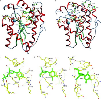
A comparison of the active site residues of PfTMK and its human counterparts revealed that the residues involved in binding and activity were highly similar, with few conservative and non-conservative substitutions. In PfTMK Tyr43, Tyr107 and Ser108 replaced residues Arg41, Phe105 and Thr106, respectively. In addition, the replacement of some residues near the proposed active site, particularly at the junction of the α2–α3, α5–α6 and α8 loops where the bulky residues (e.g. His66, Lys109, Arg150 and Phe156) are located, was observed in human TMK (Fig. 2). In contrast, the previously mentioned residues were replaced by smaller residues, namely, Glu68, Ala111, Ile152 and Thr158 in PfTMK. In the present study, 6 mutants were designed targeting these structural differences as well as conservative and non-conservative substitutions at the binding site (Fig. 2A and B). PfTMK contains the aromatic, non-polar and neutral Y43; in contrast to human, yeast and E. coli TMKs which contain the strong basic and polar arginine at the same position. Y43R was mutated to check the effect of changing the polarity at this position. In addition, the mutation of Y43 to leucine will maintain neutral and non-polar amino acid at this position; however, it will help in checking the functional importance of the phenyl ring of tyrosine. The bulky F74 was mutated to alanine to check the importance of phenyl ring stacking on the thymidylate and deoxyguanylate kinase activity of PfTMK. Y105F and S108T are conservative substitutions of the PfTMK residue based on the human TMK structure. A111K is a non-conservative substitution of the PfTMK residue based on the human TMK structure in which the small, neutral and non-polar alanine is mutated to the bulkier, basic and polar lysine residue. Fig. 3C, D and E show the putative interactions of TMP and the inhibitors thymidine and azidothymidine with PfTMK. The human TMK structure was used as a template to predict the coordinates of interaction.
Model for the structure of PfTMK. (A) Alignment of human and plasmodium TMKs showing target mutations. The selected mutations are Y43R, Y43L, F74A, Y107F, S108T and A111K. (B) Alignment of human and plasmodium TMKs showing the noticeable replacement of small-sized residues (Glu68, Ala111, Ile152 and Thr158) in PfTMK by bulky residues in human TMK (His66, Lys109, Arg150 and Phe156). PfTMK residues are displayed in green while human ones are white coloured. (A) and (B) are prepared with Molsoft ICM Browser Pro (Molsoft LLC, California, USA). (C) Model for ligand binding atoms. The residues shown are proposed to participate in binding TMP. (D) and (E) are models for interactions for thymidine and AZT. (C), (D) and (E) are prepared by ligand binding site prediction tool available with Accelrys Discovery Studio Visualizer (Accelrys Software, USA). Putative hydrogen bonding is shown as dashed lines. The hydrogen bonds mediating water molecules are presented as spheres. The coordinates of predictions are based on the human TMK structures.

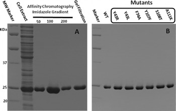
SDS-PAGE of PfTMK purification. Samples were analysed on 12·5% polyacrylamide gels and stained with Coomassie brilliant blue. (A) Affinity chromatography imidazole gradient adopted during purification (50, 100 and 200 mm imidazole) which is followed by a gel filtration polishing step. (B) Purified wild-type and mutant enzymes. PfTMKs are detectable at 24 kDa. The fraction eluted after 200 mm imidazole elution was further purified by gel filtration. About 5 mg of each protein was obtained after the purification steps.

Site-directed mutagenesis
All mutant enzymes were expressed successfully in the soluble fractions of the cells at the same level as the wild-type enzymes. Wild-type and mutant enzymes were highly purified by a single step using affinity chromatography. A stepwise imidazole gradient was adopted so that all impurities were eluted in early fractions while the latter fraction contained a highly purified protein, which was further purified by gel filtration chromatography (Fig. 3).
The results obtained from CD spectra were analysed by the K2D program, which predicts the percentage of secondary structure content. No significant difference could be detected between the far-UV CD spectra of wild-type and mutant enzymes, indicating that the mutant proteins retain the main secondary structure composition of the wild-type enzyme. We therefore conclude that the main conformation of PfTMK was not changed by site-directed mutagenesis and the difference in biochemical parameters was not due to disruption of the secondary structure content.
Y43R and Y43L
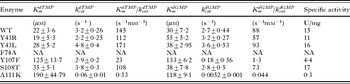
The two Tyr43 mutants showed more or less a similar activity profile with the wild-type enzyme (Table 2).
Summary of kinetic constants of wild-type and mutant PfTMK enzymes
(The kinase reaction was carried out using TMP or dGMP as substrates. Results are an average of 3 or more independent experiments and errors are expressed as mean of standard deviation. NA stands for non-applicable, the result obtained was very low and does not follow saturation kinetics. The specific activity was measured with TMP as substrate.)

F74A
Irrespective of whether dTMP and dGMP, together with their pyrimidine and larger purine rings, use the same active site, we introduced an F74A mutation. According to our model, the replacement of Phe74 with alanine will cause loss of the phenyl ring stacking with the pyrimidine ring of dTMP. We assumed that if the larger purine ring of dGMP has an effective distance with Phe74, and using the same stacking reaction, the active site of both substrates will be highly related and this point mutation could equally affect the enzyme activity with either substrate. The kinetic parameters were not measurable with either substrate. Nevertheless, we observed a very weak activity, which did not permit measurement of kinetic parameters. However, this mutation equally affected the dTMP and dGMP kinase activities.
Y107F-
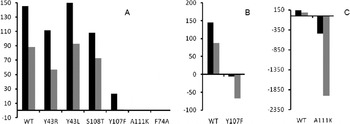
This mutant showed an interesting activity profile. The apparent K m for dTMP and dGMP was increased by 4 to 5-fold. The most striking feature of this mutant was the altered catalytic efficiency for either substrate. In the case of dTMP, the catalytic constant was slightly changed while the catalytic efficiency k catdTMP/K mdTMP was decreased 6-fold due to the increased K mdTMP. If dGMP was used as the substrate, the catalytic efficiency k catdGMP/K mdGMP was decreased by about 65-fold (Fig. 4).
Effect of mutations on thymidylate kinase and deoxyguanylate kinase activity. (A) The catalytic efficiency (k cat/K m) of wild type and mutants for TMP (black) and dGMP (grey). (B) The effect of Y107F mutation on PfTMK activity. WT represents the catalytic efficiency of wild-type PfTMK. The TMP and dGMP kinase activity was decreased by 6- and 67-fold, respectively. (C) The effect of A111K mutation on PfTMK activity. WT represents the catalytic efficiency of wild-type PfTMK. The TMP and dGMP kinase activity was decreased by 439- and 2000-fold, respectively.

S108T
This mutant is a conservative substitution of the PfTMK residue based on the human TMK structure. As expected, the S108T mutant maintained a biological activity similar to that of the wild-type enzyme.
A111K
Our goal in designing this mutant was to compare its impact on the activity of PfTMK with purine and pyrimidine NMPs. PfTMK models based on human and yeast TMK structures have revealed the close proximity of this residue to the enzyme active site. The fact that residues of human TMK are more bulky at this position led us to change PfTMK A111 to the larger lysine residue. In addition, we assumed that in order to pack the bulky purine ring in the limited space of the active site, the substrate adopts conformational changes and also clashes with bulky residues. The decrease in enzyme activity may be due to steric hindrance with the bulky purine ring. This mutant showed a profile similar to that of the Y107F mutant but demonstrated more severe impact on dGMP activity. The catalytic efficiency of A111k was 2000-fold lower for dGMP, which was about 5 times higher than that for dTMP (Fig. 4).
Enzyme inhibition
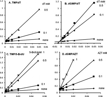
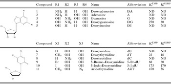
We found that PfTMK can be inhibited by nucleosides and nucleoside analogues; thus, the phosphate group is not essential for the binding of inhibitors to nucleoside monophosphate kinase (Table 3). To elucidate whether PfTMK can be inhibited by purine nucleosides, we carried out the kinase reaction in the presence of various purine nucleosides. Deoxyguanosine (dG) was found to inhibit PfTMK. Interestingly, dG was more potent than the dGMP kinase inhibitor, and it was shown to also be capable of inhibiting TMP kinase activity. Deoxythymidine showed the highest inhibition with a K i of 22 μm (Fig. 5). Moreover, substitution of the 5-methyl group of thymidine was explored. The 5-haloginated deoxyuridines showed less potent inhibitory effects. The presence of iodine or bromine at this position decreased the affinity of the compounds, while substitution with hydrogen, which is smaller than the methyl group, did not induce any inhibitory effects. From Table 3, we can deduce that dGMP kinase activity can be more efficiently inhibited than TMP kinase activity, particularly with dG. Moreover, azidothymidine (AZT) readily inhibited dGMP kinase reaction, indicating the importance of the ribose 3′OH position for proper dGMP kinase activity. No inhibition was observed with all ribonucleosides, indicating that 2′deoxysugars are essential for binding and that the presence of 2′OH of the sugar portion significantly affected binding affinity. From the inhibitory profile of PfTMK, we can deduce that specific locations are highly critical for producing inhibition. Investigation into primary purine and pyrimidine ring substituents has revealed that specific groups are critical for proper binding and inhibition. At position 4 of the pyrimidine ring, the presence of oxygen (dT and dU) was essential for binding while that of dC, which does not contain O4, produced no noticeable inhibition. Furthermore, the 2-amino-purine derivative (dG) was essential for binding, and thus dA and dI showed no inhibitory activity.
Inhibition of PfTMK kinase activity. Initial velocities were determined at various TMP or dGMP concentrations in the presence of a saturating amount of ATP. Various inhibitors were added at concentrations shown on each graph. Inhibition data indicated that thymidine is a competitive inhibitor with regard to TMP (A) with K i value of inhibition calculated to be 0·022 mm, and a mixed-type inhibitor for dGMP (B) with K i value of 0·018 mm. The 5-halogenated deoxyuridines exhibited less potent competitive inhibitory effect (C), while AZT was a competitive inhibitor for dGMP kinase activity of PfTMK (D).

Evaluation of the inhibitory properties of various nucleosides and nucleoside analogues
(K i values are expressed in μm. The K i values are K iTMP and K idGMP when the substrate is TMP and dGMP, respectively. ND stands for ‘not detected’.)

Isothermal titration microcalorimetry
In a typical ITC experiment, aliquots of a ligand solution are added in a time-controlled manner to a cell containing the macromolecule (e.g. enzyme). During addition of ligand, it reacts with the macromolecule resulting in the absorption or release of heat, which can be measured by the microcalorimetry instrument. The produced heat is directly proportional to the fraction of bound ligand as well as to the degree of interaction between the interacting populations. For the initial injections, all or most of the added ligand is bound to the enzyme resulting in a large heat pulse. After a group of subsequent injections of ligand the macromolecule becomes saturated and the heat signal gradually diminishes until only the background heat of dilution is observed. The heat associated with each injection is calculated by integrating the area under the deflection of the measured signal (amount of heat produced per unit of time, μcal/sec). The enthalpy change (ΔH) is the total heat produced or absorbed during titrations. Measurement of heat changes allows for accurate determination of the binding constant and complete thermodynamic profile of the ligand-macromolecule interaction. The observed binding isotherm is normalized as kilocalories per mole of injectant and plotted against the molar ratio of ligand to macromolecule. By calculating the free energy of binding (ΔG); as described in the Materials and Methods section, we can estimate the enthalpic and entropic contributions of the interaction. Interpretation of such terms will give an insight into the nature of the interaction between the test molecules. During the past decades ITC has been considered as a powerful tool or a specialist method for understanding molecular interactions; nowadays, ITC is becoming a method of choice for a wide range of applications (Pierce et al. Reference Pierce, Raman and Nall1999; Weber and Salemme, Reference Weber and Salemme2003; Velázquez Campoy and Freire, Reference Velázquez Campoy and Freire2005; Liang, Reference Liang2008).
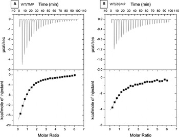
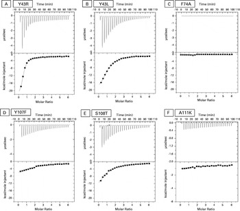
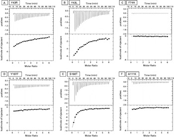
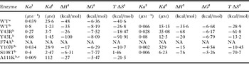
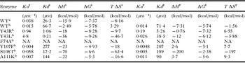
Thermodynamic characterization of the interaction between PfTMK and its substrates was investigated, and the typical thermograms obtained during titrations are shown in Figs 6, 7 and 8. Unfortunately, with ITC software it is impossible to predict the binding properties for the dimeric protein. To overcome this deficit, the PfTMK concentration (40–60 μm as a monomer) was divided by 2 to give the expected binding isotherms for dimers. By assuming the monomeric state, the best fitting of data was detected with the one set of sites model (Table 4). By checking the binding isotherms in the PfTMK dimer; with the exception of A111K, all saturation curves of the mutant enzymes fitted well with the one and two set of sites models. Interestingly, the isotherms detected with the one set sites in either monomer or dimer were highly comparable, indicating that the one set of sites is expected to be the most reliable fitting method. By assuming a single set of binding sites, we found a 1:2 stoichiometry of nucleotide and active sites, suggesting that only one nucleotide binds to one active site in a dimer or that a second nucleotide binding site with substantially different properties may exist. We obtained good fitting, as shown in Fig. 6, when we fitted the obtained data using a different fitting model assuming 1 or 2 independent binding sites. The model predicted 2 binding sites with varying degrees of binding affinities; however, most of the thermodynamic parameters were highly comparable with the one set of sites model. Irrespective of whether the enzyme can adopt equilibrium between the two conditions, if it can change from one state to another under certain circumstances, or if it can show such subtle differences under physiological conditions, further investigations are still required to fully clarify its properties. Table 5 shows that the binding constant for the first binding site K a1 is higher than that for the second site K a2, indicating that the first class of binding sites determines the binding affinity of TMP. In contrast, the two dGMP binding sites equally contribute to the binding equilibrium (Table 6).
ITC profiles of the binding of TMP and dGMP to WT PfTMK. The top panels show the raw data heat changes produced by successive injections of TMP into PfTMK. The lower panel shows the integrated binding isotherms as a function of the molar ratio of ligand to enzyme.

ITC profiles of the binding of TMP to PfTMK mutants. The top panels show the raw data heat changes produced by successive injections of TMP into PfTMK. The lower panel shows the integrated binding isotherms as a function of the molar ratio of ligand to enzyme.

ITC profiles of the binding of dGMP to PfTMK mutants. The top panels show the raw data heat changes produced by successive injections of TMP into PfTMK. The lower panel shows the integrated binding isotherms as a function of the molar ratio of ligand to enzyme.

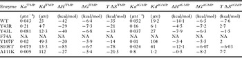
Summary of the thermodynamic parameters determined for the binding of TMP with PfTMK
(The measured ΔH is corrected for small heat changes from control experiments. The best fit of data is by single sites model assuming monomers (60 μm PfTMK).)

Summary of the thermodynamic parameters determined for the binding of TMP with PfTMK
(The measured ΔH is corrected for small heat changes from control experiments. The wild-type data are fitted with one set sites or two set sites model assuming a PfTMK dimer.)

1 The first binding site.
2 The second binding site.
a Fitting by single set of sites model.
b Fitting by two set of sites model.
e The best fitting is by single sites model. Fitting with two sites model is accompanied by high standard error.
Summary of the thermodynamic parameters determined for the binding of dGMP with PfTMK
(The measured ΔH is corrected for small heat changes from control experiments. The wild-type data represent fitting with one set sites model or two set sites model assuming a PfTMK dimer.)

1 The first binding site.
2 The second binding site.
a Fitting by single set of sites model.
b Fitting by two set of sites model.
Results show that PfTMK binds to both TMP and dGMP with relatively similar and high affinities, that is, K ddTMP and K ddGMP of 25 and 26 μm, respectively. Although the net free energy change was similar between the two binding reactions, the larger purine base caused a marked decrease in the enthalpic and entropic components of free energy.
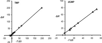
The binding of TMP was highly exothermic with a negative enthalphy change (ΔH) equal to −48 Kcal/mol. In contrast, that of dGMP showed less exothermic interaction with a ΔH of −15·9 Kcal/mol. The association of TMP with PfTMKs was enthalpy-driven, showing that H-bonds and van der Waals interactions play a major role in the binding. The enthalpic force was opposed by strong non-favourable negative entropy. The interaction of dGMP with wild-type PfTMK was not very different from those obtained for TMP but with lesser magnitude. However, that of dGMP with the mutant enzymes showed a different profile in which the binding was enthalpically driven, opposed by a weak entropic term or even accompanied, in some cases, by favourable positive entropic contributions. These changes in entropy indicate global conformational changes or changes in the ordering of water molecules or other small molecules to which the binding of dGMP, but not TMP, is adversely affected. With either substrate, the reaction with PfTMK was highly unfavourable entropically. The negative ΔS suggests the conformational restriction of functional groups in the enzyme as well as the rotational and vibrational degrees of freedom in the binding groups. The enthalpy-entropy plot of both TMP and dGMP binding indicates common enthalpy-entropy compensation, a common process involving binding to biological molecules (Fig. 9).
Enthalpy-entropy compensation for the interaction of PfTMK with TMP and dGMP. The values of ΔH and TΔS are obtained from the integrated thermodynamic data from wild type and mutants of PfTMK.

The thermodynamic parameters of the interaction between Y43R and Y43L with TMP were indistinguishable from that of the wild type as governed by the binding affinities. However, a remarkably higher binding affinity was observed with dGMP. The thermodynamic parameters of F74A (Tables 3, 4 and 5; Figs 7 and 8) confirm the findings of mutant activity, supporting the premise that both TMP and dGMP use the same plane of stacking interactions.
DISCUSSION
Based on the model of the PfTMK structure, we predict that few amino acid replacements may be the cause of the underlying ability of PfTMK to use purine NMPs. Interestingly, slight structural changes may also be the cause besides diverse substrate specificities; for example, in herpes virus thymidine kinase the replacement of two hydrogen bonds to thymidine by single-water-mediated hydrogen bonding improved the affinity to guanine nucleoside analogues (Vogt et al. Reference Vogt, Perozzo, Pautsch, Prota, Schelling, Pilger, Folkers, Scapozza and Schulz2000). The presence of smaller amino acid residues near the proposed active site in PfTMK (which is replaced by a bulkier residue in human TK) may contribute to a larger and more flexible active site in order to accommodate the purine ring. To validate these predictions, we analysed the kinetic data of several point mutant enzymes.
Mutant kinetics data revealed that the catalytic efficiency of the mutants was altered by the effect of mutations. The most interesting features were detected with Y107F and A111K. Primarily, through the analyses of kinetic data, we predicted that the interaction of Y107 with TMP is different from that of dGMP. The catalytic efficiency with TMP was reduced by 6-fold. Interestingly, the catalytic efficiency with dGMP was reduced by more than 65-fold. The markedly lower activity of this mutant with dGMP strongly suggests the importance of this position for the dGMP kinase activity. From the calorimetric data; with all fitting models, the Y107F mutant showed lower binding affinity. Thus, this suggests that position is important for both binding and activity of dGMP. The effect of F105Y mutation, corresponding to Y107F mutation in PfTMK, was previously investigated in human TMK (Brundiers et al. Reference Brundiers, Lavie, Veit, Reinstein, Schlichting, Ostermann, Goody and Konrad1999). The mutant enzyme turnover number was decreased by 5-fold. The single substitution by interfering with the water structure stabilizing the open conformation of the P-loop promotes P-loop closure (i.e. the open state is destabilized by F105Y mutation). Thus, this mutant has been shown to support the preference for closed (active) conformation (Ostermann et al. Reference Ostermann, Segura-Peña, Meier, Veit, Monnerjahn, Konrad and Lavie2003). Similarly, we predict that the Y107F mutant can affect the interaction with residues of the P-loop as well as the water molecules at this position.
The A111K mutant showed a drastic decrease in catalytic efficiency particularly with dGMP. The lengthening of the side chain at this position clearly abolished catalysis. Interestingly, PfTMK A111 is not predicted to directly interact with the substrate. Thus, we presume that this mutation affects the conformation of the active site. Furthermore, this mutation is likely to induce local folding and conformational changes, which will distort the conformation of the active site. The results confirmed that smaller residues such as A111 were necessary for adapting larger purine nucleotides.
The mutant F74A yielded a protein with practically no activity. By ITC, it was not possible to estimate the binding affinity or thermodynamic parameters with certainty; however, a very weak exothermic peak could be detected. We assume that the substrate can bind to the enzyme with a very low affinity from which the binding isotherm cannot be effectively measured. The very low binding affinity is reflected with the very low activity detected in the spectrophotometric assay. In human TMK, the equivalent residue is believed to stabilize the planar and aromatic base moiety. The catalytic activity and thermodynamic parameters indicated the impact of this mutation on TMP and dGMP. Thus, we assume that this residue stabilizes the planar base moieties in either TMP or dGMP binding. In herpes virus thymidine kinase, the binding of the guanine nucleoside analogues, acyclovir and guancyclovir, shows a similar arrangement to that found in the complex formed with dT, and their thymidine and guanine rings approximately occupy the same geometric plane (Brown et al. Reference Brown, Visse, Sandhu, Davies, Rizkallah, Melitz, Summers and Sanderson1995; Champness et al. 1995).
The inhibitory studies demonstrated that PfTMK can be inhibited by purine nucleosides. Interestingly, dG showed no inhibitory activity against human TMK at concentrations up to 2 mm (Pochet et al. Reference Pochet, Dugué, Labesse, Delepierre and Munier-Lehmann2003). Nucleoside monophosphate-based inhibitors suffer from several drawbacks, such as degradation by phosphatases, and low membrane permeability, due to the presence of phosphate. It is therefore very helpful to find a non-phosphorylated nucleoside analogue capable of inhibiting NMP kinase (Zhang, Reference Zhang2005). Fortunately, thymidine kinase activity has not been detected in P. falciparum (Reyes et al. Reference Reyes, Rathod, Sanchez, Mrema, Rieckmann and Heidrich1982). Thus, nucleosides and their analogues could not possibly be metabolized inside the parasitic cytosol. Our finding that nucleosides such as dT and dG were able to inhibit PfTMK in the low micromolar range raises the possibility of modifying such nucleosides in order to develop more specific analogues, particularly dG analogues, which will be highly selective for the parasitic enzyme. The removal of the 5-methyl group of thymidine caused complete loss of inhibitory properties, indicating that the cavity hosting this position can be filled by suitable base modifications. None of the examined ribonucleotides showed any inhibitory activity. In our structure model, the presence of Tyr107 rendered the enzyme selective for 2′-deoxynucloesides over ribonucleotides, and 2′-hydroxyl was highly vulnerable to clash with Tyr107.
The crystal structures of enzyme-substrate complexes provide direct visualization of the molecular determinants of interactions involved in substrate binding. However, the exact contribution of such interactions cannot be easily understood. Thermodynamic measurements provide information on the energetic basis of macromolecular interactions and facilitate understanding of the forces underlying molecular recognition. The design of new drugs can be improved by determining the thermodynamic parameters of binding. It has been shown that enthalpic and entropic terms are highly valuable for determining the quality of various ligands (Ladbury, Reference Ladbury2001). The binding of TMP to PfTMK is enthalpically driven and reflects the strength of the TMP-protein interaction relative to the solvent, which is primarily due to hydrogen bond formation and van der Waals interaction. The negative ΔHs for the binding of TMP indicate that the reaction is exothermic and that the binding is enthalpically favoured. The favourable enthalpy indicates correct placement of the hydrogen bond acceptor and donor groups at the binding interface. The large negative TΔS indicates entropically unfavourable binding and suggests that the hydrophobic interactions are not the driving force of binding.
The binding affinity of dGMP did not differ significantly from that of TMP, the natural substrate of TMP kinases. In addition, the obtained values for the K d of TMP and dGMP coincided with the previously determined K mTMP and K mdGMP, that is, 22 and 30 μm, respectively (Kandeel and Kitade, Reference Kandeel and Kitade2008). The nearly identical K d values from ITC assays and K m values from steady-state kinetics suggest that substrate binding rather than catalysis is likely the rate-limiting step of the enzyme. The free energy of binding of dGMP was lower than that of TMP, indicating that additional interactions occur with TMP, which might be lost with larger dGMP.
The highly favourable binding enthalpy (negative) indicates stable binding complex formation, which is compensated by entropy loss (negative entropies), that is, complex tightening. In the cases of Y107F and A111K, the mutants induced a noticeable loss in binding enthalpies and positive favourable entropic changes in dGMP binding, indicating a more unstable enzyme-ligand complex formation and lesser complex tightening. This finding, together with those of mutant catalytic parameters and inhibition by AZT, indicates the functional roles of Tyr107 in dGMP binding. Furthermore, these data highlight the effect of introducing a bulk residue instead of A111. The presence of lysine at this position affected the binding thermodynamics as well as the catalytic properties of dGMP. This may be due to binding hindrance or the unfavourable steric conditions for dGMP binding. The importance of this observation lies from the presence of such bulkier residues in the human enzyme; however, the parasite enzyme lacks such residues. Thus, the important findings and implications of this investigation include the importance of targeting these differences in developing new selective inhibitors. We assume that purine or pyrimidine nucleoside derivatives containing bulky groups can be tolerated by parasitic enzymes, although they will find unfavourable binding and clashes with bulky residues in the human enzyme. Although purine-based compounds will have a disadvantage of possible binding to human guanine-utilizing enzymes, the structure/function relationship and the lead compound optimization accompanied by thermodynamic refinement of the target drug can lead to the development of a highly selective inhibitor.
The data fitted with two set sites model agrees with that described with the single sites model. The binding of TMP to the wild type as well as mutants was enthalpically driven and opposed by unfavourable entropy. Interestingly, the isotherm of dGMP binding was shifted to give a favourable entropic term with the second binding site with Y43R, Y107f and A111K. The favourable entropy may come from release of water molecules upon binding or minimal loss of conformational degrees of freedom as a result of these mutations.
In conclusion, this work has described the implications of the development of a new antimicrobial target based on mutational, kinetic, inhibitory and thermodynamic analyses. It has also provided interesting results about the characteristics of tolerating a bulky purine and the smaller pyrimidine NMPs in the enzyme active site. We conclude that the packing of purine NMPs in the active site shares, together with the natural substrate TMP, common and different interactions with the active site residues. In this context, the purine and pyrimidine rings of nucleotides share a common binding geometric plane with nearly similar stacking interactions, whereas the deoxyribose sugar produces different configuration patterns, allowing more stabilizing interactions to occur. The enthalpic term was the main driving force for either substrate with highly comparable binding affinities. Finally, it is possible that PfTMK can be selectively inhibited by future modifications of purine-based compounds.
This research was in part supported by Grants-in-Aid for Scientific Research on Priority Area No. 16017239 (to Y.K.). We appreciate the financial support from the Egyptian government (to M.K.).