Introduction
Benign paroxysmal positional vertigo (BPPV) is a type of vertigo characterised by episodic attacks that last for seconds and are triggered by specific head positions.Reference Giommetti, Lapenna, Panichi, Mobaraki, Longari and Ricci1 Benign paroxysmal positional vertigo can occur at any age but is more common in older adults. It is thought to occur as a result of otoconia in the macula of the utricle falling into the semicircular canals. Otoconia falling into the semicircular canals can move freely in the canals (canalolithiasis) or can adhere to the crista ampulla of the semicircular canals (cupulolithiasis).Reference Hornibrook2 Because of their anatomical location, displaced otoconia often fall into the posterior canals (PC). For this reason, posterior canal-BPPV is observed in 85 per cent of cases.Reference von Brevern, Radtke, Lezius, Feldmann, Ziese and Lempert3
Canalolithiasis and cupulolithiasis create transient, abnormal and asymmetric excitations or inhibitions in the stimulated canal ampulla. Benign paroxysmal positional vertigo is diagnosed by clinical history and characteristic nystagmus observed during the provocation manoeuvre. Recurrent attacks cause significant disability, limited mobility, restriction of social life activities, anxiety and depression, and a decrease in quality of life, especially in older individuals.Reference Çetin, Çağaç, Düzenli, Bozan and Elasan4
Most BPPV patients recover with a single canalith repositioning manoeuvre. Others require multiple canalith repositioning manoeuvres for recovery. Rarely, the canalith repositioning manoeuvre is unsuccessful, and this type of BPPV is called refractory.Reference Dorigueto, Mazzetti, Gabilan and Ganança5 On the other hand, even if the canalith repositioning manoeuvre is successful, some patients may experience a non-spinning dizziness that differs from typical BPPV symptoms. This condition, called residual dizziness, includes imbalance, lightheadedness and a sense of floating.Reference Çetin, Çağaç, Düzenli, Bozan and Elasan4
Although residual dizziness is quite common, it is often overlooked after BPPV symptoms have improved. In some patients, it can become chronic.Reference Gambacorta, D’Orazio, Pugliese, Di Giovanni, Ricci and Faralli6 Studies have shown a relationship between residual dizziness, whose prevalence in the literature is reported to range from 23 to 70 per cent, and factors such as age, gender, anxiety level, duration of BPPV and the number of canalith repositioning manoeuvres applied.Reference Çetin, Çağaç, Düzenli, Bozan and Elasan4
Many daily activities involve both cognitive and motor tasks, therefore single-task balance assessments, such as the timed up and go test, functional reach and Romberg test, may be insufficient for evaluating balance and fall risk, particularly in individuals with mild symptoms.Reference Sertel, Sakızlı, Bezgin, Savcun Demirci, Şahan T and Kurtoğlu7, Reference Wollesen, Scrivener, Soles, Billy, Leung and Martin8 In addition to balance and fall risk, the dual-task paradigm, which simultaneously incorporates cognitive and motor tasks, has been increasingly used in recent years to assess attention, cognitive functions and the interaction between motor and cognitive processes.Reference Sertel, Sakızlı, Bezgin, Savcun Demirci, Şahan T and Kurtoğlu7–Reference Tsang, Miller and Pang10
Most studies in the literature have focused on the cause of residual dizzinessReference Teggi, Giordano, Bondi, Fabiano and Bussi11–Reference Soylemez, Bolat, Karakoc, Can, Basak and Aydogan14 and very few on the effects of residual dizziness.Reference Sun, Ma, Yang, He, Wang and Shen15 To our knowledge, no study has investigated cognitive functions and dual-task performance in individuals with residual dizziness. This study aimed to investigate the effects of residual dizziness after successful canalith repositioning manoeuvre on cognitive functions and dual-task performance in middle-aged and older BPPV patients.
Materials and methods
Participants
The power analysis performed with the G*Power 3.1 program determined that the sample size should be a minimum of 38 people with an alpha error of 0.05, a power of 85 per cent and an effect size of d equal to 0.9.Reference Yamout16
Both verbal and written consent was obtained from all participants in accordance with the Declaration of Helsinki. The study was approved by the Hacettepe University Health Sciences Ethics Committee (session date:02.04.2024, research no: SBA 24/327, decision no:2024/07-50).
This prospective cross-sectional study was conducted on patients who underwent a neuro-otological examination by an otolaryngologist and were referred to the hearing and balance disorders clinic. The medical histories of the patients were reviewed. All patients underwent videonystagmography, including gaze testing, oculomotor tests and positional tests (Dix-Hallpike, supine roll and deep head hanging manoeuvres).Reference Bhattacharyya, Gubbels, Schwartz, Edlow, El-Kashlan and Fife17 Patients diagnosed with posterior canal-BPPV based on positional tests were included in the study. These patients completed the Dizziness Handicap Inventory and the Beck Anxiety Inventory before undergoing the canalith repositioning manoeuvre. The type, side and symptom duration of BPPV were recorded.
Based on the characteristic nystagmus observed during positional tests, canalolithiasis and cupulolithiasis were distinguished according to the criteria defined by Imai and Inohara.Reference Imai and Inohara18 Patients diagnosed with posterior canal-BPPV underwent the canalith repositioning manoeuvre to reposition the otoliths. As recommended by Bhattacharyya et al.,Reference Bhattacharyya, Gubbels, Schwartz, Edlow, El-Kashlan and Fife17 the Epley manoeuvre was performed for posterior canal-BPPV canalolithiasis, while the Semont manoeuvre was applied for posterior canal-BPPV cupulolithiasis. All patients underwent a provocative manoeuvre again 30 minutes later for control purposes.
Individuals who did not report vertigo symptoms and showed no characteristic nystagmus during the control manoeuvre (a successful canalith repositioning manoeuvre) were included in the study. Patients for whom the canalith repositioning manoeuvre was unsuccessful were asked to return to the clinic the next day. On their return, both provocative and therapeutic manoeuvres were repeated. After 30 minutes, the success of the canalith repositioning manoeuvre was reassessed using provocative manoeuvres.
Patients for whom the canalith repositioning manoeuvre was successful were included in the study, while those who remained unsuccessful after a total of three canalith repositioning manoeuvres were excluded. Patients with secondary BPPV due to conditions such as Ménière’s disease and vestibular neuritis, as well as those with lateral and anterior canal BPPV, abnormal gaze and oculomotor test results, or neurological and orthopaedic disorders, were excluded from the study. As a result, 44 individuals who recovered from idiopathic posterior canal-BPPV following a successful canalith repositioning manoeuvre were included in the study.
To assess residual dizziness, participants were invited for a follow-up visit 1 week (7 days) after the successful canalith repositioning manoeuvre. Residual dizziness was defined as a sensation of non-spinning vertigo, imbalance or lightheadedness. Participants were asked whether they experienced residual dizziness (yes/no). Based on the presence of residual dizziness, participants were divided into two groups. Twenty-two individuals (50 per cent) who reported residual dizziness were included in the residual dizziness group, while 22 individuals (50 per cent) who did not report residual dizziness were included in the non-residual dizziness group. The cognitive functions of the participants were assessed using the digit span and Stroop test-Scientific and Technological Research Council of Turkey-Basic Sciences Research Group (TBAG) form. Participants’ dual-task performance was evaluated by administering cognitive (digit span) and motor tasks in addition to the timed up and go test.
Dizziness Handicap Inventory
The Turkish version of the Dizziness Handicap Inventory was administered to the participants.Reference Canbal, Cebeci, Çamur Duyan, Kurtaran and İ19 The inventory consists of 3 subscales (physical, emotional and functional) and 25 questions. The Cronbach alpha values of the subscales are between 0.67 and 0.82. The total score is calculated out of 100. The magnitude of the score represents the magnitude of the disability related to dizziness.
Beck Anxiety Inventory
The Turkish version of the Beck Anxiety Inventory was applied to the participants.Reference Ulusoy, Hisli sahin and Erkmen20 The inventory consists of 21 questions, with a Cronbach’s alpha value of 0.93. The total score is calculated out of 63, with higher scores indicating higher levels of anxiety.
Stroop test-Scientific and Technological Research Council of Turkey form
The Turkish adaptation of the Stroop test is called the Stroop test- (TBAG) form.Reference Karakaş, Erdoğan, Soysal, Ulusoy, Yüceyurt Ulusoy and Alkan21 This test is a neuropsychological tool that assesses attention, cognitive flexibility, interference control, information processing and parallel processing. The test consists of five sections and four cards, with each section increasing in difficulty. In the first section (Stroop 1), participants are asked to read colour names written in black ink. In the second section (Stroop 2), they are asked to read colour names written in different colours. In the third section (Stroop 3), participants are asked to state the colour of circles filled with different colours. In the fourth section (Stroop 4), participants are required to name the colour of written words (not colour names). In the fifth section (Stroop 5), the second card is used again, and this time, participants are asked to state the colour of the words. The participant’s completion time is measured with a stopwatch.
Digit span test
The visual-auditory digit span test, a subtest of the Wechsler Memory Scale-Revised, is part of a comprehensive neuropsychological test battery.Reference Karakas, Kafadar and Eski22 Participants were administered the computerised version of the visual forward and backward digit span tests. Number sequences were presented at a rate of one digit per second (using PowerPoint) and participants were asked to remember these sequences. The test started with three-digit sequences. If participants could correctly recall the sequence, the length of the sequence was increased. If they were unable to repeat the sequence in the correct order, the same-length sequence was presented again. If a participant failed to recall two consecutive sequences correctly, the test was terminated and the maximum number of digits the participant could retain was recorded.
For the forward digit span, participants were asked to recall the number sequence in the same order, while for the backward digit span, they were asked to recall the sequence in reverse order. The maximum number of digits the participant could recall for both the forward and backward digit spans was recorded. The forward digit span primarily assesses short-term memory, while the backward digit span evaluates working memory.
Evaluation of dual-task performance
The participants’ dual-task performance was tested using motor and cognitive tasks simultaneously administered with the timed up and go test. For the single-task timed up and go test, participants were instructed to walk the 3-m course as quickly as possible, turn around and return to the starting point to sit down. The time was recorded using a stopwatch. For the timed up and go motor task, participants were asked to carry a tray with a water-filled glass while performing the timed up and go test. For the cognitive task, the digit span test was administered simultaneously with the timed up and go test. The reason for using the digit span test as the cognitive task was its ease of adaptation to the dual-task paradigm, with both an easier (forward) and a more challenging (backward) version available. The maximum number of digits participants could recall was recorded along with the timed up and go test. Participants were asked to recall the number sequences they had memorised after completing the timed up and go test. The participants’ timed up and go-forward and timed up and go-backward times were recorded.
Statistical analysis
The statistical analysis of the study was performed using SPSS 21 software (IBM Corp., Armonk, NY, USA). The normality of the data distribution was tested using the Shapiro–Wilk test. Normally distributed data are presented as mean ± standard deviation (SD), while non-normally distributed data are presented as the median (minimum–maximum). To compare the groups, the normality of the data was considered. Normally distributed data were compared using the student’s t-test, while non-normally distributed data were compared using the Mann–Whitney U test. The relationships between the data were analysed using either the Spearman or Pearson correlation test. A significance level of p equal to 0.05 was accepted.
Results
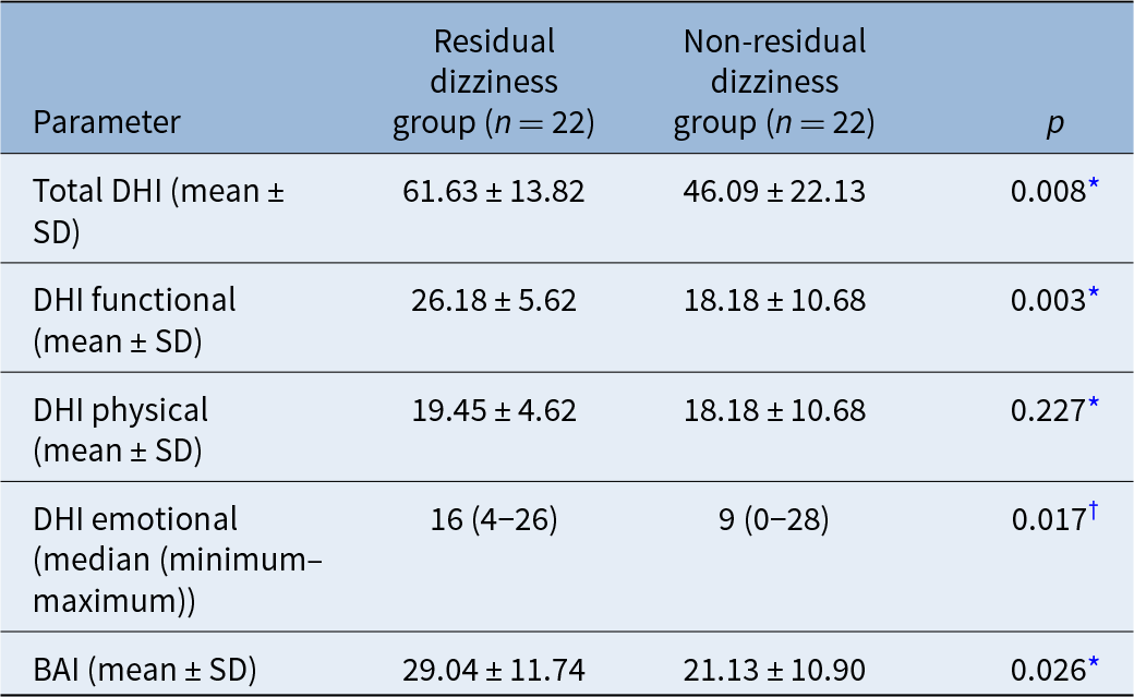
There were no differences between the groups in terms of age, gender, co-morbidities and education status (p > 0.05; Table 1). The residual dizziness group had significantly higher total Dizziness Handicap Inventory scores than the non-residual dizziness group (mean, 61.63 ± 13.82 vs 46.09 ± 22.13; p = 0.008). The functional (F) sub-dimension score was also significantly higher in the residual dizziness group than in the non-residual dizziness group (mean, 26.18 ± 5.62 vs 18.18 ± 10.68; p = 0.003), and the emotional (E) sub-dimension score was likewise higher (median, 16 (range: 4–26) vs 9 (0–28); p = 0.017). In contrast, no significant difference was observed in the physical sub-dimension (p = 0.227). In addition, the mean Beck Anxiety Inventory score was significantly higher in the residual dizziness group than in the non-residual dizziness group (mean, 29.04 ± 11.74 vs 21.13 ± 10.90; p = 0.026). These results are presented in Table 2.
Age and gender distribution by groups

* Student’s t-test. †Fisher exact test. ‡Chi-square test. SD = standard deviation
DHI, BAI and benign paroxysmal positional vertigo duration by groups

* Student t-test; †Mann–Whitney U test. DHI = Dizziness Handicap Inventory; BAI = Beck Anxiety Inventory; SD = standard deviation
The mean Beck Anxiety Inventory score of the participants was 25.09 ± 11.89, the mean E-Dizziness Handicap Inventory score was 13.18 ± 7.99 and the median F-Dizziness Handicap Inventory score was 24.00 (range: 2–36). There was a strong positive correlation between the Beck Anxiety Inventory and F-Dizziness Handicap Inventory scores (p < 0.001, r = 0.65) and between the Beck Anxiety Inventory and E-Dizziness Handicap Inventory scores (p < 0.001, r = 0.73).
Among the posterior canal-BPPV patients, 25 (56.8 per cent) had right-sided involvement, 16 (36.4 per cent) had left-sided involvement and 3 (6.8 per cent) had bilateral involvement. Of these patients, 42 (95.5 per cent) had the canalolithiasis type, while 2 (4.5 per cent) had the cupulolithiasis type. There was no significant difference between the groups in terms of the affected side or posterior canal-BPPV type (p > 0.05). Additionally, the number of canalith repositioning manoeuvres performed did not differ between residual dizziness and non-residual dizziness individuals (p > 0.05). However, BPPV duration was significantly longer in the residual dizziness group compared with the non-residual dizziness group (median, 20 days (range: 2–150) vs 7 days (range: 1–360); p = 0.003]. The affected side, BPPV type, number of manoeuvres and BPPV duration according to the groups are presented in Table 3.
The affected side, BPPV type, number of manoeuvres and BPPV duration according to the groups

* Chi-square test; †Mann–Whitney U test; ‡Fisher exact test. BPPV = benign paroxysmal positional vertigo
There was no difference between the groups in terms of single-task timed up and go and single task digit span scores (p > 0.05). Single-task timed up and go and single task digit span scores by group are presented in Table 4.
Single-task timed up and go and single task digit span scores by groups

* Mann–Whitney U-test.
There was no difference between the groups in terms of timed up and go-motor, timed up and go-forward, and timed up and go-backward scores (p > 0.05). Dual task performances by group are presented in Figure 1.
Dual task performances by group. TUG = timed up and go; RD = residual dizziness.

There was no significant difference between the groups in terms of Stroop 1, Stroop 3 and Stroop 5 scores (p > 0.05). However, Stroop 2 completion times were significantly longer in the residual dizziness group compared with the non-residual dizziness group (median, 15.14 (10.03–50.35) vs 11.94 (7.87–22.63); p < 0.031). Stroop 4 scores were worse in the residual dizziness group than in the non-residual dizziness group (median, 26.86 (16.64–54.14) vs 22.47 (11.55–48.08); p < 0.046). Stroop test scores by group are presented in Figure 2.
Stroop test-Scientific and Technological Research Council of Turkey-Basic Sciences Research Group (TBAG) form scores by group. RD = residual dizziness.

Discussion
This study investigated cognitive function and dual-task balance performance in individuals with and without residual dizziness after a successful canalith repositioning manoeuvre. Our findings reveal that individuals with residual dizziness had worse Stroop test scores. However, there were no differences between the groups in terms of digit span and dual-task performance.
The pathophysiology of residual dizziness remains unclear, but several hypotheses have been proposed in recent years.Reference Giommetti, Lapenna, Panichi, Mobaraki, Longari and Ricci1 Di Girolamo et al.Reference Di Girolamo, Ottaviani, Scarano, Picciotti and Di Nardo23 suggested that small otoconial remnants may persist in the semicircular canals following the canalith repositioning manoeuvre. Although these remnants may not stimulate the cupula enough to produce nystagmus, they could cause mild dizziness in patients. Another study investigating the effects of the Epley manoeuvre performed using a TRV chair on residual dizzinessReference Soylemez, Bolat, Karakoc, Can, Basak and Aydogan14 indicated that the correct application of the canalith repositioning manoeuvre could influence residual dizziness, supporting the hypothesis proposed by Di Girolamo et al.
Some other studies have associated residual dizziness with otolith dysfunction.Reference Suh, Oh, Chae, Lee, Chang and Mun12, Reference Seo, Shiraishi, Kobayashi, Mutsukazu, Fujita and Saito13, Reference von Brevern, Schmidt, Schönfeld, Lempert and Clarke24 Von Brevern et al.Reference von Brevern, Schmidt, Schönfeld, Lempert and Clarke24 reported that, after a successful canalith repositioning manoeuvre, individuals with BPPV exhibited smaller torsional otolith-ocular reflex responses compared with the control group. The authors suggested that residual dizziness could be attributed to otolith dysfunction resulting from utricular macular degeneration. Seo et al.Reference Seo, Shiraishi, Kobayashi, Mutsukazu, Fujita and Saito13 performed ocular vestibular evoked myogenic potential testing on individuals who underwent the canalith repositioning manoeuvre and found a link between residual dizziness and utricular dysfunction. Suh et al.Reference Suh, Oh, Chae, Lee, Chang and Mun12 proposed that the relationship between residual dizziness and otolith dysfunction occurs via osteopenia.
The Dizziness Handicap Inventory is a widely used psychometric assessment tool for evaluating the level of disability in individuals with dizziness. Through its three sub-dimensions, this inventory assesses the emotional, physical and functional disability caused by dizziness. The degree of disability induced by dizziness is subjective and is associated with an individual’s levels of fear and anxiety.Reference Omara, Basiouny, Shabrawy and Shafei25 Martellucci et al.Reference Martellucci, Pagliuca, de Vincentiis, Greco, De and Nobili26 reported that the F-Dizziness Handicap Inventory and E-Dizziness Handicap Inventory scores are related to residual dizziness, and that these two subscale scores could be used to predict the development of residual dizziness in BPPV patients after the canalith repositioning manoeuvre. Similarly, our study demonstrated that posterior canal-BPPV patients with higher F-Dizziness Handicap Inventory and E-Dizziness Handicap Inventory scores had a greater risk of developing residual dizziness.
The E-Dizziness Handicap Inventory subscale investigates the anxiety and insecurity experienced by patients as a result of dizziness, whereas the F-Dizziness Handicap Inventory subscale examines changes in daily life functionality caused by dizziness and fear of falling. Both functionally and emotionally driven impairments are closely linked to an individual’s psychological state. The strong positive correlation found in our study between Beck Anxiety Inventory scores and both F-Dizziness Handicap Inventory and E-Dizziness Handicap Inventory scores supports this assumption. Therefore, in the present study, anxiety may have played a significant role in the relationship between F-Dizziness Handicap Inventory and E-Dizziness Handicap Inventory scores and residual dizziness.
There is a bidirectional relationship between anxiety and dizziness. While symptoms caused by dizziness can increase individuals’ anxiety levels, disorders such as anxiety can also lead to dizziness by affecting the central vestibular system. The best-known example of this relationship is persistent postural perceptual dizziness.Reference Staab, Eckhardt-Henn, Horii, Jacob, Strupp and Brandt27 The hypothalamus and amygdaloid nuclei, which play a crucial role in the formation of sensory responses, are connected to the vestibular nuclei via the parabrachial nucleus,Reference Balaban28, Reference Herbert, Moga and Saper29 therefore psychiatric disorders may contribute to dizziness.
Teggi et al.Reference Teggi, Giordano, Bondi, Fabiano and Bussi11 reported that there was no relationship between residual dizziness and gender, affected canal and the number of canalith repositioning manoeuvre applications in their study investigating the cause of residual dizziness. However, increased age, symptom duration and anxiety levels were associated with a higher risk of residual dizziness. The authors concluded that residual dizziness is linked to anxiety and that long-term BPPV increases anxiety levels. Similarly, in our study, posterior canal-BPPV patients with higher pre-canalith repositioning manoeuvre anxiety levels had a greater risk of developing residual dizziness. Anxiety may therefore play a significant role in the development of residual dizziness, and assessing anxiety levels in BPPV patients may help to predict residual dizziness following a successful canalith repositioning manoeuvre.
Findings from studies investigating the effects of symptom duration and age in patients with residual dizziness are conflicting. Seo et al.Reference Seo, Shiraishi, Kobayashi, Mutsukazu, Fujita and Saito13 reported that residual dizziness was not associated with age, gender, BPPV duration or the affected side. Suh et al.Reference Suh, Oh, Chae, Lee, Chang and Mun12 stated that residual dizziness was not related to age, diabetes mellitus, hypertension or dyslipidaemia. However, the authors found a relationship between residual dizziness and BPPV duration.
Teggi et al.Reference Teggi, Giordano, Bondi, Fabiano and Bussi11 identified age over 72 years and BPPV duration more than 9 days as risk factors for residual dizziness in individuals with BPPV. Our study showed that residual dizziness in posterior canal-BPPV patients was not related to age, gender, affected side, number of canalith repositioning manoeuvre applications, education level, diabetes mellitus, hypertension or dyslipidaemia. However, residual dizziness risk was higher in individuals with prolonged BPPV duration.
The relationship between residual dizziness and BPPV duration may be explained by the compensation mechanism of the central vestibular system. The central vestibular system attempts to compensate for abnormal peripheral vestibular inputs. In individuals with BPPV, displaced otoconia create abnormal excitations or inhibitions in the semicircular canals. As the duration of BPPV increases, the central vestibular system may develop an adaptation to these abnormal inputs through compensation. After a successful canalith repositioning manoeuvre, this process is reversed. That is, the central vestibular system, which has adapted to abnormal inputs, may struggle to adjust to normal inputs.Reference Prokopakis, Lachanas, Christodoulou and Velegrakis30
Cognitive function is a broad term that refers to mental processes such as memory, attention, language skills and learning. Recent studies have provided evidence of a relationship between the vestibular system and cognitive functions. Bhattacharyya et al.Reference Bhattacharyya, Barman and Antony31 investigated cognitive abilities in individuals with BPPV and Ménière’s disease. The authors reported that individuals with BPPV and Ménière’s disease experienced more difficulties in quick thinking, decision-making and concentration compared with the control group. Another cohort studyReference Kim, Yoo, Min and Choi32 found that individuals over the age of 60 years with BPPV had a higher risk of developing neurodegenerative dementia.
Although the exact mechanism linking the vestibular system and cognitive functions has not been fully established, several hypotheses have been proposed. One such hypothesis suggests that BPPV and cognitive decline may share a similar underlying mechanism.Reference Bhattacharyya, Barman and Antony31 In BPPV patients, neuronal degeneration occurs in the saccular macula, as well as cell loss in the saccular ganglion and the superior and inferior vestibular nerve ganglion cells.Reference Gacek33, Reference Yang, Kim, Lee and Lee34 The vestibular system is the first sensory system to develop phylogenetically, and in dementia patients, neuronal degeneration has been shown to affect evolutionarily older neurons earlier.Reference Bhattacharyya, Barman and Antony31, Reference Nandi and Luxon35 Thus, it is possible that BPPV in older individuals may be associated with neuronal cell loss or could reflect an early stage of dementia. However, the current evidence is limited and longitudinal studies are needed to clarify this potential link.
There are a limited number of studies investigating the cognitive functions of individuals with residual dizziness.Reference Sun, Ma, Yang, He, Wang and Shen15 Sun et al.Reference Sun, Ma, Yang, He, Wang and Shen15 evaluated the disability and anxiety levels of patients with BPPV and applied the canalith repositioning manoeuvre to them. One week later, they divided the participants into two groups: those with residual dizziness and those without residual dizziness. They assessed the cognitive status of both groups using the Mini-Mental State Examination. The authors reported that individuals with higher levels of anxiety and disability had a greater risk of developing residual dizziness compared with those with lower levels. However, they found no difference in Mini-Mental State Examination scores between individuals with and without residual dizziness. As a result, the authors concluded that BPPV is a temporary condition and does not affect cognitive functions or that Mini-Mental State Examination may not fully reflect the cognitive impairment associated with dizziness in individuals with residual dizziness.
The Mini-Mental State Examination is a widely used screening test for detecting cognitive impairments.Reference Carnero-Pardo36 However, the test includes a limited number of questions evaluating different cognitive domains, particularly those assessing memory and executive functions, therefore its ability to distinguish mild cognitive impairments and dementia is debated.Reference Carnero-Pardo36
In our study, we used a methodology similar to that of Sun et al. We applied the canalith repositioning manoeuvre to patients with BPPV and assessed their residual dizziness status one week later. We divided the individuals into two groups, those with residual dizziness and those without residual dizziness, and compared their cognitive functions. Unlike the study by Sun et al.,Reference Sun, Ma, Yang, He, Wang and Shen15 we used the Stroop test and the digit span test to assess cognitive functions. The Stroop test is considered the ‘gold standard’ for evaluating attentionReference Macleod37 and assesses information processing speed and automatic and parallel processing in cognitive tasks. The digit span test is a useful test for evaluating short-term memory and working memory. The present study showed that individuals in the residual dizziness group had worse Stroop 2 and Stroop 4 scores compared with those in the non-residual dizziness group. Although individuals in the residual dizziness group had lower backward digit span scores than those in the non-residual dizziness group, the difference was not statistically significant. In conclusion, our findings revealed that individuals who developed residual dizziness after a successful canalith repositioning manoeuvre exhibited poorer cognitive performance (Stroop test) compared with those who did not develop residual dizziness.
There are reciprocal pathways between the central vestibular system and central structures responsible for cognitive functions. Central vestibular degeneration caused by vestibular dysfunction may therefore lead to neuronal atrophy and cell loss in structures such as the hippocampus, which are responsible for cognitive functions.Reference Guo, Wang, Liang, Tian, Liu and Guo38 Looking at this mechanism from the opposite perspective, the impairment of cognitive structures may affect the central vestibular system, potentially leading to dizziness. Thus, the development of residual dizziness after a successful canalith repositioning manoeuvre may result from the interaction between the cognitive and central vestibular systems. However, further studies are needed that incorporate BPPV, cognitive functions, residual dizziness and the central vestibular system.
Functional mobility is essential for individuals to perform daily living activities and participate in society. However, mobility often involves the simultaneous execution of two tasks. These tasks typically include a secondary motor or cognitive task in addition to a primary motor task. The ability to perform these tasks concurrently is crucial for maintaining independence. However, any sensory impairment or cognitive decline that disrupts motor performance and postural stability may restrict functional movement. Such conditions can limit individuals’ daily activities, increase the risk of falls and contribute to a fear of falling.Reference Brustio, Magistro, Zecca, Rabaglietti and Liubicich39 Assessing dual-task performance is therefore important for understanding an individual’s functional mobility. Dual-task tests simulate real-life situations and evaluate motor performance.Reference Brustio, Magistro, Zecca, Rabaglietti and Liubicich39
To the best of our knowledge, no studies in the literature have investigated dual-task performance in individuals with residual dizziness. Some studies have assessed single-task balance skills in individuals with residual dizziness.Reference Assal, Morsy, Almagassbi and Eldeeb40 Assal et al.Reference Assal, Morsy, Almagassbi and Eldeeb40 examined the sensory organisation test results of BPPV patients before and after the canalith repositioning manoeuvre. The authors reported that individuals without residual dizziness showed improvements in sensory organisation test values following a successful canalith repositioning manoeuvre. However, they found that sensory organisation test values did not improve in patients with residual dizziness. Similarly, Di Girolamo et al.Reference Di Girolamo, Ottaviani, Scarano, Picciotti and Di Nardo23 suggested that despite a successful canalith repositioning manoeuvre, small otoconia may remain in the semicircular canals of BPPV patients, which could explain why postural control does not fully recover in these individuals.
We assessed individuals’ dynamic functional balance skills using the timed up and go test. The timed up and go test is a functional assessment with high test–retest reliability that evaluates walking and dynamic balance abilities without requiring specialised equipment.Reference Kear, Guck and McGaha41 It has been reported to provide significant insights, particularly in predicting fall risk in individuals with lower functional capacity.Reference Kear, Guck and McGaha41 To evaluate participants’ dual-task performance, we administered additional motor and cognitive tasks alongside the timed up and go test. Although the single-task timed up and go score of the residual dizziness group was worse than that of the non-residual dizziness group, the difference was not statistically significant.
Similarly, the dual-task performance scores (timed up and go-motor, timed up and go-backward, timed up and go-forward) of the residual dizziness group were lower than those of the non-residual dizziness group, but again, the differences were not statistically significant. These findings suggest that residual dizziness, which is characterised by relatively mild lightheadedness symptoms, does not significantly impact timed up and go performance or timed up and go-based dual-task performance. On the other hand, despite the lack of statistical significance, the fact that residual dizziness patients had worse scores in both single-task and dual-task timed up and go tests compared with non-residual dizziness patients may indicate that the timed up and go test lacks the sensitivity to detect such subtle symptoms.
Future studies should assess dual-task performance in individuals with residual dizziness using postural tasks combined with motor or cognitive tasks through computerised dynamic post-urography and similar objective measurement systems
Limitations
First, although the sample size was deemed statistically adequate based on the power analysis, its relatively small number may limit the generalisability of the findings. Second, the cross-sectional design of the study does not allow for establishing a causal relationship between residual dizziness and cognitive functions. Longitudinal studies are required to clarify the directionality and temporal sequence of this relationship. Third, only a single follow-up assessment was conducted one week after the canalith repositioning manoeuvre. This limited follow-up period may not adequately capture the long-term course of residual dizziness, its natural resolution or the persistence of symptoms. Future research with larger cohorts, longitudinal designs and extended follow-up periods would provide a more comprehensive understanding of the prognosis of residual dizziness, the persistence of symptoms and the response to treatment.
• Although patients with benign paroxysmal positional vertigo (BPPV) typically recover easily with the canalith repositioning manoeuvre, some individuals develop residual dizziness following treatment
• Most studies in the literature have focused on the causes of residual dizziness, identifying factors such as anxiety levels and the duration of BPPV as potential risk factors
• This study is the first to examine the effects of residual dizziness, emerging after a successful canalith repositioning manoeuvre, on cognitive functions and dual-task performance
• Individuals with residual dizziness performed worse on the Stroop test compared with those without residual dizziness
• The findings suggest a bidirectional relationship in which residual dizziness may impair cognition, while pre-existing cognitive deficits may increase susceptibility to residual dizziness
Conclusion
Prolonged BPPV duration, high anxiety levels and increased disability in individuals with BPPV may elevate the risk of residual dizziness. Patients with residual dizziness demonstrated poorer Stroop test performance compared with those without residual dizziness. However, no significant difference was observed between the groups in terms of dual-task performance. Our findings suggest a strong interaction between cognitive functions and the vestibular system. While residual dizziness may impair cognitive performance, particularly in executive functions, it is also possible that pre-existing cognitive deficits contribute to the development of residual dizziness.
Data availability statement
The data collected and presented in the current study are not available publicly or by request because of privacy and ethical concerns. Further enquiries can be directed to the corresponding author.
Competing interests
The authors declare no conflicts of interest.